International Journal of
Reproductive Biomedicine
Mon, Oct 19, 2026
[Archive]
Volume 24, Issue 8 (August 2026)
IJRM 2026, 24(8): 721-738 |
Back to browse issues page



Ethics code: IR.SSU.SPH.REC.1400.165


Download citation:
BibTeX | RIS | EndNote | Medlars | ProCite | Reference Manager | RefWorks
Send citation to:


BibTeX | RIS | EndNote | Medlars | ProCite | Reference Manager | RefWorks
Send citation to:
Zamani E, Kamali K, Seifi Ashkezari A A, Sodaizadeh H, Yaeghoobi M, Haghiralsadat B F. Comparative study of niosomal nanoparticles encapsulating Artemisia aucheri and Artemisia sieberi extracts on Michigan Cancer Foundation-7 breast cancer cell proliferation: An in vitro study. IJRM 2026; 24 (8) :721-738
URL: http://ijrm.ir/article-1-3556-en.html
URL: http://ijrm.ir/article-1-3556-en.html
Elaheh Zamani1 

 , Kazem Kamali1 , Ali Akbar Seifi Ashkezari2 , Hamid Sodaizadeh1 , Marzieh Yaeghoobi3 , Bibi Fatemeh Haghiralsadat *4
1- Natural Resources Faculty, Yazd University, Yazd, Iran.
2- Iran University of Medical Sciences, Tehran, Iran.
3- Biotechnology Research Center, Yazd Reproductive Sciences Institute, Shahid Sadoughi University of Medical Sciences, Yazd, Iran.
4- Biotechnology Research Center, Yazd Reproductive Sciences Institute, Shahid Sadoughi University of Medical Sciences, Yazd, Iran. & School of Advanced Medical Technologies, Shahid Sadoughi University of Medical Sciences, Yazd, Iran. ,fhaghirosadat@gmail.com; fhaghirosadat@ut.ac.ir
2- Iran University of Medical Sciences, Tehran, Iran.
3- Biotechnology Research Center, Yazd Reproductive Sciences Institute, Shahid Sadoughi University of Medical Sciences, Yazd, Iran.
4- Biotechnology Research Center, Yazd Reproductive Sciences Institute, Shahid Sadoughi University of Medical Sciences, Yazd, Iran. & School of Advanced Medical Technologies, Shahid Sadoughi University of Medical Sciences, Yazd, Iran. ,
Keywords: Niosomes, Nanoparticles, Plant preparations, Breast neoplasms, Cell line, Tumor, Drug delivery systems.
Full-Text [PDF 3368 kb]
(219 Downloads)
| Abstract (HTML) (271 Views)
3.3. FTIR results analysis
Full-Text: (41 Views)
1. Introduction
Cancer is recognized as a major public health challenge worldwide and is the second leading cause of death. Common treatments for cancer include surgery, chemotherapy, radiotherapy, targeted therapy, immunotherapy, and hormonal therapy (1). Breast cancer is the most common type of cancer among women worldwide, with approximately one in 8 women developing the disease during their lifetime (2). Due to various strategies, the overall survival rate of breast cancer patients has increased. However, challenges such as tumor growth, drug resistance, and tumor heterogeneity remain important factors that can prevent a complete cure. Combination therapy, using 2 or more therapeutic agents with different mechanisms, is a fundamental strategy to overcome drug resistance, reduce toxicity, and achieve synergistic effects. This approach is particularly relevant when integrating natural bioactive compounds with nanotechnology-based delivery systems (3). The human breast cancer cell line Michigan Cancer Foundation-7 (MCF-7) is frequently used to investigate potential therapeutic agents. MCF-7 provides a valuable model for studying hormone-responsive breast cancer and evaluating novel anticancer treatments. Due to the drawbacks of conventional chemotherapy, it is imperative to seek alternative and effective treatments (4).
Medicinal plants containing natural bioactive compounds with antioxidant and cytotoxic properties have shown significant potential. The antioxidant capacity of plant extracts is crucial as it can mitigate oxidative stress, a key contributor to cancer progression, and may enhance the efficacy of chemotherapeutic agents (5). Artemisia is one of the largest genera in the Asteraceae family, comprising 200 to more than 500 species (6). These species produce numerous compounds, including flavonoids, alkaloids, phenols, quinones, terpenoids, and coumarins, which exhibit diverse biological activities such as anticancer, antidepressant, antidiabetic, hepatoprotective, antihypertensive, and antiparasitic effects (6, 7). Among them, A. aucheri and A. sieberi are known for their bioactive potential, yet a comparative analysis of their antioxidant and anticancer efficacy, especially when delivered via nanocarriers, remains underexplored.
Nanotechnology is the latest effort to develop and target many cancers. The small size of delivery systems allows them to pass through weak blood vessels, thus increasing drug potency. Modern delivery systems include polymeric micelles, liposomes, nanodispersions, nanorods, dendrimers, and cyclodextrins. The main role of these systems is to provide controlled and sustained drug release with longer circulation times and reduced complications related to drug resistance. In addition, nanotechnology encapsulation enhances solubility, targeted delivery, and stabilization of the physicochemical state, while preventing drug elimination by excessive immune system activity. Among these, niosomes made of nonionic surfactants such as tween 80 (Polysorbate 80) have shown great potential as drug carriers. Compared to other vesicles, such as liposomes, niosomes have improved bioavailability, greater stability, higher encapsulation efficiency, low toxicity due to their nonionic nature, and lower preparation cost (8).
This study aimed to investigate and compare the antioxidant properties of A. aucheri and A. sieberi extracts obtained by different methods (Maceration and Soxhlet) and solvent concentrations, as well as to develop, characterize, and evaluate the cytotoxic effect of optimized niosomal nanoparticles containing these extracts on the MCF-7 breast cancer cell line and the non-tumorigenic MCF-10A cell line.
2. Materials and Methods
2.1. Study design
This in vitro study was conducted at Shahid Sadoughi University of Medical Sciences, Yazd, Iran, from 2023-2024. In addition to the preparation of A. aucheri and A. sieberi plant extracts, the following materials were used: cholesterol (Sigma-Aldrich, ≥ 95% purity), phosphate-buffered saline (PBS) (Sigma-Aldrich, cell culture grade), 3-(4, 5-dimethylthiazol-2-yl)-2,5-diphenyltetrazolium bromide (MTT) powder (Sigma-Aldrich, ≥ 98% purity), dimethyl sulfoxide (Sigma-Aldrich, ≥ 99.9% purity), polysorbate 80 (tween 80, nonionic surfactant) and solvent chloroform were obtained from Merck. Dulbecco’s modified Eagle’s culture medium, fetal bovine serum, and penicillin-streptomycin were obtained from Gibco. MCF-7 and MCF-10A cells, in the form of 2 separated 75 ml cell flasks containing approximately 9 × 10⁵ cells, were also obtained from the Pasteur Institute of Iran cell bank.
2.2. Experiment steps
The aerial parts of A. aucheri and A. sieberi were collected from Yazd province, Iran. The plant species were authenticated by a botanist at the Herbarium of Yazd University. The plant material was shade-dried at room temperature and ground into a fine powder using an electric grinder. Hydroalcoholic extraction containing ethanol from the Artemisia plant was performed using the Soxhlet and Maceration methods. For both methods, 50 gr of the obtained dried Artemisia powder was used with 250 ml of a hydroalcoholic solution (water and ethanol at varying concentrations of 0%, 60%, and 80% v/v ethanol). For the Soxhlet method, the powder was placed in a Soxhlet apparatus. The extraction process was carried out for 6 hr under reflux. After the end of the process, the resulting extract was separated with a paper filter, and the solvent was evaporated using a rotary evaporator at 40°C.
For the Maceration method, the powder was soaked in the hydroalcoholic solution for 14 days at room temperature with occasional shaking. The mixture was then filtered through filter paper. Finally, the solvent was evaporated in an oven at 40°C. The dried extracts were stored in dark glass containers at 4°C until further use.
2.3. Standard curve plotting
A spectrophotometer (Epoch, BioTek, USA) was used to determine the maximum absorption wavelength (λmax) and plot standard curves. Standard solutions of A. aucheri and A. sieberi extracts were prepared in isopropanol and PBS, pH 7.4, across a concentration range of 1-100 µg/mL. Absorbance was measured at the respective λmax, and calibration curves were plotted (absorbance vs. concentration) with R² > 0.99. All measurements were performed in triplicate.
2.4. Antioxidant properties investigation
In this method, first the plant extract was prepared, and then the reaction was carried out using Folin-Ciocalteu reagent and sodium carbonate. Optical absorption was measured at a wavelength of 750 nm, and the total phenolic content was calculated in terms of milligrams of gallic acid equivalent per gram of dry extract (mg GA/g DE). Also, the total flavonoid content was measured using the aluminium chloride method, and the results were reported as milligrams of quercetin equivalent per gram of dry extract (mg QE/gr DE).
2.5. 2,2-diphenyl-1-picrylhydrazyl (DPPH) free radical assay
The antioxidant potential was investigated using the DPPH free radical assay. A stock solution of each dried extract was prepared at 1 mg/mL in methanol. Different concentrations (10-100 µg/mL) of the extract (50 µL) were mixed with 450 µL of a 0.1 mM DPPH solution in methanol. The mixture was vortexed and incubated in the dark for 20 min at room temperature. The reduction of DPPH free radicals was measured at a wavelength of 517 nm. A standard curve of quercetin (5-50 µM) was used for comparison, and the results were expressed as percentage inhibition.
2.6. 2,2'-azino-bis (3-ethylbenzothiazoline-6-sulfonic acid) (ABTS) test
The ABTS radical cation (ABTS•⁺) was generated by reacting 7 mM ABTS solution with 2.45 mM potassium persulfate and allowing the mixture to stand in the dark for 12-16 hr. The ABTS•⁺ solution was diluted with ethanol to an absorbance of 0.70 ± 0.02 at 734 nm. Then, 50 µl of the diluted extract or ascorbic acid standard (100-700 µM) was added to 1 ml of the ABTS•⁺ working solution. The mixture was vortexed and incubated at 28°C for 60 min. The absorbance was recorded at 734 nm.
2.7. Cupric reducing antioxidant capacity (CUPRAC) test
The antioxidant test using the CUPRAC method is based on the technique developed by Irawan and colleagues (9). This work was carried out on crude hydroalcoholic extracts from different treatments obtained during the extraction process. Sample concentrations ranging 40-160 mg/L were prepared from a stock solution with a concentration of 1000 mg/L of hydroalcoholic extract. Each solution was placed in five 5 mL flasks, and then 1 mL of CUPRAC solution was added to it. After that, each solution was homogenized with alcohol. The solution was incubated for 30 min at 37°C, and then the light absorbance of the solution was measured at a wavelength of 459 nm using an optical spectrophotometer.
2.8. Ferric reducing antioxidant power (FRAP) test
The ability of the extracts to reduce ferric ions was estimated using a modified version of the Oyaizu method. The FRAP reagent was prepared immediately before use by mixing 0.3 M acetate buffer (pH 3.6), 10 mM 2,4,6-tripyridyl-s-triazine in 40 mM hydrochloric acid, and 20 mM FeCl3·6H2O solution in a 10:1:1 volume ratio. Then, 100 µL of the extract was combined with 3 mL of the freshly prepared FRAP reagent. The mixture was vortexed and incubated at 37°C in a water bath for 30 min. After incubation, the absorbance of the solution was measured at 593 nm using a spectrophotometer. A standard curve was constructed using known concentrations of FeSO4·7H2O, and the results were calculated and expressed as micromoles of Ferrous (Fe2+) equivalent per gram of dry extract (µmol Fe2+/gr extract).
2.9. Synthesis and optimization of niosomal nanoparticles
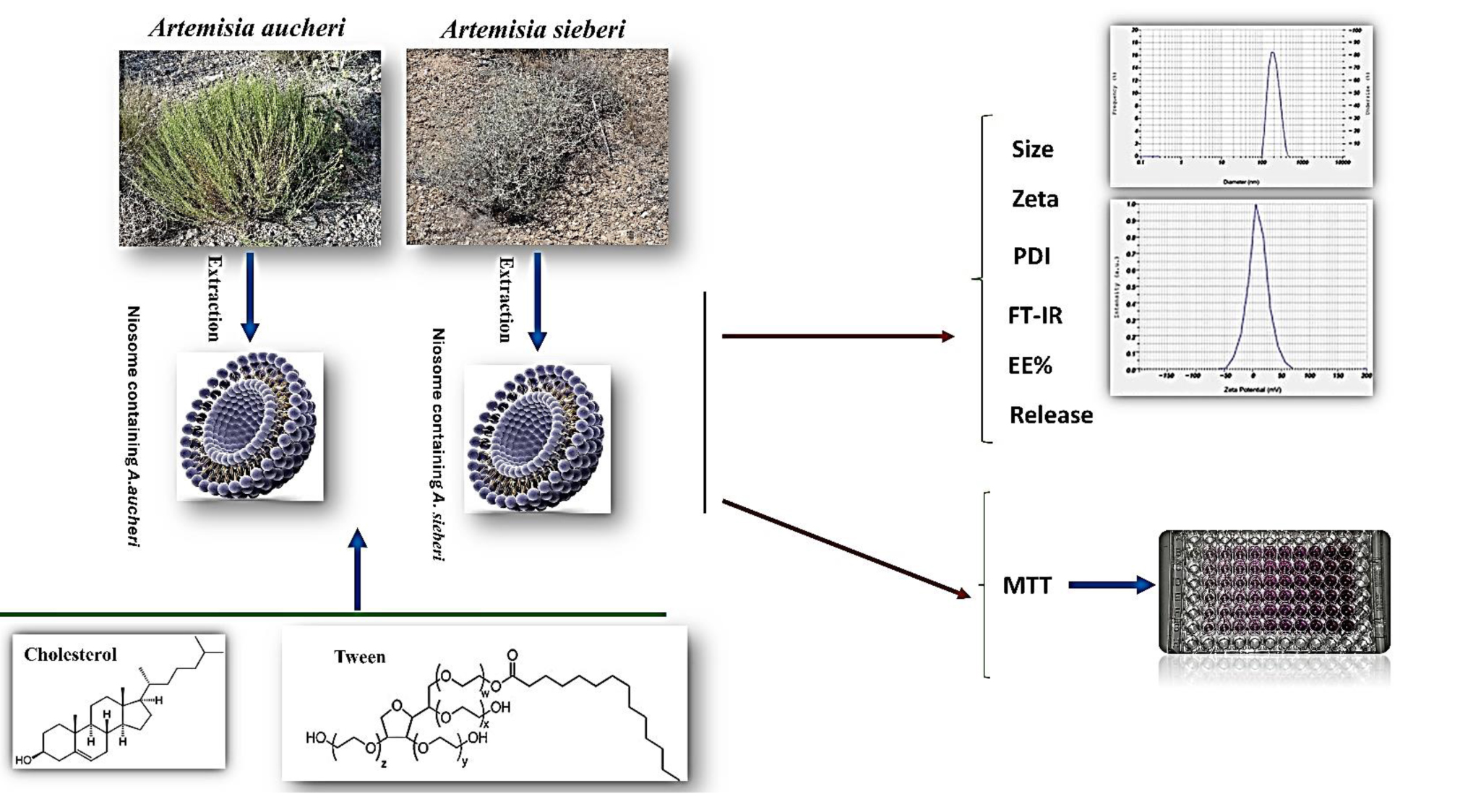
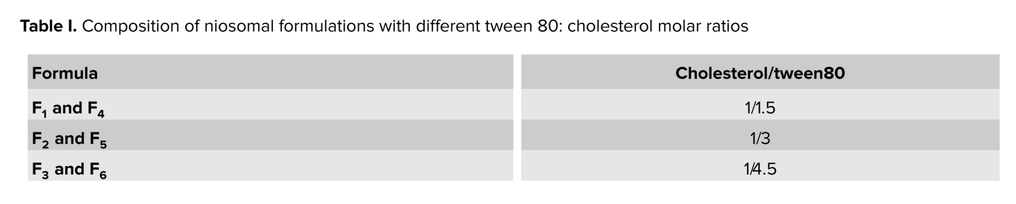
Niosomes were prepared using the thin-film hydration method as follows: cholesterol and the nonionic surfactant tween 80 were dissolved in 10 mL of chloroform in a round-bottom flask at different molar ratios (Table I). The organic solvent was evaporated under reduced pressure at 45°C and 150 rpm for 45 min using a rotary evaporator (Heidolph, Germany), resulting in a thin lipid film on the flask wall. The film was hydrated by adding 5 mL of an aqueous phase containing a known concentration of the Artemisia extract (1 mg/mL) in sterile distilled water under continuous stirring at 50°C and 150 rpm for 30 min. The resulting niosomal suspension was then sonicated using a probe sonicator (Ultrasonic Homogenizer, 100 W) for 5 min to reduce vesicle size and achieve a homogeneous dispersion. In table I, formulations F1 to F3 relate to the niosomal system containing A. sieberi extract, while formulations F4-F6 pertain to the niosomal system containing A. aucheri extract.
2.10. Investigation of the encapsulation rate of niosomal nanoparticles
To determine the efficiency of drug encapsulation in different niosomal formulations, 100 µg of extract-containing nanoparticles were mixed with isopropyl alcohol at a volume ratio of 1:10 to destroy the niosomal membrane and release the loaded drug. Then, using an ultraviolet-visible spectrometer at a specific maximum wavelength, the amount of drug encapsulated in each formulation was determined. The percentage of drug encapsulation in different niosomal formulations was calculated using the following formula (10).

2.11. Kinetics of niosome release studies
Release studies were performed using the dialysis bag method. 2 mm of the niosomal formulation were placed in a cellulose acetate dialysis bag (molecular weight cut-off: 12 kDa). The bag was suspended in 100 mL of PBS, pH = 7.4, as the release medium in a graduated cylinder. The container was placed on a magnetic stirrer at 50 rpm, and the temperature was maintained at 37 ± 0.5°C. At predetermined time intervals (0.5, 1, 2, 3, 4, 5, 6, 7 hr), 1 mL of the release medium was withdrawn and replaced with an equal volume of fresh PBS to maintain sink conditions. The absorbance of the withdrawn samples was measured at the respective λmax using a UV-Vis spectrophotometer (Shimadzu, Japan), and the cumulative percentage of drug released was calculated (11).
2.12. Characterization of niosomal nanoparticles
The average size and size distribution of niosomes were measured using the dynamic light scattering technique using Zeta Sizer. Briefly, the niosomal formulations were diluted 1:100 with deionized water to avoid multiple diffraction phenomena due to particle interactions. Fourier-transform infrared spectroscopy (FTIR) analysis was performed to characterize the niosomes containing Artemisia extract. Niosome samples were placed directly on the attenuated total reflectance plate of the FTIR instrument. The instrument was set in the range of 4000 to 650 cm⁻¹ with a resolution of 4 cm⁻¹. The samples were exposed to infrared radiation for a few seconds, and the recorded absorption data were normalized and analyzed using the relevant software. Specific absorption regions representing the functional groups present in the niosomes and Artemisia extract were identified. This method allowed us to obtain detailed information about the chemical structure and interactions present in the niosomes and to investigate the changes caused by Artemisia extract loading.
2.13. Cell viability assay
The cytotoxic effects were evaluated on the MCF-7 breast cancer cell line and the non-tumorigenic MCF-10A cell line. Cells were seeded in 96-well plates at a density of 1 × 10⁴ cells per well and incubated for 24 hr at 37°C in a humidified atmosphere with 5% CO₂ to allow attachment. Subsequently, the culture medium was replaced with fresh medium containing various concentrations (0.5, 2.5, 12.5, 62.5, 312.5, 1562.5, and 7812.5 µg/mL) of the following test groups: 1) free extracts of A. sieberi and A. aucheri, 2) niosomal formulations containing the extracts (at equivalent extract concentrations), and 3) blank niosomes (without extract). After 48 hr of incubation, 20 µL of MTT solution (5 mg/mL in PBS) was added to each well, and the plates were incubated for another 4 hr. The formazan crystals formed were dissolved by adding 100 µL of dimethyl sulfoxide to each well. The absorbance was measured at 570 nm using a microplate reader (BioTek, USA). Cell viability was calculated as a percentage relative to untreated control cells. The IC₅₀ was determined using nonlinear regression analysis (GraphPad Prism 8).
2.14. Ethical Considerations
This research project was approved by the ethics committee of the School of Public Health, Shahid Sadoughi University of Medical Sciences, Yazd, Iran (Code: IR.SSU.SPH.REC.1400.165).
2.15. Statistical Analysis
In this study, 2 types of data were utilized. The numerical data consists of continuous measurements, which include variables such as [the rate of encapsulation and release of the system], presented as mean ± standard deviation. The categorical data comprises information that is divided into specific groups or categories. To analyze the data, the ANOVA test was employed to examine the differences in means among various groups. The statistical significance level for this study was set at 0.05. All statistical analysis were conducted using GraphPad Prism version 8. This software provides powerful tools for data analysis and scientific graphing.
3. Results
3.1. Investigation and analysis of the antioxidant properties of A. aucheri and A. sieberi
The antioxidant activities of the extracts, assessed by ABTS, CUPRAC, DPPH, and FRAP assays, are presented in figure 1. Overall, values ranged from 22.66-50.93 mg TE/g DE across all methods. For both species, Soxhlet extraction with 60% ethanol consistently yielded the highest activities. Under these optimal conditions, A. aucheri exhibited markedly higher values for ABTS, CUPRAC, DPPH, and FRAP (50.93, 47.54, 48.99, and 46.48, respectively) compared to A. sieberi (47.07, 43.75, 39.91, and 39.98, respectively).
Similarly, total phenolic content, total flavonoid content, and extraction yield (Figure 2) ranged from 17-40 mg GAE/g DE, 2.8-13.9 mg QE/g DE, and 13-25%, respectively. The highest values for all 3 parameters were again obtained with Soxhlet extraction using 60% ethanol, with A. aucheri (40.05, 13.9, and 25.2%) outperforming A. sieberi (37.26, 10.16, and 19.65%).
3.2. Physicochemical characterization and encapsulation performance of niosomal nanoparticles
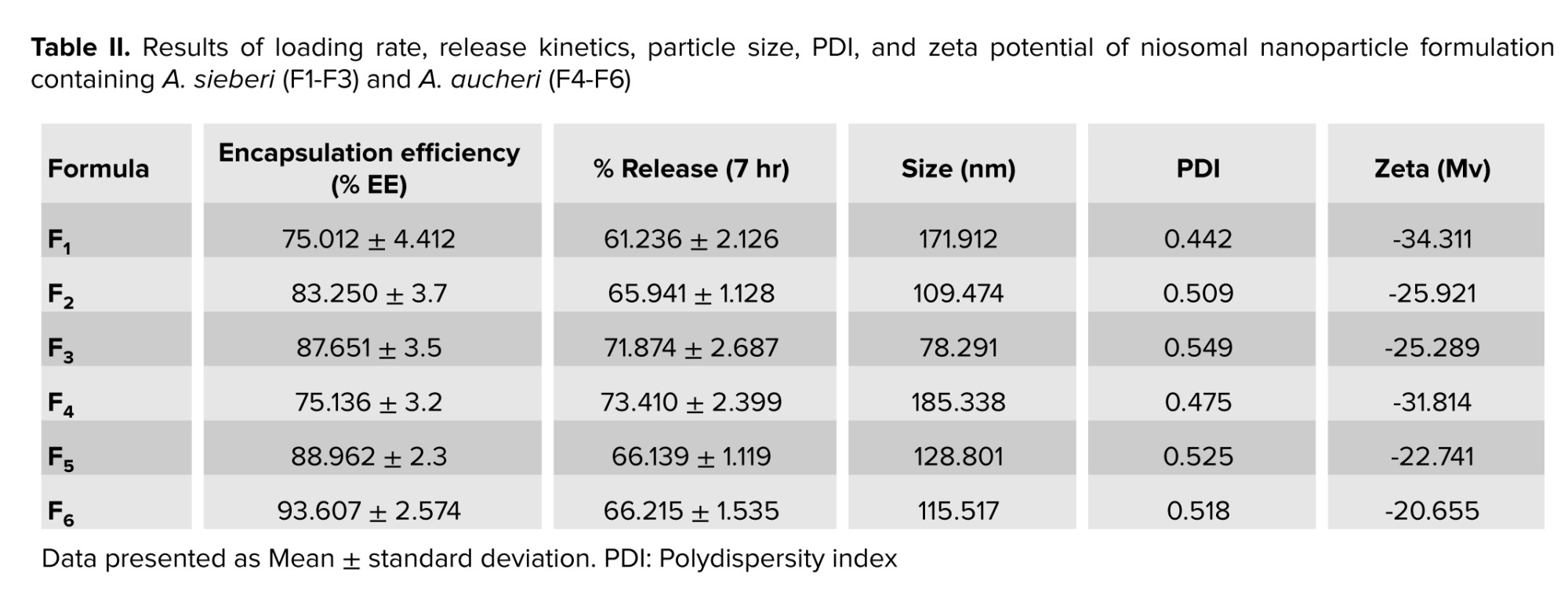
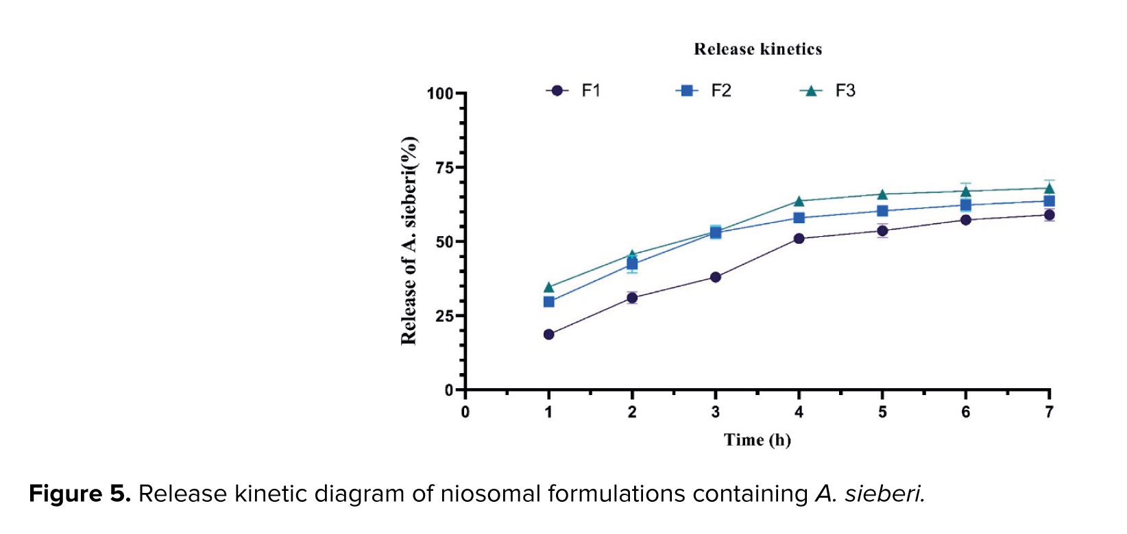
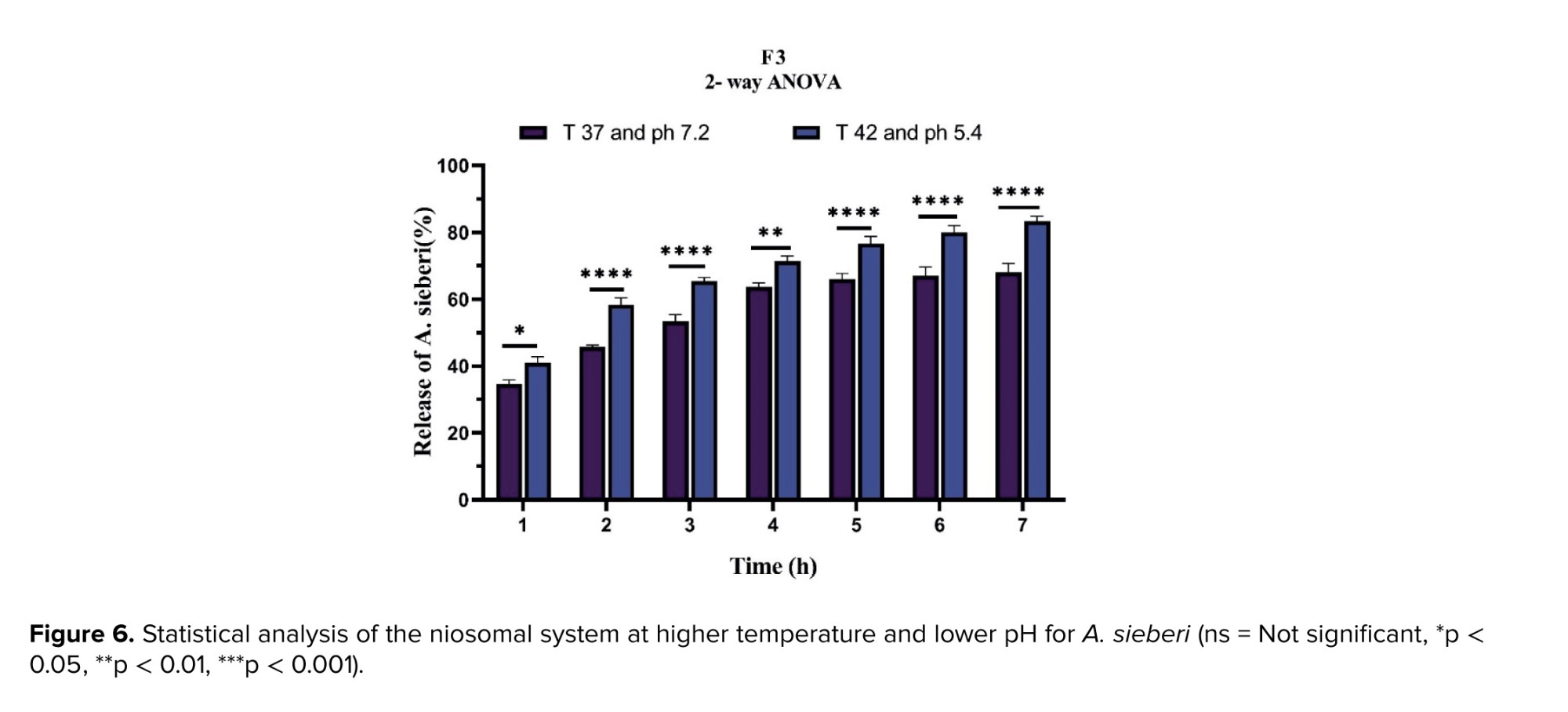
The maximum absorption wavelengths were 356 nm for A. sieberi and 292 nm for A. aucheri (1 mg/mL). The tween-to-cholesterol ratio significantly influenced both the physicochemical properties and encapsulation performance of the niosomal formulations. For A. sieberi, increasing this ratio from F1-F3 reduced particle size from 171.9-78.29 nm, shifted zeta potential from 34.3 mV to -25.2 mV, and increased polydispersity index (PDI) from 0.442 to 0.549. Concurrently, uptake and release improved from 75% and 61% (F1) to 83% and 65% (F2), with F3 achieving the best overall performance at 87% uptake and 71% release. For A. aucheri, similar trends were observed: particle size decreased from 185.3 nm (F4) to 115.5 nm (F6), zeta potential changed from -31.8 mV to -20.6 mV, and PDI rose from 0.475 to 0.518. In terms of performance, F4 showed 75% uptake and 73% release, F5 increased uptake to 88% with 66% release, and F6 provided the highest entrapment (93%), while F4 remained superior for release (73%). Overall, higher tween-to-cholesterol ratios reduced particle size, altered surface charge, and broadened size distribution, while generally enhancing encapsulation efficiency. These findings indicate that formulation optimization can significantly tune nanoparticle stability and drug delivery performance. All experiments were performed in triplicate and analyzed by ANOVA (Figures 3-6 and Table II).
Cancer is recognized as a major public health challenge worldwide and is the second leading cause of death. Common treatments for cancer include surgery, chemotherapy, radiotherapy, targeted therapy, immunotherapy, and hormonal therapy (1). Breast cancer is the most common type of cancer among women worldwide, with approximately one in 8 women developing the disease during their lifetime (2). Due to various strategies, the overall survival rate of breast cancer patients has increased. However, challenges such as tumor growth, drug resistance, and tumor heterogeneity remain important factors that can prevent a complete cure. Combination therapy, using 2 or more therapeutic agents with different mechanisms, is a fundamental strategy to overcome drug resistance, reduce toxicity, and achieve synergistic effects. This approach is particularly relevant when integrating natural bioactive compounds with nanotechnology-based delivery systems (3). The human breast cancer cell line Michigan Cancer Foundation-7 (MCF-7) is frequently used to investigate potential therapeutic agents. MCF-7 provides a valuable model for studying hormone-responsive breast cancer and evaluating novel anticancer treatments. Due to the drawbacks of conventional chemotherapy, it is imperative to seek alternative and effective treatments (4).
Medicinal plants containing natural bioactive compounds with antioxidant and cytotoxic properties have shown significant potential. The antioxidant capacity of plant extracts is crucial as it can mitigate oxidative stress, a key contributor to cancer progression, and may enhance the efficacy of chemotherapeutic agents (5). Artemisia is one of the largest genera in the Asteraceae family, comprising 200 to more than 500 species (6). These species produce numerous compounds, including flavonoids, alkaloids, phenols, quinones, terpenoids, and coumarins, which exhibit diverse biological activities such as anticancer, antidepressant, antidiabetic, hepatoprotective, antihypertensive, and antiparasitic effects (6, 7). Among them, A. aucheri and A. sieberi are known for their bioactive potential, yet a comparative analysis of their antioxidant and anticancer efficacy, especially when delivered via nanocarriers, remains underexplored.
Nanotechnology is the latest effort to develop and target many cancers. The small size of delivery systems allows them to pass through weak blood vessels, thus increasing drug potency. Modern delivery systems include polymeric micelles, liposomes, nanodispersions, nanorods, dendrimers, and cyclodextrins. The main role of these systems is to provide controlled and sustained drug release with longer circulation times and reduced complications related to drug resistance. In addition, nanotechnology encapsulation enhances solubility, targeted delivery, and stabilization of the physicochemical state, while preventing drug elimination by excessive immune system activity. Among these, niosomes made of nonionic surfactants such as tween 80 (Polysorbate 80) have shown great potential as drug carriers. Compared to other vesicles, such as liposomes, niosomes have improved bioavailability, greater stability, higher encapsulation efficiency, low toxicity due to their nonionic nature, and lower preparation cost (8).
This study aimed to investigate and compare the antioxidant properties of A. aucheri and A. sieberi extracts obtained by different methods (Maceration and Soxhlet) and solvent concentrations, as well as to develop, characterize, and evaluate the cytotoxic effect of optimized niosomal nanoparticles containing these extracts on the MCF-7 breast cancer cell line and the non-tumorigenic MCF-10A cell line.
2. Materials and Methods
2.1. Study design
This in vitro study was conducted at Shahid Sadoughi University of Medical Sciences, Yazd, Iran, from 2023-2024. In addition to the preparation of A. aucheri and A. sieberi plant extracts, the following materials were used: cholesterol (Sigma-Aldrich, ≥ 95% purity), phosphate-buffered saline (PBS) (Sigma-Aldrich, cell culture grade), 3-(4, 5-dimethylthiazol-2-yl)-2,5-diphenyltetrazolium bromide (MTT) powder (Sigma-Aldrich, ≥ 98% purity), dimethyl sulfoxide (Sigma-Aldrich, ≥ 99.9% purity), polysorbate 80 (tween 80, nonionic surfactant) and solvent chloroform were obtained from Merck. Dulbecco’s modified Eagle’s culture medium, fetal bovine serum, and penicillin-streptomycin were obtained from Gibco. MCF-7 and MCF-10A cells, in the form of 2 separated 75 ml cell flasks containing approximately 9 × 10⁵ cells, were also obtained from the Pasteur Institute of Iran cell bank.
2.2. Experiment steps
The aerial parts of A. aucheri and A. sieberi were collected from Yazd province, Iran. The plant species were authenticated by a botanist at the Herbarium of Yazd University. The plant material was shade-dried at room temperature and ground into a fine powder using an electric grinder. Hydroalcoholic extraction containing ethanol from the Artemisia plant was performed using the Soxhlet and Maceration methods. For both methods, 50 gr of the obtained dried Artemisia powder was used with 250 ml of a hydroalcoholic solution (water and ethanol at varying concentrations of 0%, 60%, and 80% v/v ethanol). For the Soxhlet method, the powder was placed in a Soxhlet apparatus. The extraction process was carried out for 6 hr under reflux. After the end of the process, the resulting extract was separated with a paper filter, and the solvent was evaporated using a rotary evaporator at 40°C.
For the Maceration method, the powder was soaked in the hydroalcoholic solution for 14 days at room temperature with occasional shaking. The mixture was then filtered through filter paper. Finally, the solvent was evaporated in an oven at 40°C. The dried extracts were stored in dark glass containers at 4°C until further use.
2.3. Standard curve plotting
A spectrophotometer (Epoch, BioTek, USA) was used to determine the maximum absorption wavelength (λmax) and plot standard curves. Standard solutions of A. aucheri and A. sieberi extracts were prepared in isopropanol and PBS, pH 7.4, across a concentration range of 1-100 µg/mL. Absorbance was measured at the respective λmax, and calibration curves were plotted (absorbance vs. concentration) with R² > 0.99. All measurements were performed in triplicate.
2.4. Antioxidant properties investigation
In this method, first the plant extract was prepared, and then the reaction was carried out using Folin-Ciocalteu reagent and sodium carbonate. Optical absorption was measured at a wavelength of 750 nm, and the total phenolic content was calculated in terms of milligrams of gallic acid equivalent per gram of dry extract (mg GA/g DE). Also, the total flavonoid content was measured using the aluminium chloride method, and the results were reported as milligrams of quercetin equivalent per gram of dry extract (mg QE/gr DE).
2.5. 2,2-diphenyl-1-picrylhydrazyl (DPPH) free radical assay
The antioxidant potential was investigated using the DPPH free radical assay. A stock solution of each dried extract was prepared at 1 mg/mL in methanol. Different concentrations (10-100 µg/mL) of the extract (50 µL) were mixed with 450 µL of a 0.1 mM DPPH solution in methanol. The mixture was vortexed and incubated in the dark for 20 min at room temperature. The reduction of DPPH free radicals was measured at a wavelength of 517 nm. A standard curve of quercetin (5-50 µM) was used for comparison, and the results were expressed as percentage inhibition.
2.6. 2,2'-azino-bis (3-ethylbenzothiazoline-6-sulfonic acid) (ABTS) test
The ABTS radical cation (ABTS•⁺) was generated by reacting 7 mM ABTS solution with 2.45 mM potassium persulfate and allowing the mixture to stand in the dark for 12-16 hr. The ABTS•⁺ solution was diluted with ethanol to an absorbance of 0.70 ± 0.02 at 734 nm. Then, 50 µl of the diluted extract or ascorbic acid standard (100-700 µM) was added to 1 ml of the ABTS•⁺ working solution. The mixture was vortexed and incubated at 28°C for 60 min. The absorbance was recorded at 734 nm.
2.7. Cupric reducing antioxidant capacity (CUPRAC) test
The antioxidant test using the CUPRAC method is based on the technique developed by Irawan and colleagues (9). This work was carried out on crude hydroalcoholic extracts from different treatments obtained during the extraction process. Sample concentrations ranging 40-160 mg/L were prepared from a stock solution with a concentration of 1000 mg/L of hydroalcoholic extract. Each solution was placed in five 5 mL flasks, and then 1 mL of CUPRAC solution was added to it. After that, each solution was homogenized with alcohol. The solution was incubated for 30 min at 37°C, and then the light absorbance of the solution was measured at a wavelength of 459 nm using an optical spectrophotometer.
2.8. Ferric reducing antioxidant power (FRAP) test
The ability of the extracts to reduce ferric ions was estimated using a modified version of the Oyaizu method. The FRAP reagent was prepared immediately before use by mixing 0.3 M acetate buffer (pH 3.6), 10 mM 2,4,6-tripyridyl-s-triazine in 40 mM hydrochloric acid, and 20 mM FeCl3·6H2O solution in a 10:1:1 volume ratio. Then, 100 µL of the extract was combined with 3 mL of the freshly prepared FRAP reagent. The mixture was vortexed and incubated at 37°C in a water bath for 30 min. After incubation, the absorbance of the solution was measured at 593 nm using a spectrophotometer. A standard curve was constructed using known concentrations of FeSO4·7H2O, and the results were calculated and expressed as micromoles of Ferrous (Fe2+) equivalent per gram of dry extract (µmol Fe2+/gr extract).
2.9. Synthesis and optimization of niosomal nanoparticles
Niosomes were prepared using the thin-film hydration method as follows: cholesterol and the nonionic surfactant tween 80 were dissolved in 10 mL of chloroform in a round-bottom flask at different molar ratios (Table I). The organic solvent was evaporated under reduced pressure at 45°C and 150 rpm for 45 min using a rotary evaporator (Heidolph, Germany), resulting in a thin lipid film on the flask wall. The film was hydrated by adding 5 mL of an aqueous phase containing a known concentration of the Artemisia extract (1 mg/mL) in sterile distilled water under continuous stirring at 50°C and 150 rpm for 30 min. The resulting niosomal suspension was then sonicated using a probe sonicator (Ultrasonic Homogenizer, 100 W) for 5 min to reduce vesicle size and achieve a homogeneous dispersion. In table I, formulations F1 to F3 relate to the niosomal system containing A. sieberi extract, while formulations F4-F6 pertain to the niosomal system containing A. aucheri extract.
2.10. Investigation of the encapsulation rate of niosomal nanoparticles
To determine the efficiency of drug encapsulation in different niosomal formulations, 100 µg of extract-containing nanoparticles were mixed with isopropyl alcohol at a volume ratio of 1:10 to destroy the niosomal membrane and release the loaded drug. Then, using an ultraviolet-visible spectrometer at a specific maximum wavelength, the amount of drug encapsulated in each formulation was determined. The percentage of drug encapsulation in different niosomal formulations was calculated using the following formula (10).
2.11. Kinetics of niosome release studies
Release studies were performed using the dialysis bag method. 2 mm of the niosomal formulation were placed in a cellulose acetate dialysis bag (molecular weight cut-off: 12 kDa). The bag was suspended in 100 mL of PBS, pH = 7.4, as the release medium in a graduated cylinder. The container was placed on a magnetic stirrer at 50 rpm, and the temperature was maintained at 37 ± 0.5°C. At predetermined time intervals (0.5, 1, 2, 3, 4, 5, 6, 7 hr), 1 mL of the release medium was withdrawn and replaced with an equal volume of fresh PBS to maintain sink conditions. The absorbance of the withdrawn samples was measured at the respective λmax using a UV-Vis spectrophotometer (Shimadzu, Japan), and the cumulative percentage of drug released was calculated (11).
2.12. Characterization of niosomal nanoparticles
The average size and size distribution of niosomes were measured using the dynamic light scattering technique using Zeta Sizer. Briefly, the niosomal formulations were diluted 1:100 with deionized water to avoid multiple diffraction phenomena due to particle interactions. Fourier-transform infrared spectroscopy (FTIR) analysis was performed to characterize the niosomes containing Artemisia extract. Niosome samples were placed directly on the attenuated total reflectance plate of the FTIR instrument. The instrument was set in the range of 4000 to 650 cm⁻¹ with a resolution of 4 cm⁻¹. The samples were exposed to infrared radiation for a few seconds, and the recorded absorption data were normalized and analyzed using the relevant software. Specific absorption regions representing the functional groups present in the niosomes and Artemisia extract were identified. This method allowed us to obtain detailed information about the chemical structure and interactions present in the niosomes and to investigate the changes caused by Artemisia extract loading.
2.13. Cell viability assay
The cytotoxic effects were evaluated on the MCF-7 breast cancer cell line and the non-tumorigenic MCF-10A cell line. Cells were seeded in 96-well plates at a density of 1 × 10⁴ cells per well and incubated for 24 hr at 37°C in a humidified atmosphere with 5% CO₂ to allow attachment. Subsequently, the culture medium was replaced with fresh medium containing various concentrations (0.5, 2.5, 12.5, 62.5, 312.5, 1562.5, and 7812.5 µg/mL) of the following test groups: 1) free extracts of A. sieberi and A. aucheri, 2) niosomal formulations containing the extracts (at equivalent extract concentrations), and 3) blank niosomes (without extract). After 48 hr of incubation, 20 µL of MTT solution (5 mg/mL in PBS) was added to each well, and the plates were incubated for another 4 hr. The formazan crystals formed were dissolved by adding 100 µL of dimethyl sulfoxide to each well. The absorbance was measured at 570 nm using a microplate reader (BioTek, USA). Cell viability was calculated as a percentage relative to untreated control cells. The IC₅₀ was determined using nonlinear regression analysis (GraphPad Prism 8).
2.14. Ethical Considerations
This research project was approved by the ethics committee of the School of Public Health, Shahid Sadoughi University of Medical Sciences, Yazd, Iran (Code: IR.SSU.SPH.REC.1400.165).
2.15. Statistical Analysis
In this study, 2 types of data were utilized. The numerical data consists of continuous measurements, which include variables such as [the rate of encapsulation and release of the system], presented as mean ± standard deviation. The categorical data comprises information that is divided into specific groups or categories. To analyze the data, the ANOVA test was employed to examine the differences in means among various groups. The statistical significance level for this study was set at 0.05. All statistical analysis were conducted using GraphPad Prism version 8. This software provides powerful tools for data analysis and scientific graphing.
3. Results
3.1. Investigation and analysis of the antioxidant properties of A. aucheri and A. sieberi
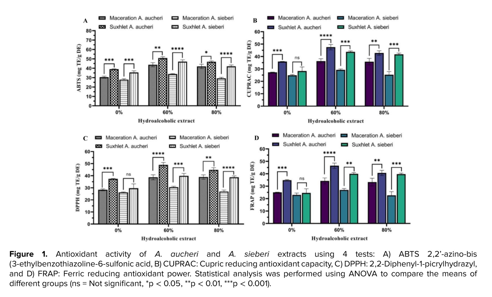
The antioxidant activities of the extracts, assessed by ABTS, CUPRAC, DPPH, and FRAP assays, are presented in figure 1. Overall, values ranged from 22.66-50.93 mg TE/g DE across all methods. For both species, Soxhlet extraction with 60% ethanol consistently yielded the highest activities. Under these optimal conditions, A. aucheri exhibited markedly higher values for ABTS, CUPRAC, DPPH, and FRAP (50.93, 47.54, 48.99, and 46.48, respectively) compared to A. sieberi (47.07, 43.75, 39.91, and 39.98, respectively).
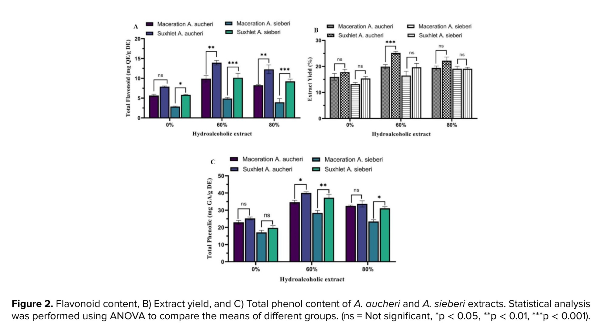
Similarly, total phenolic content, total flavonoid content, and extraction yield (Figure 2) ranged from 17-40 mg GAE/g DE, 2.8-13.9 mg QE/g DE, and 13-25%, respectively. The highest values for all 3 parameters were again obtained with Soxhlet extraction using 60% ethanol, with A. aucheri (40.05, 13.9, and 25.2%) outperforming A. sieberi (37.26, 10.16, and 19.65%).
3.2. Physicochemical characterization and encapsulation performance of niosomal nanoparticles
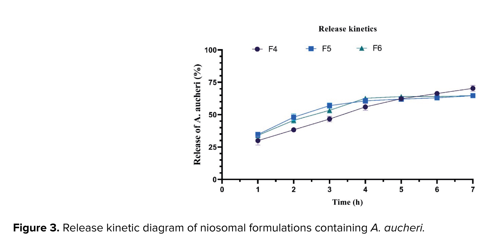
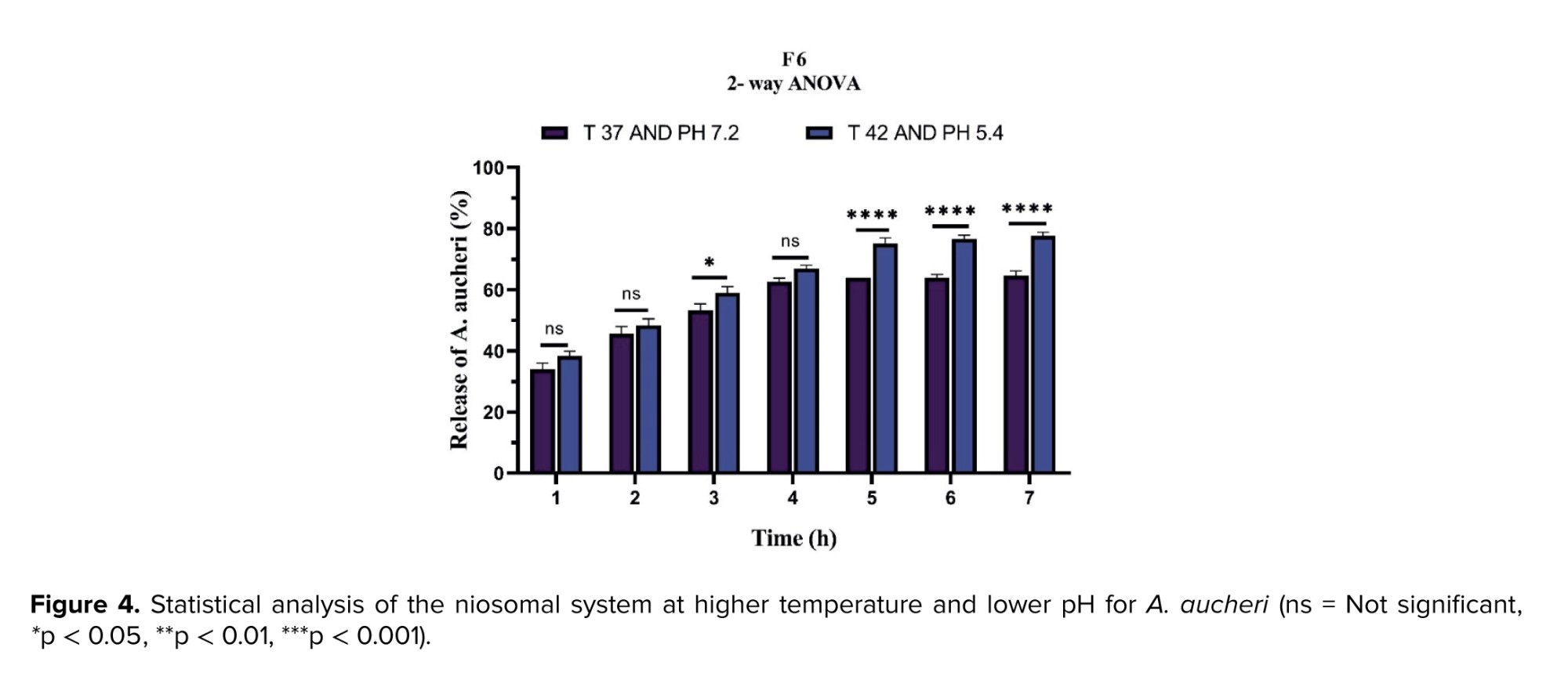
The maximum absorption wavelengths were 356 nm for A. sieberi and 292 nm for A. aucheri (1 mg/mL). The tween-to-cholesterol ratio significantly influenced both the physicochemical properties and encapsulation performance of the niosomal formulations. For A. sieberi, increasing this ratio from F1-F3 reduced particle size from 171.9-78.29 nm, shifted zeta potential from 34.3 mV to -25.2 mV, and increased polydispersity index (PDI) from 0.442 to 0.549. Concurrently, uptake and release improved from 75% and 61% (F1) to 83% and 65% (F2), with F3 achieving the best overall performance at 87% uptake and 71% release. For A. aucheri, similar trends were observed: particle size decreased from 185.3 nm (F4) to 115.5 nm (F6), zeta potential changed from -31.8 mV to -20.6 mV, and PDI rose from 0.475 to 0.518. In terms of performance, F4 showed 75% uptake and 73% release, F5 increased uptake to 88% with 66% release, and F6 provided the highest entrapment (93%), while F4 remained superior for release (73%). Overall, higher tween-to-cholesterol ratios reduced particle size, altered surface charge, and broadened size distribution, while generally enhancing encapsulation efficiency. These findings indicate that formulation optimization can significantly tune nanoparticle stability and drug delivery performance. All experiments were performed in triplicate and analyzed by ANOVA (Figures 3-6 and Table II).
3.3. FTIR results analysis
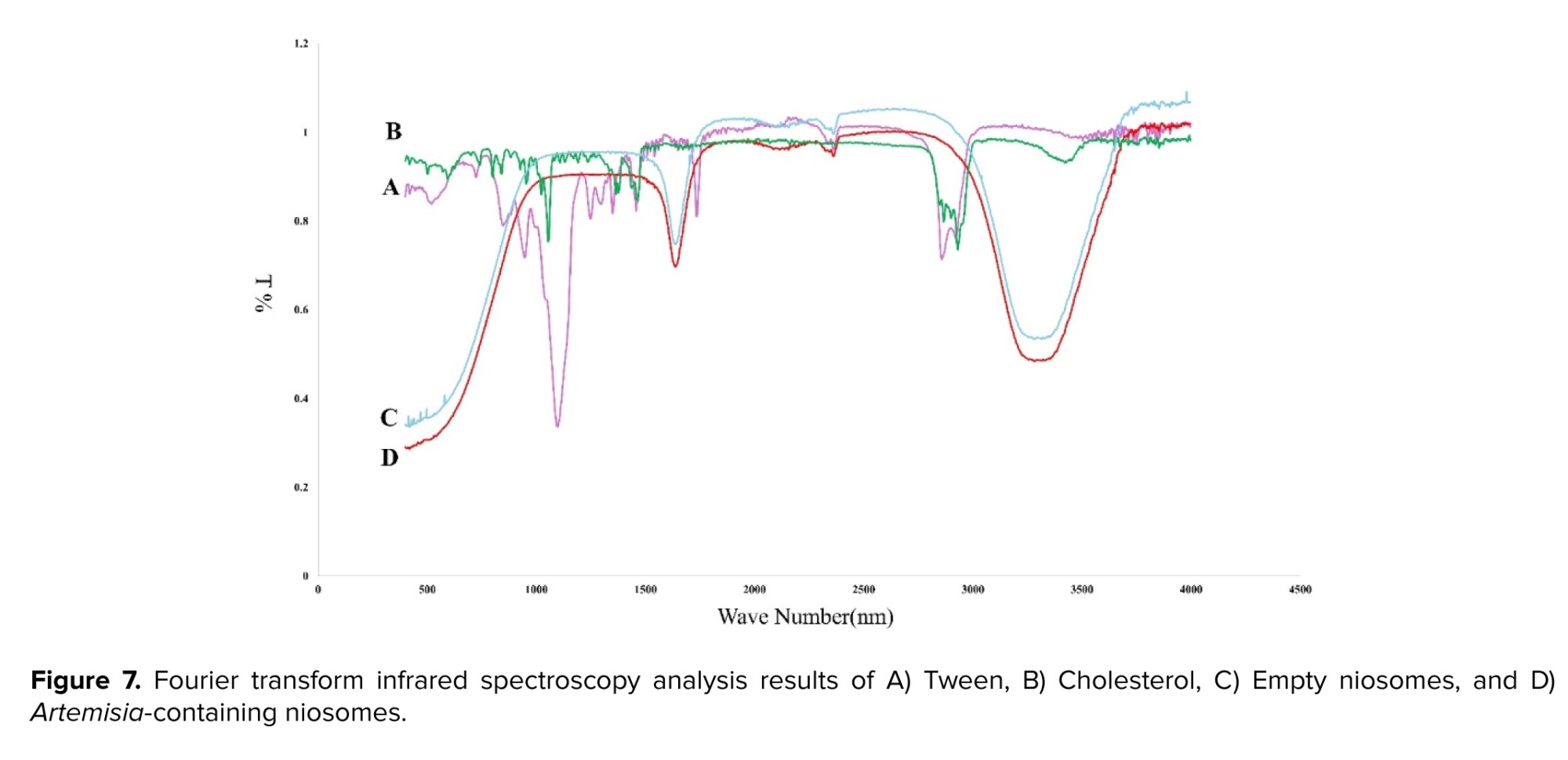
In this study, FTIR analysis was performed to investigate the chemical properties of empty niosomal nanoparticles and niosomal nanoparticles containing Artemisia extracts and key compounds such as tween and cholesterol. This analysis helps to identify the functional groups and chemical bonds present in these compounds. In the FTIR spectrum of empty niosomal nanoparticles, distinct peaks related to alcohol and amine bonds are observed.
In particular, C-O stretching bonds and C-N stretching bonds can be identified. In this study, an increase in the depth of FTIR peaks was detected in drug-loaded nanoparticles compared to empty nanoparticles, which could indicate significant changes in the chemical structure and molecular interactions. These changes are caused by drug loading and their effects on the chemical bonds in the nanoparticles. In empty nanoparticles, FTIR peaks usually represent the central bonds, such as C-O and C-N bonds, which are related to the basic structure of the nanoparticles.
However, when drugs are added to these nanoparticles, new interactions are created between the drug and the nanoparticle matrix, which can lead to an increase in the peak depth. This increase in peak depth means that more energy is absorbed by the chemical bonds that have been affected by drug loading. Changes in the intensity and position of the peaks may also be observed, indicating changes in the chemical environment surrounding these bonds. In contrast, in the FTIR spectrum of tween, peaks related to alcohol bonds (C-O) and C-H stretching bonds can be observed. These peaks indicate the presence of tween as a surfactant in the formulation of niosomal nanoparticles, which can affect the stability and drug loading. Conversely, the FTIR spectrum of cholesterol also includes peaks related to carbon-carbon (C-C) and carbon-oxygen (C = O) bonds. These bonds indicate the role of cholesterol in creating a suitable structure for niosomal nanoparticles. In general, FTIR analysis allows us to obtain detailed information about the compounds present in empty niosomal nanoparticles and those containing different extracts (Figure 7).
3.4. Cytotoxicity assessment
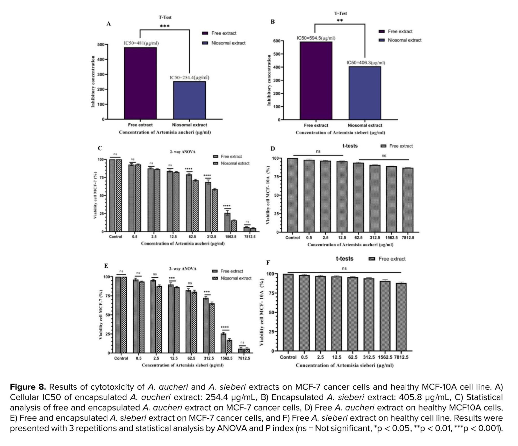
The cytotoxic effects of free and niosomal-encapsulated A. aucheri and A. sieberi extracts were evaluated on MCF-7 breast cancer cells using the MTT assay over a concentration range of 0.5-7812.5 μg/mL. Empty niosomes were included as controls. For both extracts, maximum cell viability occurred at 5 μg/mL and minimum at 7812.5 μg/mL in both free and encapsulated forms. For A. aucheri, free extract showed 92.75% (max) and 4.99% (min) viability, while the niosomal form gave 92.94% and 6.43%, respectively. For A. sieberi, free extract yielded 96.37% and 5.96%, and the niosomal form 93.71% and 5.74%. Notably, increasing nanoparticle concentration significantly reduced cell viability (p < 0.05) as shown in figure 8.
The IC₅₀ values for the optimized niosomal formulations against MCF-7 cells were 254.4 µg/mL for A. aucheri (F4/F6) and 405.8 µg/mL for A. sieberi (F3). Statistical comparisons (ANOVA, 3 replicates) revealed that encapsulated extracts exhibited significantly higher toxicity than their free counterparts (Figure 8A-F). Empty niosomes showed no cytotoxic effect, confirming their safety. Furthermore, free extracts tested on healthy MCF-10A cells demonstrated low toxicity, indicating a favourable safety profile.
Overall, encapsulation in niosomal nanoparticles enhanced the anti-cancer efficacy of both extracts while maintaining low toxicity toward normal cells, supporting their potential as safe and effective therapeutic agents.
4. Discussion
In particular, C-O stretching bonds and C-N stretching bonds can be identified. In this study, an increase in the depth of FTIR peaks was detected in drug-loaded nanoparticles compared to empty nanoparticles, which could indicate significant changes in the chemical structure and molecular interactions. These changes are caused by drug loading and their effects on the chemical bonds in the nanoparticles. In empty nanoparticles, FTIR peaks usually represent the central bonds, such as C-O and C-N bonds, which are related to the basic structure of the nanoparticles.
However, when drugs are added to these nanoparticles, new interactions are created between the drug and the nanoparticle matrix, which can lead to an increase in the peak depth. This increase in peak depth means that more energy is absorbed by the chemical bonds that have been affected by drug loading. Changes in the intensity and position of the peaks may also be observed, indicating changes in the chemical environment surrounding these bonds. In contrast, in the FTIR spectrum of tween, peaks related to alcohol bonds (C-O) and C-H stretching bonds can be observed. These peaks indicate the presence of tween as a surfactant in the formulation of niosomal nanoparticles, which can affect the stability and drug loading. Conversely, the FTIR spectrum of cholesterol also includes peaks related to carbon-carbon (C-C) and carbon-oxygen (C = O) bonds. These bonds indicate the role of cholesterol in creating a suitable structure for niosomal nanoparticles. In general, FTIR analysis allows us to obtain detailed information about the compounds present in empty niosomal nanoparticles and those containing different extracts (Figure 7).
3.4. Cytotoxicity assessment
The cytotoxic effects of free and niosomal-encapsulated A. aucheri and A. sieberi extracts were evaluated on MCF-7 breast cancer cells using the MTT assay over a concentration range of 0.5-7812.5 μg/mL. Empty niosomes were included as controls. For both extracts, maximum cell viability occurred at 5 μg/mL and minimum at 7812.5 μg/mL in both free and encapsulated forms. For A. aucheri, free extract showed 92.75% (max) and 4.99% (min) viability, while the niosomal form gave 92.94% and 6.43%, respectively. For A. sieberi, free extract yielded 96.37% and 5.96%, and the niosomal form 93.71% and 5.74%. Notably, increasing nanoparticle concentration significantly reduced cell viability (p < 0.05) as shown in figure 8.
The IC₅₀ values for the optimized niosomal formulations against MCF-7 cells were 254.4 µg/mL for A. aucheri (F4/F6) and 405.8 µg/mL for A. sieberi (F3). Statistical comparisons (ANOVA, 3 replicates) revealed that encapsulated extracts exhibited significantly higher toxicity than their free counterparts (Figure 8A-F). Empty niosomes showed no cytotoxic effect, confirming their safety. Furthermore, free extracts tested on healthy MCF-10A cells demonstrated low toxicity, indicating a favourable safety profile.
Overall, encapsulation in niosomal nanoparticles enhanced the anti-cancer efficacy of both extracts while maintaining low toxicity toward normal cells, supporting their potential as safe and effective therapeutic agents.
4. Discussion
The present study investigated the potential of niosomal nanoparticles for delivering A. aucheri and A. sieberi extracts, focusing on antioxidant capacity, physicochemical characteristics, and cytotoxic efficacy against breast cancer cells. Initial findings revealed that the antioxidant potential and phytochemical content (phenolics and flavonoids) of both Artemisia species were significantly influenced by the extraction method and solvent concentration.
Superior results were obtained with the Soxhlet method using 60% ethanol, attributable to the efficiency of this method in extracting thermostable compounds and the optimal polarity of 60% ethanol for solubilizing a wide range of antioxidants. Furthermore, the consistently higher antioxidant activity and phytochemical yield of A. aucheri compared to A. sieberi suggest species‑specific differences in bioactive compound profiles, warranting further phytochemical investigation.
In recent decades, drug delivery via vesicles has emerged as an innovative approach to encapsulating drugs to achieve more favorable release, higher solubility, and delivery of both hydrophobic and hydrophilic compounds, as well as longer circulation times. Niosomal carriers acquire their lipid bilayer structure through self‑assembly of nonionic surfactants in an aqueous environment. Niosomes offer several advantages, including industrial‑scale production, lower cost, greater stability, biodegradability, biocompatibility, and lower toxicity. Due to their unique structure and composition, niosomes have shown greater physical and chemical stability than liposomes (3).
Findings related to antioxidant and total phenol tests showed that the hydroalcoholic extracts of this plant have significant antioxidant properties; A. aucheri extract exhibited more antioxidant activity and phenolic compounds than A. sieberi. Also, extraction by the Soxhlet method was significantly different from the Maceration method, indicating the importance of the extraction method and solvent percentage for medicinal plants. The encapsulation efficiency of niosomal nanoparticles containing A. sieberi extract was 75%, 83%, and 85% in 3 different formulations, respectively. For A. aucheri extract, encapsulation efficiency was 75%, 88%, and 93% in 3 formulations (F4, F5, F6). This variation clearly shows the effect of the surfactant ratio (tween-to-cholesterol) and preparation conditions on the ability of nanoparticles to load plant extracts effectively. In particular, the F3/F6 formula was the best option with the highest encapsulation rate. This may be due to the high loading capacity and specific physical properties of niosomal nanoparticles with an optimal surfactant‑to‑cholesterol ratio, which allows more effective drug loading.
Characterization of nanoparticles showed that the size of optimized niosomal nanoparticles containing A. aucheri and A. sieberi was 115 nm and 79 nm, respectively, and their zeta potentials were -20 mV and -25 mV for the optimized nanoparticles of formula F3/F6, indicating appropriate stability of these systems. Moreover, intermolecular interactions assessed by FTIR showed no interference of molecules with the components of the encapsulated nanoparticles. Overall, these results indicate the high potential of niosomal nanoparticles loaded with A. aucheri and A. sieberi extracts for targeted drug delivery to cancer cells.
Also, the effect of environmental factors such as pH and temperature on drug release can provide new strategies for treating cancers. The type of surfactant and different percentages used in niosome synthesis can have significant effects on stability, entrapment rate, and release kinetics. The thin‑film hydration method is one of the most common passive loading methods (12). In this study, different formulations were investigated by the thin‑film synthesis method to optimize niosomal nanoparticles by changing the tween‑to‑cholesterol ratio. Niosomes and liposomes, as vesicle‑based drug delivery systems, are composed of bilayers. The hydrophilicity and type of surfactant, as well as the length of the carbon chain, affect the particle size of the vesicles (13).
Niosomes are usually combined with sorbitans (span) or polysorbates (tween), making them more resistant to oxidation and hydrolysis. Also, niosome formulation is cheaper and simpler than liposomes, and they have good long‑term stability. These surfactants are biocompatible, have low toxicity, and are readily available. In contrast, liposomes, made from phospholipids, undergo rapid oxidation and hydrolysis, resulting in poor stability and high formulation costs. Also, purification of phospholipids is difficult and costly due to the natural resources. Much research has been done on the synthesis and characterization of niosomes to improve drug entrapment. Understanding the molecular properties of surfactants, their molecular structure, and dynamic behavior is crucial for developing macromolecular properties such as fluidity, elasticity, and shear strength (14). In general, niosomes and liposomes are vesicle‑based systems consisting of bilayers. These layers are naturally unstable, and cholesterol is used to stabilize them. Cholesterol helps to reduce leakage and penetration of solutes into the aqueous core of the vesicles. Research has shown that the alkyl chain length of fatty alcohols and the type of surfactant affect the drug release rate from niosomes. Niosomes containing fatty alcohols show a slower release rate than those containing cholesterol. Also, the length of the fatty acid chain of surfactants affects entrapment efficiency and release rate. Cholesterol has been studied extensively as the only membrane stabilizer. Its addition to phospholipid bilayers reduces permeability and increases entrapment efficiency. Fatty alcohols showed a similar release pattern to cholesterol, but their release rates were slower, and the shorter the chain length, the higher the release rate. These findings highlight the importance of cholesterol and the appropriate selection of compounds in the design of niosomes and liposomes for pharmaceutical applications (15). Cholesterol also plays an important role in the physical properties of lipid membranes, and its addition to bilayers increases stability (14). Increasing the cholesterol content in niosomes greatly affects vesicle stability and permeability. In general, increasing drug loading by increasing cholesterol up to a certain point may increase stability and permeability, but entrapment efficiency may decrease (16). In contrast, increasing the ratio of cholesterol to surfactants increases the drug release rate from the dialysis bag, indicating that these nanoparticles have high size, zeta potential, and entrapment efficiency (17). In the present study, 3 formulations with different tween‑to‑cholesterol ratios were used, and the uptake and release of the niosomal system increased with increasing surfactant‑to‑cholesterol percentage, probably due to the increase in the carbon chain percentage of the nonionic surfactant, which significantly affects niosomal nanoparticle characterization. However, a retrospective study showed that a 1:1 molar ratio between nonionic surfactant and cholesterol provided the most optimal state for drug entrapment, and excess cholesterol may damage the niosomal membrane structure (18). This was not consistent with our results; in other words, increasing the surfactant‑to‑cholesterol ratio increased uptake and release of the niosomal systems. Nanoparticle size, zeta potential, and polydispersity index were determined using dynamic light scattering. Vesicles larger than 1 μm were unstable. Retrospective studies have shown that reducing the amount of surfactants, low temperature, and high drug dose will increase size, while increasing the surfactant‑to‑lipid ratio and using nonionic surfactants will reduce particle size (19). Another study showed that the particle size of niosomal formulations decreases with increasing lipid content. A possible explanation is that the hydrophobicity of the lipid bilayer increases, causing disorder in the vesicle membrane and leading to an increase in vesicle radius to create a more thermodynamically stable shape. Another explanation is that the space occupied by lipid molecules increases with higher lipid content, resulting in increased membrane fluidity and a decrease in niosomal size (20).
In the present study, by increasing the amount of tween surfactant relative to cholesterol, the size of niosomal nanoparticles decreased significantly in all cases, consistent with previous studies. Zeta potential is a key parameter in investigating interactions between colloidal particles, including niosomes. It indicates the intensity of the electric charge on the particle surface and can greatly impact the stability and efficiency of drug delivery systems. Surface charge is influenced by acidic and basic groups on the particle surface. When acidic groups dissociate, a negative charge is created, while basic surfaces acquire a positive charge. The magnitude depends on the acidic or basic strength of surface groups and the pH of the solution. By changing pH or reducing surface ionization, the surface charge can be brought to zero; for negatively charged particles, lowering pH can lead to charge neutralization. Zeta potential of niosomes typically varies between -100 mV and 0 mV. If particles in suspension have a zeta potential greater than +30 mV or less than -30 mV, they tend to repel each other and aggregation is reduced. If zeta potential is low, repulsive forces are insufficient, leading to flocculation. Thus, zeta potential is critical for determining niosome stability and efficacy. By carefully adjusting the molar ratios of surfactants and other ingredients, desired stability can be achieved, and unwanted aggregation prevented (17).
According to our results, the zeta potential of niosomal nanoparticles is within an acceptable and stable range, consistent with retrospective studies. The optimal niosomal nanoparticles (F3/F6) containing A. aucheri and A. sieberi extracts were reported as -20 mV and -25 mV, respectively. Lower PDI values indicate a tendency for nanoparticles to have a single peak on the PDI scale (0-1.0). High PDI values indicate significant separation between large and small droplets. Also, if particles are more homogeneous, the polydispersity value is closer to zero (19). In this study, the PDI of optimal A. aucheri and A. sieberi niosomal nanoparticles was 0.518 and 0.549, respectively, which is consistent with previous studies. These points are of great importance in designing niosomal‑based drug delivery systems and can significantly impact the effectiveness of drug treatments.
The antioxidant and anticancer properties of many effective herbal ingredients have been proven. Artemisia is also considered a plant with strong medicinal and antioxidant properties.
In this study, the cytotoxic effect of these extracts on MCF‑7 cancer cells was investigated. In general, encapsulating active ingredients in niosomal structures can increase the cytotoxic effect on cancer cell lines. Accordingly, many studies have been conducted. For example, Dabbagh Moghaddam et al. studied melittin, a peptide from honeybee venom, as a therapeutic option for breast cancer. They optimized melittin‑containing niosomes and empty niosomes and evaluated their anticancer effects on 4T1 and SKBR3 cell lines and on BALB/c mice. The results showed that melittin‑containing nanoparticles had greater anticancer effects than free melittin (21). Also, Nasirian et al. investigated the effectiveness of a niosomal formulation loaded with enciprazine hydrochloride (Nio‑USAN) in preventing colon cancer. Results showed that this formulation was highly effective in reducing apoptosis and increasing transcription of cell death‑related genes in HT‑29 cells. In particular, BCL2 gene transcription was significantly reduced in the treatment group, indicating the high potential of encapsulation to enhance drug efficacy in cancer treatment. These findings emphasize that encapsulation can protect and improve the performance of drug compounds against environmental factors and increase their bioavailability (22). In addition, Dawoud and colleagues investigated the effect of niosomes loaded with Withania somnifera extract on breast cancer cells. Results showed that ASH niosomes loaded with this extract had suitable zeta potential, small particle size, and low PDI. Treatment of MCF‑7 cells with ASH‑NIO resulted in concentration‑dependent suppression of cell growth, with IC50 values of 24.13 μM for ASH‑NIO and 10.45 μM for olaparib. Gene expression analysis showed a significant decrease in Bcl2 mRNA expression and an increase in P53, Bax, caspase‑9, and caspase‑3 gene expression in ASH‑NIO‑treated cells. These results provide further evidence of the potential anticancer effects of ASH‑NIO and suggest that this compound may be useful as an adjunct therapy for breast cancer. In other words, encapsulating the drug in nanoparticles showed a greater effect on cancer cells (23). In the present study, the cytotoxicity of niosomal nanoparticles containing Artemisia at some concentrations had a greater effect on cancer cells than the free extract at the same concentration, confirming that encapsulation of active substances increases the effect on cancer cells, which is consistent with the results of previous studies.
4.1. Strengths and Limitations
While this study provides promising in vitro results, several limitations should be acknowledged. First, the lack of in vivo validation limits the translation of these findings to clinical applications. Second, a standard chemotherapeutic agent was not included as a positive control for direct efficacy comparison. Third, detailed phytochemical analysis of the extracts using techniques such as gas chromatography-mass spectrometry or high-performance liquid chromatography was not performed, which would have provided more precise identification of the specific bioactive compounds responsible for the observed antioxidant and cytotoxic effects. Fourth, detailed mechanistic studies on apoptosis induction pathways were not performed. Fifth, the long-term stability of the formulations under different storage conditions was not evaluated.
5. Conclusion
This study set out to determine whether the tween-to-cholesterol ratio could serve as a critical formulation parameter to optimize the physicochemical properties, encapsulation efficiency, and anticancer activity of niosomal nanoparticles loaded with A. sieberi and A. aucheri extracts. The findings confirm that increasing this ratio consistently reduces particle size, alters zeta potential, and enhances both drug loading and release profiles across all formulations. Importantly, the optimized niosomes particularly F3 for A. sieberi and F4/F6 for A. aucheri demonstrated markedly improved cytotoxic effects against MCF‑7 breast cancer cells compared to the free extracts, as evidenced by significantly lower IC₅₀ values. This enhanced efficacy is attributed not only to improved encapsulation but also to the physicochemical modifications induced by formulation tuning, which likely facilitate better cellular internalization. Beyond efficacy, the results carry substantial implications for therapeutic safety and selectivity. The complete absence of toxicity from empty niosomes, together with the minimal cytotoxicity of both free and encapsulated extracts toward healthy MCF‑10A cells, underscores the favorable safety profile of this nano-delivery system. This dual advantage potent tumor cell killing alongside preserved normal cell viability addresses a major limitation of conventional phytotherapy, where non-specific toxicity and poor bioavailability often hinder clinical translation. Furthermore, the antioxidant profiling revealed that A. aucheri consistently outperformed A. sieberi across all assays, suggesting that species selection can further influence the overall therapeutic potential. The FTIR analyses confirmed successful drug loading and molecular interactions between the extracts and the niosomal matrix, supporting the structural integrity of the formulations. Collectively, these findings directly answer the research question by demonstrating that rational manipulation of the tween-to-cholesterol ratio is a powerful strategy to design niosomal nanoparticles with tailored physicochemical properties and optimized biological performance. The study provides a robust preclinical foundation for developing plant-based nanoformulations as safe, effective, and selectively targeted therapeutic agents for breast cancer treatment. Future work should focus on in vivo validation and mechanistic exploration of the enhanced antitumor activity observed here.
Data Availability
Data supporting the findings of this study are available upon reasonable request from the corresponding author.
Author Contributions
E. Zamani: Made substantial contributions to the acquisition of data (primary data collection) and conducting experiments; participated in drafting the work; gave final approval of the version to be published; agrees to be accountable for all aspects of the work related to data collection and experimental procedures. K. Kamali: Made substantial contributions to the analysis and interpretation of data (statistical data analysis); participated in drafting and writing the manuscript; gave final approval of the version to be published; agrees to be accountable for all aspects of the work related to statistical analysis. A.A. Seifi Ashkezari: Made substantial contributions to the acquisition (assisted in data collection) and analysis/interpretation of data (statistical analysis); participated in writing the manuscript; gave final approval of the version to be published; agrees to be accountable for all aspects of the work related to data collection and statistical analysis. H. Sodaizadeh: Made substantial contributions to the design of the project; critical revision of the manuscript for important intellectual content (editing and finalizing); gave final approval of the version to be published; agrees to be accountable for all aspects of the work related to manuscript editing and finalization. M. Yaeghoobi: Made substantial contributions to the research methodology; interpretation of data (provided technical support in data interpretation); contributed to scientific editing of the manuscript (critical review); gave final approval of the version to be published; agrees to be accountable for all aspects of the work related to data interpretation and scientific editing. BF. Haghiralsadat: Made substantial contributions to the conception and design of the work (overall project management and experimental design); critically revised and finalized the manuscript for submission; gave final approval of the version to be published; agrees to be accountable for all aspects of the work and ensures that questions related to accuracy or integrity are appropriately investigated and resolved (corresponding author).
Acknowledgments
The authors appreciate Yazd University, Yazd, Iran and the Paramedical Research Center, Shahid Sadoughi University of Medical Sciences, Yazd, Iran, for providing resources and facilities for this research. We also thank the technical staff for their cooperation in the laboratory. This research received no specific grant from any funding agency in the public, commercial, or not-for-profit sectors.
The authors acknowledge the assistance of DeepSeek AI for language polishing and proofreading. The authors are fully responsible for the final manuscript.
Conflict of Interest
The authors declare that there is no conflict of interest.
Type of Study: Original Article |
Subject:
Cellular and Molecular Biology of Reproduction
Send email to the article author
| Rights and permissions | |
 |
This work is licensed under a Creative Commons Attribution-NonCommercial 4.0 International License. |