International Journal of
Reproductive Biomedicine
Tue, Sep 29, 2026
[Archive]
Volume 24, Issue 8 (August 2026)
IJRM 2026, 24(8): 721-738 |
Back to browse issues page



Ethics code: IR.SSU.SPH.REC.1400.165


Download citation:
BibTeX | RIS | EndNote | Medlars | ProCite | Reference Manager | RefWorks
Send citation to:


BibTeX | RIS | EndNote | Medlars | ProCite | Reference Manager | RefWorks
Send citation to:
Zamani E, Kamali K, Seifi Ashkezari A A, Sodaizadeh H, Yaeghoobi M, Haghiralsadat B F. Comparative study of niosomal nanoparticles encapsulating Artemisia aucheri and Artemisia sieberi extracts on Michigan Cancer Foundation-7 breast cancer cell proliferation: An in vitro study. IJRM 2026; 24 (8) :721-738
URL: http://ijrm.ir/article-1-3556-en.html
URL: http://ijrm.ir/article-1-3556-en.html
Elaheh Zamani1 

 , Kazem Kamali1 , Ali Akbar Seifi Ashkezari2 , Hamid Sodaizadeh1 , Marzieh Yaeghoobi3 , Bibi Fatemeh Haghiralsadat *4
, Kazem Kamali1 , Ali Akbar Seifi Ashkezari2 , Hamid Sodaizadeh1 , Marzieh Yaeghoobi3 , Bibi Fatemeh Haghiralsadat *4
1- Natural Resources Faculty, Yazd University, Yazd, Iran.
2- Iran University of Medical Sciences, Tehran, Iran.
3- Biotechnology Research Center, Yazd Reproductive Sciences Institute, Shahid Sadoughi University of Medical Sciences, Yazd, Iran.
4- Biotechnology Research Center, Yazd Reproductive Sciences Institute, Shahid Sadoughi University of Medical Sciences, Yazd, Iran. & School of Advanced Medical Technologies, Shahid Sadoughi University of Medical Sciences, Yazd, Iran. ,fhaghirosadat@gmail.com; fhaghirosadat@ut.ac.ir
2- Iran University of Medical Sciences, Tehran, Iran.
3- Biotechnology Research Center, Yazd Reproductive Sciences Institute, Shahid Sadoughi University of Medical Sciences, Yazd, Iran.
4- Biotechnology Research Center, Yazd Reproductive Sciences Institute, Shahid Sadoughi University of Medical Sciences, Yazd, Iran. & School of Advanced Medical Technologies, Shahid Sadoughi University of Medical Sciences, Yazd, Iran. ,
Abstract: (38 Views)
Background: Breast cancer is the second leading cause of cancer-related deaths among women, accounting for approximately 30% of all new cancer diagnoses in women annually.
Objective: Given the prevalence of breast cancer among women, the primary objective of this study was to develop an effective drug delivery system for treating this disease using niosome containing Artemisia plants.
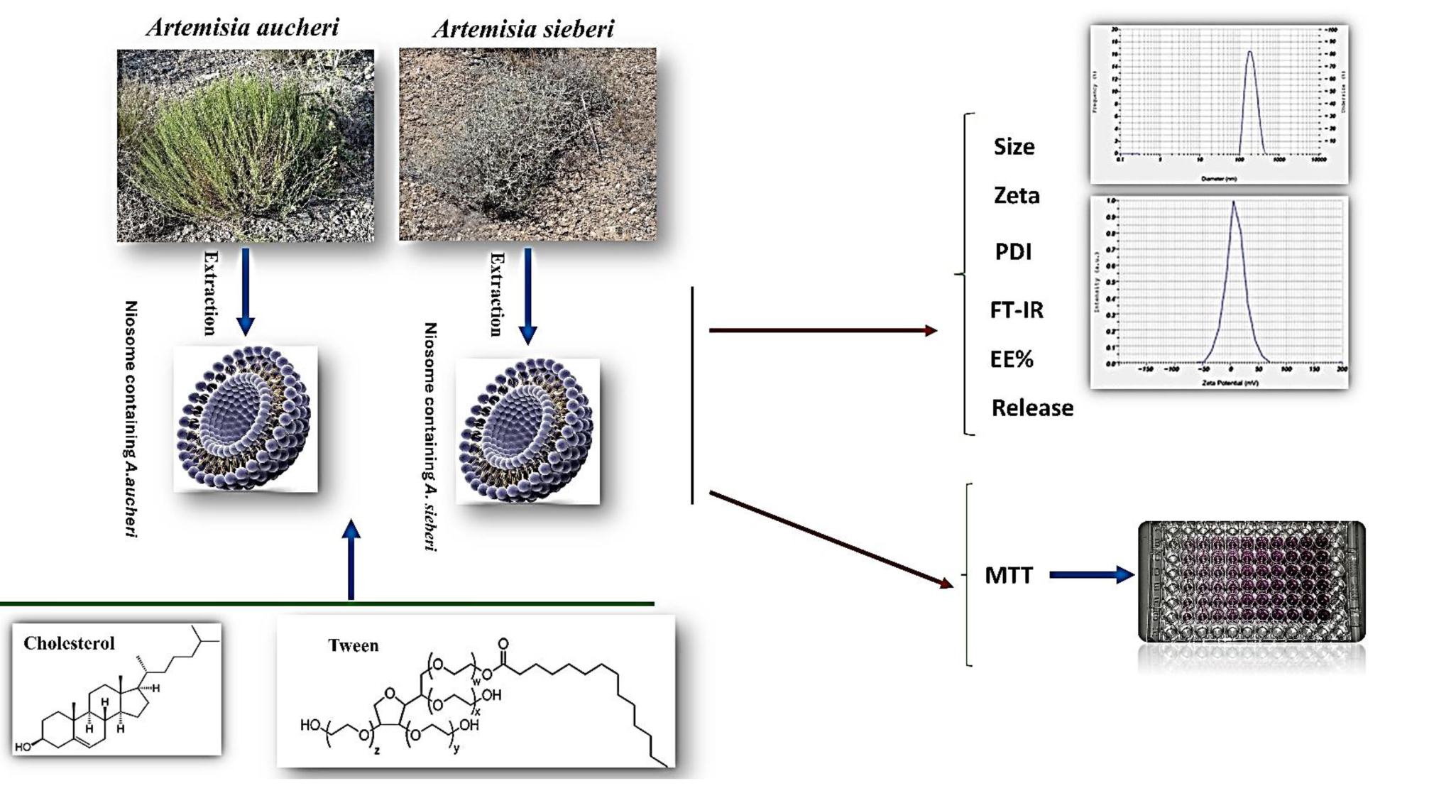
Materials and Methods: This in vitro study involved the hydroalcoholic extraction of Artemisia using both the Maceration and Soxhlet methods, followed by the evaluation of its antioxidant properties. Niosomal nanoparticles were synthesized via the thin-film hydration technique. Characterization of the nanoparticles, including particle size and zeta potential, was conducted using dynamic light scattering (Zeta Sizer) and fourier-transform infrared spectroscopy. Cytotoxicity assessment was performed employing the MTT (3-(4,5-dimethylthiazol-2-yl)-2,5-diphenyltetrazolium bromide) assay, with statistical analysis carried out using one-way ANOVA and determination of significance.
Results: This study demonstrated that Artemisia aucheri extract, particularly when obtained via the Soxhlet method using 60% ethanol, exhibited superior antioxidant activity and higher total phenolic/flavonoid content compared to Artemisia sieberi. The optimized niosomal nanoparticles exhibited an encapsulation efficiency of 87% for A. sieberi (F3) and 93% for A. aucheri (F6). Cumulative release rates over 7 hr were 71% for A. sieberi (F3) and 73% for A. aucheri (F4) extracts. The optimized nanoparticles displayed sizes of 115 nm for A. aucheri (F6) and 79 nm for A. sieberi (F3), along with zeta potentials of -20.6 mV and -25.2 mV, respectively. Fourier-transform infrared spectroscopy analysis confirmed successful loading and favorable intermolecular interactions. The cytotoxicity of the encapsulated nanoparticles against Michigan Cancer Foundation-7 cells showed a significant increase compared to their free form (p < 0.05), with blank niosomes showing no notable toxicity. The half-maximal inhibitory concentration values were 254.4 µg/mL for encapsulated A. aucheri and 405.8 µg/mL for encapsulated A. sieberi.
Conclusion: Based on the results, the use of plant extracts and niosomal nanoparticles can be considered a positive step for the treatment of breast cancer.
Objective: Given the prevalence of breast cancer among women, the primary objective of this study was to develop an effective drug delivery system for treating this disease using niosome containing Artemisia plants.
Materials and Methods: This in vitro study involved the hydroalcoholic extraction of Artemisia using both the Maceration and Soxhlet methods, followed by the evaluation of its antioxidant properties. Niosomal nanoparticles were synthesized via the thin-film hydration technique. Characterization of the nanoparticles, including particle size and zeta potential, was conducted using dynamic light scattering (Zeta Sizer) and fourier-transform infrared spectroscopy. Cytotoxicity assessment was performed employing the MTT (3-(4,5-dimethylthiazol-2-yl)-2,5-diphenyltetrazolium bromide) assay, with statistical analysis carried out using one-way ANOVA and determination of significance.
Results: This study demonstrated that Artemisia aucheri extract, particularly when obtained via the Soxhlet method using 60% ethanol, exhibited superior antioxidant activity and higher total phenolic/flavonoid content compared to Artemisia sieberi. The optimized niosomal nanoparticles exhibited an encapsulation efficiency of 87% for A. sieberi (F3) and 93% for A. aucheri (F6). Cumulative release rates over 7 hr were 71% for A. sieberi (F3) and 73% for A. aucheri (F4) extracts. The optimized nanoparticles displayed sizes of 115 nm for A. aucheri (F6) and 79 nm for A. sieberi (F3), along with zeta potentials of -20.6 mV and -25.2 mV, respectively. Fourier-transform infrared spectroscopy analysis confirmed successful loading and favorable intermolecular interactions. The cytotoxicity of the encapsulated nanoparticles against Michigan Cancer Foundation-7 cells showed a significant increase compared to their free form (p < 0.05), with blank niosomes showing no notable toxicity. The half-maximal inhibitory concentration values were 254.4 µg/mL for encapsulated A. aucheri and 405.8 µg/mL for encapsulated A. sieberi.
Conclusion: Based on the results, the use of plant extracts and niosomal nanoparticles can be considered a positive step for the treatment of breast cancer.
Keywords: Niosomes, Nanoparticles, Plant preparations, Breast neoplasms, Cell line, Tumor, Drug delivery systems.
Type of Study: Original Article |
Subject:
Cellular and Molecular Biology of Reproduction
Send email to the article author
| Rights and permissions | |
 |
This work is licensed under a Creative Commons Attribution-NonCommercial 4.0 International License. |