
International Journal of
Reproductive Biomedicine
Mon, Oct 26, 2026
[Archive]
Volume 24, Issue 6 (June 2026)
Int J Reprod BioMed. 2026, 24(6): 547-549 |
Back to browse issues page




Download citation:
BibTeX | RIS | EndNote | Medlars | ProCite | Reference Manager | RefWorks
Send citation to:


BibTeX | RIS | EndNote | Medlars | ProCite | Reference Manager | RefWorks
Send citation to:
Afkhami E, Movahedin M, Datli Beigi S A, Aflatoonian B. Extracellular vesicles in assisted reproductive technology: A double-edged sword in fertility enhancement. Int J Reprod BioMed. 2026; 24 (6) :547-549
URL: http://ijrm.ir/article-1-3891-en.html
URL: http://ijrm.ir/article-1-3891-en.html

1- Anatomical Sciences Department, Faculty of Medical Sciences, Tarbiat Modares University, Tehran, Iran.
2- Anatomical Sciences Department, Faculty of Medical Sciences, Tarbiat Modares University, Tehran, Iran. ,movahed.m@modares.ac.ir
3- Abortion Research Center, Yazd Reproductive Sciences Institute, Shahid Sadoughi University of Medical Sciences, Yazd, Iran.
4- Stem Cell Biology Research Center, Yazd Reproductive Sciences Institute, Shahid Sadoughi University of Medical Sciences, Yazd, Iran.
2- Anatomical Sciences Department, Faculty of Medical Sciences, Tarbiat Modares University, Tehran, Iran. ,
3- Abortion Research Center, Yazd Reproductive Sciences Institute, Shahid Sadoughi University of Medical Sciences, Yazd, Iran.
4- Stem Cell Biology Research Center, Yazd Reproductive Sciences Institute, Shahid Sadoughi University of Medical Sciences, Yazd, Iran.
Full-Text [PDF 748 kb]
(256 Downloads)
| Abstract (HTML) (325 Views)
Full-Text: (53 Views)
Dear Editor
Extracellular vesicles (EVs), including exosomes and microvesicles, are essential for cell communication in the context of reproductive health (1, 2). Oocytes, granulosa cells, endometrial cells, and sperm are the hosts for these vesicles, which carry essential molecules such as proteins, fats, or genetic material (2). Assisted reproductive technologies (ART) have been receiving significant attention for their ability to facilitate the maturation, fertilization, and embryonic development of eggs through these small vesicles. The presence of these small vesicles can lead to fertility benefits, but there are also potential drawbacks (3). EVs have become a hopeful prospect for improving the success of in vitro fertilization (IVF).
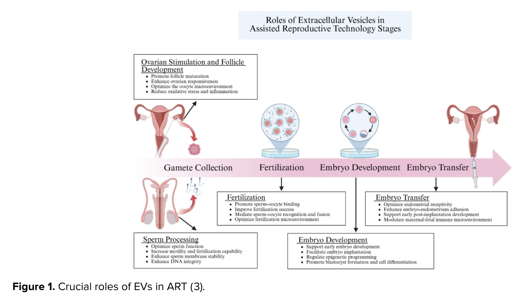
EVs found in the cumulus or follicular fluid have been shown to facilitate oocyte maturation by transporting microRNAs and proteins that enhance embryo development. In the laboratory, this support can facilitate early embryo growth and fertilization (4). Such EV-mediated support can translate into improved fertilization rates and early embryonic development in vitro (Figure 1) (3). Furthermore, the transmission of signals through endometrial EVs can enhance the flexibility of the uterus's internal walls by controlling implantation processes (5). Prostasomes, which are derived from the seminal EVs, also aid in sperm movement and readiness, thus contributing to the overall ART process (6).
Nevertheless, EVs can cause harm. Why? Damaged materials or signals released during oxidative stress in the ovarian follicle may lead to cell death and lower egg quality (3). Women with endometriosis have been found to contain inflammation-causing proteins and microRNAs that are associated with embryonic implant issues (7). EVs from old granulosa cells could be responsible for the lower success rates of oocytes produced by older mothers. EVs are not all-purpose and not necessarily good. They have a significant impact on the body depending on their environment and the conditions of production. Some of the challenges faced when working with EVs are:
Extracellular vesicles (EVs), including exosomes and microvesicles, are essential for cell communication in the context of reproductive health (1, 2). Oocytes, granulosa cells, endometrial cells, and sperm are the hosts for these vesicles, which carry essential molecules such as proteins, fats, or genetic material (2). Assisted reproductive technologies (ART) have been receiving significant attention for their ability to facilitate the maturation, fertilization, and embryonic development of eggs through these small vesicles. The presence of these small vesicles can lead to fertility benefits, but there are also potential drawbacks (3). EVs have become a hopeful prospect for improving the success of in vitro fertilization (IVF).
EVs found in the cumulus or follicular fluid have been shown to facilitate oocyte maturation by transporting microRNAs and proteins that enhance embryo development. In the laboratory, this support can facilitate early embryo growth and fertilization (4). Such EV-mediated support can translate into improved fertilization rates and early embryonic development in vitro (Figure 1) (3). Furthermore, the transmission of signals through endometrial EVs can enhance the flexibility of the uterus's internal walls by controlling implantation processes (5). Prostasomes, which are derived from the seminal EVs, also aid in sperm movement and readiness, thus contributing to the overall ART process (6).
Nevertheless, EVs can cause harm. Why? Damaged materials or signals released during oxidative stress in the ovarian follicle may lead to cell death and lower egg quality (3). Women with endometriosis have been found to contain inflammation-causing proteins and microRNAs that are associated with embryonic implant issues (7). EVs from old granulosa cells could be responsible for the lower success rates of oocytes produced by older mothers. EVs are not all-purpose and not necessarily good. They have a significant impact on the body depending on their environment and the conditions of production. Some of the challenges faced when working with EVs are:
- It is challenging to identify and utilize EVs in ART due to technical constraints.
- Ultracentrifugation, size exclusion chromatography, and immunoaffinity capture are among the methods used to produce EV groups with different purity and amounts, which can pose challenges for reproducibility (8).
- Due to differences in EV group sizes, carrying materials, and markers on their surface, there are challenges in developing standard methods (8).
- Trying to compare the outcomes of various experiments or treatments using different amounts and quantities of EVs is difficult (8).
- Safety is a concern, too. The transmission of diseases like cancer-causing agents or infections could occur with EVs due to their contamination by dirty sources (9).
- When considering EVs for human treatment, rules and ethics must be considered (8).
Deciphering the contents of EVs is also challenging. The analysis of protein and RNA shows that EVs contain numerous molecules, but it is still uncertain how they affect fertility (3). Efforts are just beginning to link specific components of EVs to better egg quality or embryo growth, often using experimental lab models with their own problems. It is not clear whether hormones, age, or ovarian changes affect the EVs (10). The safety of EVs can be checked through animal research and small-scale human trials, which can then lead to larger trials (11).
In conclusion, recent research on EVs in ART provides valuable insights into the complex mechanisms underlying fertility and pregnancy. By elucidating the role of EVs in regulating important aspects of reproductive physiology, researchers can pave the way for the development of innovative diagnostic tools and therapeutic interventions to improve ART. EVs in ART represent a double-edged sword: they have the potential to enhance the reproductive system and its quality, but their origins can also cause problems in terms of handling. In the new area of reproductive medicine, EV usage requires deep knowledge about how they work, as well as consistent methods and thorough safety checks before routine use in ART.
Acknowledgements
Grammarly Ink's AI grammar checker (2019) was used for grammatical editing.
Conflict of Interest
The authors declare that they have no known competing financial interests or personal relationships that could have appeared to influence the work reported in this paper.
In conclusion, recent research on EVs in ART provides valuable insights into the complex mechanisms underlying fertility and pregnancy. By elucidating the role of EVs in regulating important aspects of reproductive physiology, researchers can pave the way for the development of innovative diagnostic tools and therapeutic interventions to improve ART. EVs in ART represent a double-edged sword: they have the potential to enhance the reproductive system and its quality, but their origins can also cause problems in terms of handling. In the new area of reproductive medicine, EV usage requires deep knowledge about how they work, as well as consistent methods and thorough safety checks before routine use in ART.
Acknowledgements
Grammarly Ink's AI grammar checker (2019) was used for grammatical editing.
Conflict of Interest
The authors declare that they have no known competing financial interests or personal relationships that could have appeared to influence the work reported in this paper.
Type of Study: Letter to Editor |
Subject:
Assisted Reproductive Technologies
Send email to the article author
| Rights and permissions | |
 |
This work is licensed under a Creative Commons Attribution-NonCommercial 4.0 International License. |