
International Journal of
Reproductive Biomedicine
Fri, Jun 12, 2026
[Archive]
Volume 4, Issue 1 (7-2006)
IJRM 2006, 4(1): 13-17 |
Back to browse issues page

Download citation:
BibTeX | RIS | EndNote | Medlars | ProCite | Reference Manager | RefWorks
Send citation to:


BibTeX | RIS | EndNote | Medlars | ProCite | Reference Manager | RefWorks
Send citation to:
Dehghani Firoozabadi R, Klantar S M, Seyed-Hasani S M, Ghasemi N, Asgharnia M, Sheikhha M H. Cytogenetic analysis in couples with recurrent spontaneous abortion. IJRM 2006; 4 (1) :13-17
URL: http://ijrm.ir/article-1-46-en.html
URL: http://ijrm.ir/article-1-46-en.html
Razieh Dehghani Firoozabadi1 
 , Seyed Mehdi Klantar *2 , Seyed Mohammad Seyed-Hasani1 , Nasrin Ghasemi1 , Maryam Asgharnia1 , Mohammad Hasan Sheikhha1
, Seyed Mehdi Klantar *2 , Seyed Mohammad Seyed-Hasani1 , Nasrin Ghasemi1 , Maryam Asgharnia1 , Mohammad Hasan Sheikhha1
1- Research and Clinical Centre for Infertility, Shahid Sadougui University of Medical Sciences and Health Services, Yazd, Iran
2- Research and Clinical Centre for Infertility, Shahid Sadougui University of Medical Sciences and Health Services, Yazd, Iran ,smkalantar@yahoo.com
2- Research and Clinical Centre for Infertility, Shahid Sadougui University of Medical Sciences and Health Services, Yazd, Iran ,
Full-Text [PDF 53 kb]
(979 Downloads)
| Abstract (HTML) (4081 Views)
Full-Text: (606 Views)
Introduction
Human reproduction is terribly incompetent. One third of conceptions do not result in the delivery of a baby. But this inefficient process produces amazingly good outcomes. The vast majority of continuing pregnancies result in the birth of a healthy human being who will, eventually, pass his or her genes on to the next generation. Miscarriages are clinically detectable pregnancies that fail to progress. They are common and often remain unexplained, even after investigation. It causes distress for women and their partners. When a woman has had two or more miscarriages, she is likely to seek professional help in the hope that a cause and a cure will be found (1).
Most women with a history of recurrent abortion will be under the care of a gynaecologist, who will have already searched for a gynaecological cause and will have excluded most serious maternal disorders (2). Recurrent miscarriages have a range of possible causes including genetic, anatomic, endocrine, immune, infective, thrombophilic, and unexplained causes (1). Maternal problems consist of uteral malformations, immunological factors, endocrine problems and so on (1). However, most spontaneous miscarriages are caused by chromosomal abnormalities in the embryo or fetus (3,4). Results of the numerous studies showed that approximately 50% to 80% of all pregnancy losses, depend on the maternal and gestational age at the time of loss, caused by chromosomal abnormalities (5-10). In 4-8% of couples with recurrent pregnancy loss, at least one of the partners has chromosomal abnormality that probably contains balance chromosomal abnormalities (1).
The overwhelming majority of chromosomally abnormal conceptions result from the chance :union: of one normal and one aneuploid gamete or from non disjunction during embryonic development. Over 90% of the chromosomal abnormalities observed among abortuses are numerical (11), the reminder are split between structural abnormalities and mosaicism. Autosomal trisomies are the most common abnormalities (12), usually involving chromosome 13-16, 21 or 22, which followed by monosomy X and polyploidies. However, abortuses aneuploidy in recurrent abortion are significantly less than in sporadic pregnancy loss (13). Balance translocations, reciprocal and Robertsonian, are the most common structural abnormalities (14,15), and sex chromosome mosaicism, and chromosome inversion can occasionally be observed.
Chromosomal abnormalities usually are seen in karyotypes of embryo or fetus, after abortion (3). Chromosomal content of couples with recurrent abortion usually are normal. However, their gametocytes showed chromosomal abnormality, which might form during spermatogenesis or oogenesis. In addition, high rate of sperm chromosomal abnormalities was observed in recurrent abortion couples (16,17).
The aim of this study was to determine the chromosomal abnormality in couples with recurrent abortion refer to Research and Clinical Centre for Infertility of the Shahid Sadoughi University of Medical Sciences, Yazd, Iran.
Materials and Methods
In this descriptive case series study all couples (n=165 couples) with recurrent spontaneous abortion from September 2003 till September 2004 were studied. On epidemiological evidence, the definition of recurrent miscarriage should be three or more consecutive pregnancy losses (18). Women meeting the definition can be subdivided into primary and secondary groups, respectively consisting of those who have lost all previous pregnancies and those who have had one successful pregnancy followed by consecutive losses. In the present study, the patients had a history of three or more abortions and did not have any children. Their managements were started with clinical examination by gynaecologist and urologist and then by a genetic counsellor. The anatomical problems were rule out by gynaecologist and urologists.
Regarding their age, the patients were classified into four groups; 18 to 24, 25 to 29, 30 to 34 and over 35 years old. Different age groups were used to show the importance of relation between advance maternal age and chromosomal abnormality. The degree of relationship between couple was determined as consanguinity till four degree and others as no family inbreeding.
A questionnaire, include demographic characteristic, and medical and familial history, was completed for each couple. Antibody against toxsoplasmose, rubella and cytomegalovirus (CMV) were analysed for all the patients, by ELISA Trinitin 99% kits. For autoimmune study, antibody against cardiolipin and phspholipin were checked (19).
After genetic counselling, family pedigree was drawn by genetic counsellor. Cytogenetic analysis was needed for 88 couples. Karyotyping was conducted by analysis of G and/or C banding using 10ml heparinized peripheral blood sample. Metaphase spreads were made from phytohaemaglutinin-stimulated peripheral lymphocytes using standard cytogenetic techniques. Cultures were harvested and Karyotyping was performed on GTW and/or C banded chromosome preparations. The chromosomal status was analyzed using CytoVision Ultra ver.4.0 from Applied Imaging. At least 25 metaphases were analysed for each patient. All chromosomal abnormalities were reported in accordance with the current international standard nomenclature (20).
The X2-test and ANOVA were used for statistical evaluation. The level of p<0.05 was considered as significance.
Results
In this study 165 couples were studied, which classified in 10 three groups according to the number of previous abortion. In group one, couples had three abortions, in group two, had four or five and in group three had six or more abortions. The highest number of patients was seen in group one (61.2%) (Table I).
The women ages were between 18 to 43 years old (mean 27.4 years old). The highest frequency of abortion was seen in women who belonged to age group 25-29 year old. The results showed the number of abortion increased in older age, and the relation was significant (ANOVA test, p=0.047, Fig.1).


Consanguinity was seen in 32/101 couples with three abortions, 29/45 couples with four or five abortions, and 15/19 couples with six or more abortions. The significant relation was found between number of abortion and consanguinity (ANOVA test, p= 0.000). Frequency of consanguinity in these patients was 46%. Coefficient of inbreeding in 4 couples was 1/8 (double first cousins), in 70 couples was 1/16 (first cousins), and in 2 couples was 1/32 (fourth degree of relationship). The rest had fifth degree of relationship (7 couples) and no consanguinity (82 couples). The majority of these couples had third degree relationship.
The family history of recurrent abortion was found in 30 couples, which 21 of them had family inbreeding. The relation between recurrent abortion and its family history was significant (chi-square test, p=0.000).
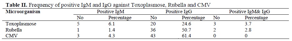
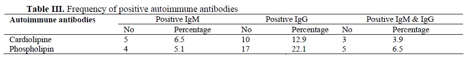
The results of the level of antibody against toxoplasmos, rubella and CMV are shown in table II. The results of autoimmune study (anti cardiolipin and anti phospholipin) are shown in table III.
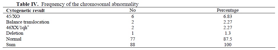
Karyotyping was preformed on peripheral blood sample of 88 couples. Eleven couples had an abnormal karyotype (12.5%), 2 in men and 9 in women, 54.5% numerical abnormality and 45.5% structural abnormality. Six women (6.82%) had monosomy X (45/XO), 2 patients (one man and one woman) had balance translocation, 2 women had 46XX/1qh+, and 1 man had deletion (table IV).
Discussion
Recurrent abortion is a difficult medical problem happening in about 1-2% of fertile women (21). Numerical chromosomal abnormalities were observed among over 90% of the abortion dues to chromosomal abnormality. Autosomal trisomies are the most common abnormality followed by monosomy X and polyploidies. In fact, autosomal monosomy is rarely found in spontaneous abortion and is thought to be responsible for preclinical abortion (22). The results of the present study showed the majority of chromosomal abnormality in these patients were monosomy X (45/XO) followed by structural abnormality, but trisomies were not seen at all. Trisomy usually accompanies with low IQ and these people in our region usually will not marry or their marriage will not continue to have children (23). However other studies showed that structural chromosomal abnormality is the most common chromosomal abnormality in couples with recurrent abortions especially couples undergoes Assisted Reproductive Technology (ART) (24,25). Invitro fertilization plus Preimplantation Genetic Diagnosis (PGD) is an important step in the management of these couples (4,26). One in 500 people carries a balanced translocation. When one member of a couple carries a balanced chromosome translocation, the risk of having a miscarriage is approximately doubled. In 3-5% of couples with recurrent miscarriage, one partner has a balanced translocation (1).
Consanguinity was higher in these couples than normal population. The higher rate of the recurrent abortion in consanguineous marriage could be the result of genetic abnormality of fetus, such as fetal autosomal recessive disorders (27). However, some other researches showed that the rate of spontaneous abortion in consanguineous and non-consanguineous mating is the same (28,29). In conclusion, present study discusses the significance of sex chromosome aneuploidy, balance translocation and deletion in couples with recurrent abortions, therefore it would be reasonable to recommend chromosome analysis to these couples (30).
Antiphospholipid syndrome, in which anticardiolipin antibodies and lupus anticoagulant are present, is detectable in 15% of women with recurrent miscarriage. Its identification is important, because treatment with aspirin or heparin or both significantly improves the likelihood of a live birth (31). The result of this study showed 5.1% of the women had positive IgM antiphospholipid and 22.1% had positive IgG antiphospholipid.
In the future to complete this study, cytogenetic analysis of the abortuses should be done, which help the family in other pregnancies. Peripheral blood karyotyping of both partners should be considered a required examination of couples with recurrent miscarriage but, the influence of other factors, such as family history of miscarriages, should be considered when deciding who should be karyotyped. When a balanced translocation is recognized, it should be recommended to have a triple screening test followed by prenatal diagnosis to see if they are the result of unbalanced translocations.
Acknowledgment
The authors wish to thank Mr. H. Fazli for his assistance in genetics laboratory.
Human reproduction is terribly incompetent. One third of conceptions do not result in the delivery of a baby. But this inefficient process produces amazingly good outcomes. The vast majority of continuing pregnancies result in the birth of a healthy human being who will, eventually, pass his or her genes on to the next generation. Miscarriages are clinically detectable pregnancies that fail to progress. They are common and often remain unexplained, even after investigation. It causes distress for women and their partners. When a woman has had two or more miscarriages, she is likely to seek professional help in the hope that a cause and a cure will be found (1).
Most women with a history of recurrent abortion will be under the care of a gynaecologist, who will have already searched for a gynaecological cause and will have excluded most serious maternal disorders (2). Recurrent miscarriages have a range of possible causes including genetic, anatomic, endocrine, immune, infective, thrombophilic, and unexplained causes (1). Maternal problems consist of uteral malformations, immunological factors, endocrine problems and so on (1). However, most spontaneous miscarriages are caused by chromosomal abnormalities in the embryo or fetus (3,4). Results of the numerous studies showed that approximately 50% to 80% of all pregnancy losses, depend on the maternal and gestational age at the time of loss, caused by chromosomal abnormalities (5-10). In 4-8% of couples with recurrent pregnancy loss, at least one of the partners has chromosomal abnormality that probably contains balance chromosomal abnormalities (1).
The overwhelming majority of chromosomally abnormal conceptions result from the chance :union: of one normal and one aneuploid gamete or from non disjunction during embryonic development. Over 90% of the chromosomal abnormalities observed among abortuses are numerical (11), the reminder are split between structural abnormalities and mosaicism. Autosomal trisomies are the most common abnormalities (12), usually involving chromosome 13-16, 21 or 22, which followed by monosomy X and polyploidies. However, abortuses aneuploidy in recurrent abortion are significantly less than in sporadic pregnancy loss (13). Balance translocations, reciprocal and Robertsonian, are the most common structural abnormalities (14,15), and sex chromosome mosaicism, and chromosome inversion can occasionally be observed.
Chromosomal abnormalities usually are seen in karyotypes of embryo or fetus, after abortion (3). Chromosomal content of couples with recurrent abortion usually are normal. However, their gametocytes showed chromosomal abnormality, which might form during spermatogenesis or oogenesis. In addition, high rate of sperm chromosomal abnormalities was observed in recurrent abortion couples (16,17).
The aim of this study was to determine the chromosomal abnormality in couples with recurrent abortion refer to Research and Clinical Centre for Infertility of the Shahid Sadoughi University of Medical Sciences, Yazd, Iran.
Materials and Methods
In this descriptive case series study all couples (n=165 couples) with recurrent spontaneous abortion from September 2003 till September 2004 were studied. On epidemiological evidence, the definition of recurrent miscarriage should be three or more consecutive pregnancy losses (18). Women meeting the definition can be subdivided into primary and secondary groups, respectively consisting of those who have lost all previous pregnancies and those who have had one successful pregnancy followed by consecutive losses. In the present study, the patients had a history of three or more abortions and did not have any children. Their managements were started with clinical examination by gynaecologist and urologist and then by a genetic counsellor. The anatomical problems were rule out by gynaecologist and urologists.
Regarding their age, the patients were classified into four groups; 18 to 24, 25 to 29, 30 to 34 and over 35 years old. Different age groups were used to show the importance of relation between advance maternal age and chromosomal abnormality. The degree of relationship between couple was determined as consanguinity till four degree and others as no family inbreeding.
A questionnaire, include demographic characteristic, and medical and familial history, was completed for each couple. Antibody against toxsoplasmose, rubella and cytomegalovirus (CMV) were analysed for all the patients, by ELISA Trinitin 99% kits. For autoimmune study, antibody against cardiolipin and phspholipin were checked (19).
After genetic counselling, family pedigree was drawn by genetic counsellor. Cytogenetic analysis was needed for 88 couples. Karyotyping was conducted by analysis of G and/or C banding using 10ml heparinized peripheral blood sample. Metaphase spreads were made from phytohaemaglutinin-stimulated peripheral lymphocytes using standard cytogenetic techniques. Cultures were harvested and Karyotyping was performed on GTW and/or C banded chromosome preparations. The chromosomal status was analyzed using CytoVision Ultra ver.4.0 from Applied Imaging. At least 25 metaphases were analysed for each patient. All chromosomal abnormalities were reported in accordance with the current international standard nomenclature (20).
The X2-test and ANOVA were used for statistical evaluation. The level of p<0.05 was considered as significance.
Results
In this study 165 couples were studied, which classified in 10 three groups according to the number of previous abortion. In group one, couples had three abortions, in group two, had four or five and in group three had six or more abortions. The highest number of patients was seen in group one (61.2%) (Table I).
The women ages were between 18 to 43 years old (mean 27.4 years old). The highest frequency of abortion was seen in women who belonged to age group 25-29 year old. The results showed the number of abortion increased in older age, and the relation was significant (ANOVA test, p=0.047, Fig.1).
Consanguinity was seen in 32/101 couples with three abortions, 29/45 couples with four or five abortions, and 15/19 couples with six or more abortions. The significant relation was found between number of abortion and consanguinity (ANOVA test, p= 0.000). Frequency of consanguinity in these patients was 46%. Coefficient of inbreeding in 4 couples was 1/8 (double first cousins), in 70 couples was 1/16 (first cousins), and in 2 couples was 1/32 (fourth degree of relationship). The rest had fifth degree of relationship (7 couples) and no consanguinity (82 couples). The majority of these couples had third degree relationship.
The family history of recurrent abortion was found in 30 couples, which 21 of them had family inbreeding. The relation between recurrent abortion and its family history was significant (chi-square test, p=0.000).
The results of the level of antibody against toxoplasmos, rubella and CMV are shown in table II. The results of autoimmune study (anti cardiolipin and anti phospholipin) are shown in table III.
Karyotyping was preformed on peripheral blood sample of 88 couples. Eleven couples had an abnormal karyotype (12.5%), 2 in men and 9 in women, 54.5% numerical abnormality and 45.5% structural abnormality. Six women (6.82%) had monosomy X (45/XO), 2 patients (one man and one woman) had balance translocation, 2 women had 46XX/1qh+, and 1 man had deletion (table IV).
Discussion
Recurrent abortion is a difficult medical problem happening in about 1-2% of fertile women (21). Numerical chromosomal abnormalities were observed among over 90% of the abortion dues to chromosomal abnormality. Autosomal trisomies are the most common abnormality followed by monosomy X and polyploidies. In fact, autosomal monosomy is rarely found in spontaneous abortion and is thought to be responsible for preclinical abortion (22). The results of the present study showed the majority of chromosomal abnormality in these patients were monosomy X (45/XO) followed by structural abnormality, but trisomies were not seen at all. Trisomy usually accompanies with low IQ and these people in our region usually will not marry or their marriage will not continue to have children (23). However other studies showed that structural chromosomal abnormality is the most common chromosomal abnormality in couples with recurrent abortions especially couples undergoes Assisted Reproductive Technology (ART) (24,25). Invitro fertilization plus Preimplantation Genetic Diagnosis (PGD) is an important step in the management of these couples (4,26). One in 500 people carries a balanced translocation. When one member of a couple carries a balanced chromosome translocation, the risk of having a miscarriage is approximately doubled. In 3-5% of couples with recurrent miscarriage, one partner has a balanced translocation (1).
Consanguinity was higher in these couples than normal population. The higher rate of the recurrent abortion in consanguineous marriage could be the result of genetic abnormality of fetus, such as fetal autosomal recessive disorders (27). However, some other researches showed that the rate of spontaneous abortion in consanguineous and non-consanguineous mating is the same (28,29). In conclusion, present study discusses the significance of sex chromosome aneuploidy, balance translocation and deletion in couples with recurrent abortions, therefore it would be reasonable to recommend chromosome analysis to these couples (30).
Antiphospholipid syndrome, in which anticardiolipin antibodies and lupus anticoagulant are present, is detectable in 15% of women with recurrent miscarriage. Its identification is important, because treatment with aspirin or heparin or both significantly improves the likelihood of a live birth (31). The result of this study showed 5.1% of the women had positive IgM antiphospholipid and 22.1% had positive IgG antiphospholipid.
In the future to complete this study, cytogenetic analysis of the abortuses should be done, which help the family in other pregnancies. Peripheral blood karyotyping of both partners should be considered a required examination of couples with recurrent miscarriage but, the influence of other factors, such as family history of miscarriages, should be considered when deciding who should be karyotyped. When a balanced translocation is recognized, it should be recommended to have a triple screening test followed by prenatal diagnosis to see if they are the result of unbalanced translocations.
Acknowledgment
The authors wish to thank Mr. H. Fazli for his assistance in genetics laboratory.
Type of Study: Original Article |
References
1. Kavalier F. Investigation of recurrent miscarriages. BMJ 2005; 331: 121-122. [DOI:10.1136/bmj.331.7509.121]
2. Harper PS. "Endocrine and reproductive disorders": Practical genetic counselling. Butterworth-Heinemann, Oxford 2003.
3. Rubio C, Simon C, Vidal F, Rodrigo L, Pehlivan T, Remohi J. Pellicer A. Chromosomal abnormalities and embryo development in recurrent miscarriage couples. Hum Reprod 2003; 18(1): 182-188. [DOI:10.1093/humrep/deg015]
4. Rubio C, Pehlivan T, Rodrigo L, Simon C, Remohi J, Pellicer A. Embryo aneuploidy screening for unexplained recurrent miscarriage. Am J Reprod Immunol 2005; 53(4): 159-165. [DOI:10.1111/j.1600-0897.2005.00260.x]
5. Hogge WA, Byrnes Al, Lanasa NC, Surti U. The clinical use of karyotypeing spontaneous abortions. Am J Obstet Gynecol 2003; 189(2): 397-400. [DOI:10.1067/S0002-9378(03)00700-2]
6. Nybo-Andersen M, Wohlfahrt J, Christens P, Olsen J, Melbye M. Maternal age and fetal loss: population based register linkage study. BMJ 2000;320: 1708-1712. [DOI:10.1136/bmj.320.7251.1708]
7. Stein Z, Susser M. The risks of having children in later life. BMJ 2000 320: 1681-1682. [DOI:10.1136/bmj.320.7251.1681]
8. Hassold T, Chiu D. Maternal age-specific rates of numerical chromosome abnormalities with special reference to trisomy. Hum Genet 1985;70: 11-17. [DOI:10.1007/BF00389450]
9. Cowchock FS, Gibas Z, Jackson LG. Chromosome errors as a cause of spontaneous abortion: the relative importance of maternal age and obstetric history. Fertil Steril 1993;59: 1101-1104. [DOI:10.1016/S0015-0282(16)55920-2]
10. Nybo-Andersen M Hansen D, Andersen K, Davey Smith G. Advanced Paternal Age and Risk of Fetal Death: A Cohort Study. Am J Epidemiol 2004; 160(12): 1214 - 1222. [DOI:10.1093/aje/kwh332]
11. Pehlivan T, Rubio C, Vidal F, Minguez Y, Gimenez C, Egozcue J, et al. In vitro fertilization plus preimplantation genetic diagnosis in patients with recurrent miscarriage: an analysis of chromosomal abnormalities in human preimplantation embryos. Fertil Steril 1999; 71(160): 1033-1039. [DOI:10.1016/S0015-0282(99)00143-0]
12. Morton NE, Chiu D, Holland C, Jacobs PA, Pettay D. Chromosome anomalies as predictors of recurrence risk for spontaneous abortion. Am J Med Genet 1987; 28(2):353-360. [DOI:10.1002/ajmg.1320280213]
13. Sullivan AE, Silver RM, La Coursiere DY, Porter TF, Branch DW. Recurrent fetal aneuploidy and recurrent miscarriage. Obstet Gynecol 2004; 104(4): 784-788. [DOI:10.1097/01.AOG.0000137832.86727.e2]
14. Campana M, Serra A, Neri G. Role of chromosome aberrations in recurrent abortion: a study of 269 balanced translocations. Am J Med Genet 1986; 24(2): 341-356. [DOI:10.1002/ajmg.1320240214]
15. Castle D, Bernstein R. Cytogenetic analysis of 688 couples experiencing multiple spontaneous abortions. Am J Med Genet 1988; 29(3): 549-556. [DOI:10.1002/ajmg.1320290312]
16. Egozcue J, Blanco J, Antin E, Egozcue S, Sarrate Z, Vidal F. Genetic analysis of sperm and implications of severe male infertility. Placenta 2003; 24 (Suppl B): 62-65. [DOI:10.1016/S0143-4004(03)00186-3]
17. Stirrat GM. Recurrent miscarriage; definition and epidemiology. Lancet 1990; 336: 673-675. [DOI:10.1016/0140-6736(90)92159-F]
18. Royal College of Obstetricians and Gynaecologists. The investigation and treatment of recurrent miscarriage. Guideline No 17. London: RCOG Press, 2003.
19. Al-Hassan S, Hellani A, Al-Shahrani A, Al-Deery M, Jaroudi K, Coskun S. Sperm chromosomal abnormalities in patients with unexplained recurrent abortions. Arch Androl 2005; 51(1): 69-76. [DOI:10.1080/014850190518062]
20. Mitelman F. An International System for Human Cytogenetic Nomenclature (ISCN). Kareger, Basel. Switzerland. 1995.
21. Wuu KD, Chiu PC, Li SY, Chen JY, Chao MC, Ko FJ, et al. Chromosomal and biochemical screening on mentally retarded school children in Taiwan. Jinrui Idengaku Zasshi 1991; 36(3): 267-274. [DOI:10.1007/BF01910545]
22. Stephenson MD, Awartani KA, Robinson WP. Cytogenetic analysis of miscarriage from couples with recurrent abortions: a case-control study. Hum Reprod 2002; 17: 446-451. [DOI:10.1093/humrep/17.2.446]
23. Kahraman S, Benkhalifa M, Donmez E, Biricik A, Sertyel S, Findikli N, Berkil H. The results of aneuploidy screening in 276 couples undergoing assisted reproductive techniques. Prenat Diagn 2004; 24(4): 307-311. [DOI:10.1002/pd.842]
24. Caglar GS, Asimakopoulos B, Nikolettos N, Diedrich K, Al-Hasani S. Preimplantation genetic diagnosis for aneuploidy screening in repeated implantation failure. Reprod Biomed Online 2005; 10(3): 381-388. [DOI:10.1016/S1472-6483(10)61800-7]
25. Gutierrez-Mateo C, Gadea L, Benet J, Wells D, Munne S, Navarro J. Aneuploidy 12 in a Robertsonian (13;14) carrier: case report. Hum Reprod 2005; 20(5): 1256-1260. [DOI:10.1093/humrep/deh751]
26. Jaber L, Halpern GJ, Shohat M. The Impact of consanguinity worldwide. Community Genet 1998; 1(1): 12-17. [DOI:10.1159/000016130]
27. Ohno M, Maeda T, Funato T, Yabe N, Matsunobu A, Yoshihara K. Cytogenetic studies in couples with repeated spontaneous abortions. Nippon Sanka Fujinka Gakkai Zasshi 1989; 41(9): 1387-1393.
28. Donbak L. Consanguinity in Kahramanmaras city, turkey, and its medical impact. Saudi Med J 2004; 25(12): 1991-1994. [DOI:10.14507/epaa.v12n28.2004]
29. Saad FA, Jauniaux E. Recurrent early pregnancy loss and consanguinity. Reprod Biomed Online 2002; 5(2): 167-170. [DOI:10.1016/S1472-6483(10)61620-3]
30. Rai R, Cohen H, Dave M, Regan L. Randomised controlled trial of aspirin and aspirin plus heparin in pregnant women with recurrent miscarriage associated with phospholipid antibodies (or antiphospholipid antibodies). BMJ 1997;314: 253. [DOI:10.1136/bmj.314.7076.253]
Send email to the article author
| Rights and permissions | |
 |
This work is licensed under a Creative Commons Attribution-NonCommercial 4.0 International License. |
