
International Journal of
Reproductive Biomedicine
Tue, Apr 30, 2024
[Archive]
Volume 20, Issue 5 (May 2022)
IJRM 2022, 20(5): 347-356 |
Back to browse issues page


![]()
![]()
![]()
Download citation:
BibTeX | RIS | EndNote | Medlars | ProCite | Reference Manager | RefWorks
Send citation to:


BibTeX | RIS | EndNote | Medlars | ProCite | Reference Manager | RefWorks
Send citation to:
Syahrizal D, Mustika C, Puspita N A, Suryokusumo M G, Hendarto H. The effect of hyperbaric oxygen therapy in the inflammatory response in a mouse model of endometriosis: An experimental study. IJRM 2022; 20 (5) :347-356
URL: http://ijrm.ir/article-1-2076-en.html
URL: http://ijrm.ir/article-1-2076-en.html
Dedy Syahrizal * 

 1, Cut Mustika2 , Nanda Ayu Puspita3 , Mohammad Guritno Suryokusumo4 , Hendy Hendarto5
1, Cut Mustika2 , Nanda Ayu Puspita3 , Mohammad Guritno Suryokusumo4 , Hendy Hendarto5
1- Department of Biochemistry, Medical Faculty, Universitas Syiah Kuala, Banda Aceh, Indonesia. , dedysyahrizal@unsyiah.ac.id
2- Department of Public Health, Medical Faculty, Universitas Syiah Kuala, Banda Aceh, Indonesia.
3- Department of Biochemistry, Medical Faculty, Universitas Syiah Kuala, Banda Aceh, Indonesia.
4- Medical Hyperbaric Study Program, Faculty of Medicine, Universitas Airlangga, Surabaya, Indonesia.
5- Department of Obstetrics and Gynaecology, Faculty of Medicine, Universitas Airlangga, Surabaya, Indonesia.
Full-Text [PDF 2053 kb]
(875 Downloads)
| Abstract (HTML) (1235 Views)
1. Introduction
Inflammation theory is one of the molecular mechanisms in the pathophysiology of endometriosis, which is demonstrated by the alteration of the physiological activities and infiltration of immune cells in the endometriosis sites (1, 2). Inflammation in endometriosis is also evident from the increase of secretion of pro-inflammatory mediators, such as cytokines and prostaglandin, in the site of lesion and peritoneal cavity (3). This condition can be stimulated by the lack, if not absence, of oxygen in the cell’s environment, known as hypoxia condition.
Hypoxia stress potentiates the pro-inflammatory pathways in endometriosis by facilitating the accumulation of inflammatory molecules such as hypoxia-inducible factor-1 alpha (HIF-1α) and nuclear factor kappa beta, which lead to the downstream cascade effect of prostaglandin production (4-6). The deprivation of oxygen level also induces the imbalance of estrogen receptors resulting in the accumulation of HIF-1α in the endometriosis lesions (4, 5). Despite the inevitable role of hypoxia in endometriosis inflammatory response, only a few studies have been conducted to elucidate the molecular basis of hypoxia and identify the suitable treatment for the hypoxic condition in endometriosis.
Hyperbaric oxygen therapy (HBOT) is a medical tested to treat hypoxia-related medical conditions, including inflammatory diseases (6). A previous study demonstrated the positive effect of HBOT in reducing the pro-inflammatory cytokine HIF-1α in endometriosis lesions, possibly due to the clearance of HIF-1α molecules in an oxygen-rich environment (7). The effect of HBOT may lead to the alteration of another inflammatory cascade involving estrogenic receptors and the corresponding inflammatory mediator such as nuclear factor kappa (NFκB), which decreases the inflammatory state in the lesions. NFκB is an important factor in endometriosis pathophysiology, constitutively activated and highly expressed in endometrial lesions. This transcription factor serves a complex interaction with the steroid receptor, resulting in the maintenance of the inflammatory reaction in the lesion.
To show the role of HBOT in lowering the inflammatory state in endometriosis, we considered it necessary to evaluate the effect of HBOT in peritoneal inflammation and changes of the expression of NFκB on the peritoneal tissue of mouse model endometriosis.
2. Materials and Methods
2.1. Chemical products
All chemical substances used in this study were purchased from Sigma-Aldrich (St. Louise, MO, USA).
2.2. Experimental animals
24 healthy adult female swiss albino mice (average age of 4 wk), with a weight range of 25-30 gr, were obtained from the Veteriner Farma Centre, Surabaya, Indonesia. Before starting the experiment, the animals were acclimatized for 1 wk for the adaptation process. The animal with more than 10% weight loss was excluded from the study.
2.3. Experimental design
Using a randomized controlled study design, the animals were designated into groups I, II, and III by simple random sampling. Each group consists of 8 animals. Group I was the pre-test group, group II was the post-test group receiving the HBOT, while group III was the post-test without HBOT. All animals were subjected to induction of apoptosis by xenotransplantation for 15 days. The pre-test group (I) and post-test group (II) were not given the HBOT treatment, while the group III was given HBOT treatment for 10 days. After the experiment, the group I was examined after endometriosis induction. The other 2 groups, II and III, were examined simultaneously after HBOT treatment for group III was completed.
2.4. Preparation of endometrial cells
The endometrial cells were collected during the surgery from uterine adenomyosis women. The tissue was then washed using phosphate buffer saline (PBS) and homogenized using mortar and pestle. The ground tissue was washed in PBS and centrifuged at 2500 rpm twice to obtain the cell pellets. Subsequently, PBS containing 200 μg/mL of streptomycin and 200 IU/mL of penicillin were added into the cell pellets and incubated overnight.
2.5. Induction of endometriosis
The procedures for the xenotransplantation of endometrial cells on the mice were performed according to the previously published protocol (8). After the adaptation, each mouse was injected with 0.1 ml (10 mg/kg body weight) of cyclosporin A to suppress the animal's immune system. Then the mice were injected with 0.2 ml ethinyl estradiol at a dose of 0.2 μg/mouse by intramuscular injection. Subsequently, all mice were injected with the 0.1 ml endometrial cell suspension by intraperitoneal injection. On day 5, after the xenotransplantation, another dose of ethinyl estradiol was repeated. On day 15, the induction of endometriosis is completed.
2.6. HBOT
One day after the endometriosis induction, the mice in group I was placed inside the hyperbaric chamber to receive the HBOT. The oxygen dose in the chamber was 100% O2, with a flow rate of 8-10 L/min. The hyperbaric oxygen treatment was given as 2.4 atm pressure for 3 × 30 min with a 5-min air break. The treatment was given for 10 days in a row.
2.7. Histological evaluation
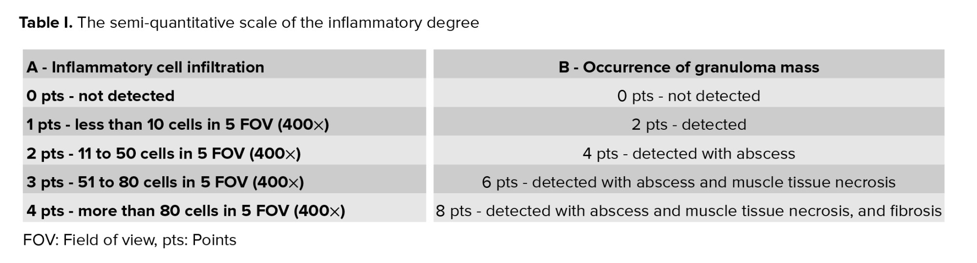
After the experiment, each group was anesthetized and euthanized to collect the peritoneum tissue's histopathological samples from the peritoneum tissue. The areas on the peritoneum showing inflammatory signs (reddest part on the peritoneum) were sliced and preserved in 10% formalin before the staining process. Tissue slides were prepared by embedding the tissue in paraffin and sliced 4-6 µm thickness. Subsequently, the slides were stained in hematoxylin-eosin. Briefly, the tissue was deparaffinized in xylol for 5 min. This step was repeated twice. Then, the slides were immersed in a gradually reduced ethanol concentration for 1 min at each concentration; 100% ethanol twice, 95% ethanol twice, and 70% ethanol twice. Afterward, the slides were washed in water for 10 min before staining Mayer hematoxylin solution for 1 min. Then, the slides were counterstained in Eosin solution for 2 min and rehydrated in 95% alcohol and 100% ethanol for 3 min in each concentration of ethanol. Subsequently, xylol was added to the slides for 3 × 3 min, then air-dried and covered with cover glass. The examination of the slides was performed under a light microscope with 400× magnification. The observation of the inflammation degree was using a modified Klopfleisch semiquantitative scoring system, which determines the inflammatory level according to the infiltration of inflammatory cells and the occurrence of granuloma mass (8, 9). As seen in table I, the scoring system summarizes the percentage of inflammatory cells (A) and the granuloma mass (B).
2.8. Immunostaining for NFκB
The tissue slides for immunohistochemistry staining were prepared following protocol described previously (10). The antibody and staining kits were purchased from Santa Cruz Biotechnology, CA, and the staining was performed according to the manufacturer's instructions. Briefly, the slides were fixed in acetone and blocked with 0.1% BSA. Peroxidase activities were depleted by using 0.3% hydrogen peroxide. Then, the slides were incubated in NFκB antibody (ab16502) at room temperature for 3 hr. Following antibody staining, the slides were washed in PBS 3 times and incubated in the biotinylated secondary. The reaction visualization was done using a diaminobenzidine kit. The microscopic examination was performed to observe the expression of NFκB expressed on the peritoneum tissue, and the score was measured semiquantitative according to the modified immunoreactive score (Index Remmele Scale) (11). The scoring system measures the percentage of positive cells (A) and the color intensity of the positive cells (Table II).

2.9. Ethical considerations
All experimental procedures followed the National guidelines for the experimental animal (Airlangga University, Indonesia) and were approved by the Animal Care and Use Committee of Airlangga University Surabaya, Indonesia (Code: ACUC no 497-KE).
2.10. Statistical analysis
Data analysis was conducted by statistical analysis using a one-way ANOVA test. The correlation between the NFκB level and the inflammation level was evaluated using Spearman's correlation test. Data were presented as a mean value with a standard deviation from 8 samples of each group. The statistical analysis used the statistical package for the social sciences (SPSS) software, version 19 (IBM Corp. Released 2010. IBM SPSS Statistics for Windows, Version 19.0. Armonk, NY: IBM Corp). A p-value < 0.05 was considered as significance.
3. Results
3.1. Histopathology degree of peritoneal inflammation
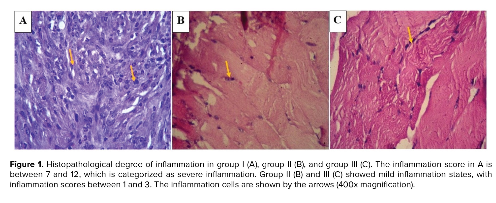
The implantation of endometrial cells has induced the inflammatory reaction in the mice peritoneal. The histopathological examination showed that the endometriosis lesions formed in all experimental animals within all groups, with various levels of inflammation degree (Figure 1).
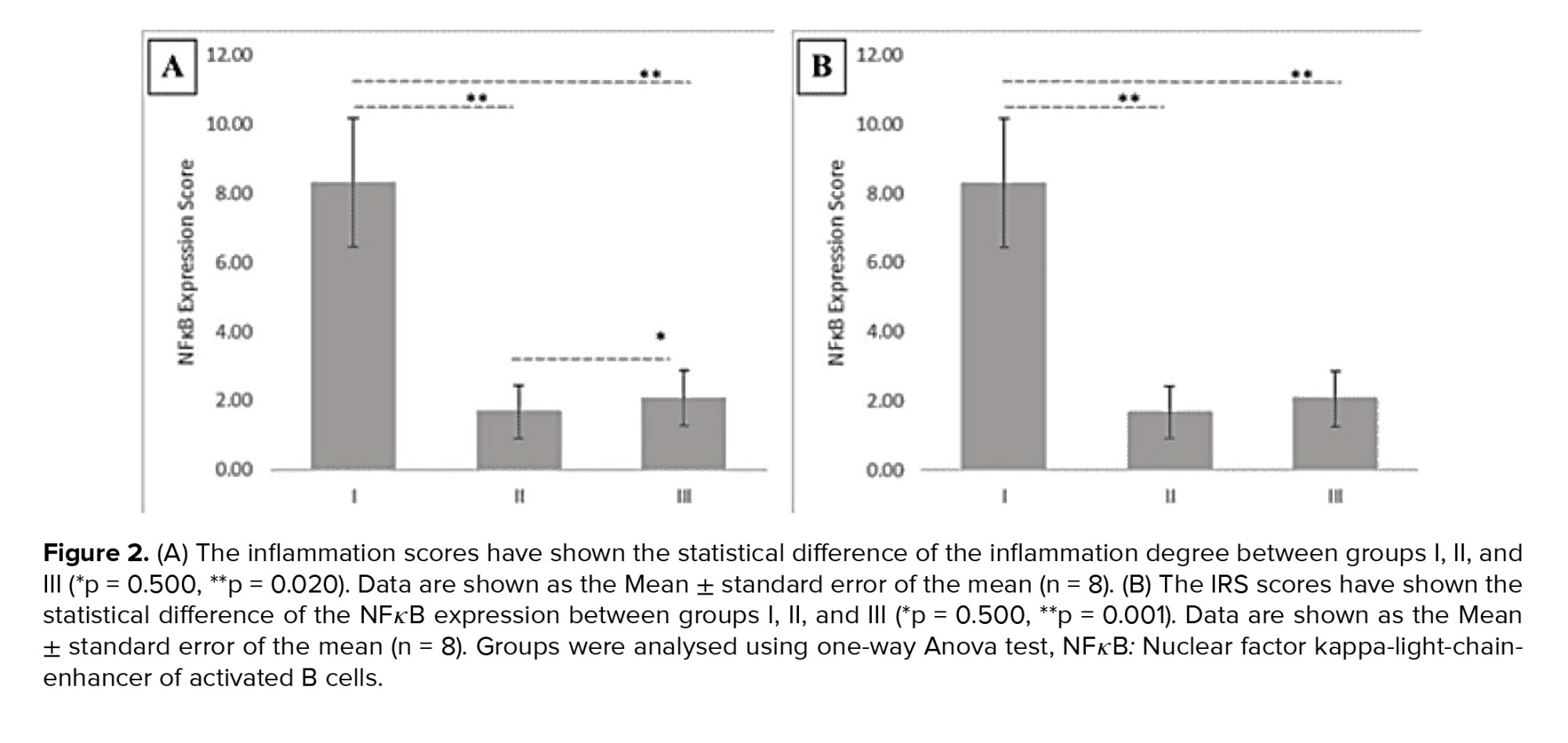
According to the Klopfleisch semiquantitative scores, the pre-test group (I) demonstrated the highest level of inflammation degree (9.41 ± 1.99) compared to the post-test groups (II: 1.60 ± 0.53; III: 2.42 ± 0.53). The group with HBOT (II) was found to have the lowest inflammation level compared to I (p = 0.020) and III (p = 0.020). The statistical difference between all groups is shown in figure 2. Overall, the results have demonstrated that HBOT has lowered the peritoneal inflammation degree caused by the endometrial lesion in mice.
3.2. NFκB expression
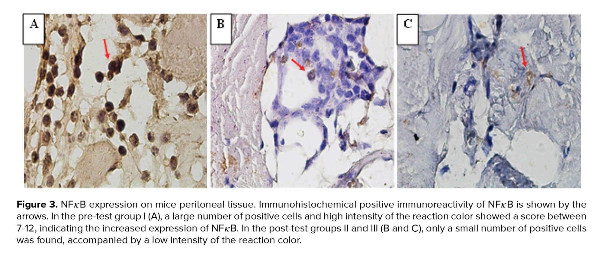
The expression of NFκB on the peritoneal tissue using immunohistochemical staining is shown in arrows (Figure 3). Based on the immuno reactive score (IRS) scoring method, the pre-test group (I) demonstrated the highest level of NFκB expression (8.8 ± 2.32) compared to the post-test group (I and II). However, there is no significant difference between II and III (2.5 ± 2.66 and 2.7 ± 2.51, respectively).
3.3. The correlation between NFκB expression and the degree of inflammation
The extent of NFκB expression with the inflammation degree disclosed a pronounced positive correlation between the studied parameter (4). The correlation analysis resulted (Figure 4) in a coefficient value amounted to r = 0.670 (spearman’s correlation).

4. Discussion
Our results demonstrated that a red and active lesion occurred as a sign of inflammatory reaction upon endometriosis induction in the mice peritoneum. Histopathological examination also demonstrated the high level of inflammation degree showed on the formed endometriosis lesions. The administration of HBOT for 2 wk on the endometriosis-induced mice significantly reduced the inflammation state of the formed lesion, compared to the group that did not receive HBOT (Klopfleisch scores 1.6 ± 0.53 and 2.4 ± 0.53, respectively). The results indicated a significant inflammation event occurred during the development of endometriosis lesions. The HBOT aimed at correcting the hypoxic environment within the cell environment increases the dissolved oxygen content in the circulation. It escalates the oxygen transfer to the inflamed cells (12, 13). It is well known that wound healing, particularly in inflammatory-related lesions, requires an adequate supply of oxygen tension because adequate oxygen supply is essential for the formation of collagen matrix and subsequent angiogenesis (10, 13). HBOT also reduces and eliminates the retention of CO2 in the alveolar, which may help in increasing the oxygen content in the cells (14). In addition to this, HBOT has demonstrated a positive effect in reviving ischemic tissue by inhibiting the adhesion of neutrophil and post-ischemic vasoconstriction (15).
In the human endometrium, NFκB protein can be expressed constitutively and play an inevitable role in the physiological changes on endometrial cells; the expression is exceptionally high during the secretion phase of the endometrial cycle (15). Activation of the NFκB pathway is known as a response to several stimuli, including inflammatory stimuli Interleukin-1 (IL-1) and tumor necrosis factor alpha (TNF-α), ROS, and hypoxia (16). In endometriosis pathogenesis, hypoxia occurs due to the disturbance of immune response, which leads to the aberrant secretion of inflammatory mediators from the endometrial tissue (17). On the other hand, the occurrence of hypoxia in a lesion will result in the exaggeration of the inflammatory response in the affected area, which may cause clinical manifestation such as chronic pain and other inflammatory-related symptoms in endometriosis (1, 18). Therefore, in the modality of endometriosis therapy, reducing the hypoxia condition may play a key role in inhibiting the progression of endometriosis lesion development by reducing the secretion and activation of inflammatory mediators and, more importantly, helping to ease the symptoms due to the inflammatory responses.
HBOT has been used in treating various medical conditions and has been proved to reduce the inflammatory mediator level (5, 19). Administration of HBOT for 2 hr a day in 6 wk on endometriosis-induced rats has demonstrated a significant lowering effect of the TNF-α level, leading to a remission of the endometriosis lesion due to the inhibition of NFκB (19). The finding from the previous study also emphasized the positive effect of HBOT in reducing the NFκB expression and other inflammatory cytokines in acute pancreatitis in rats. The same study showed that the level of nuclear factor of kappa light polypeptide gene enhancer in B-cells inhibitor, alphaIκBα, the co-activator of NFκB, was notably reduced, suggesting that the mechanism of action of HBOT involving the NFκB pathway (20). In our study, the administration of HBOT was given for 30 min 3 times a day for 10 days. The results demonstrated a significant decrease of NFκB expression after the HBOT (IRS score 2.50 ± 2.66) compared to the expression before the treatment (IRS score 8.80 ± 2.320). However, the NFκB expression showed no significant difference compared to the untreated group (IRS score 2.70 ± 2.51). The finding may show that in both groups receiving non-receiving HBOT, the low expression of NFκB has given a clear sign of remission of the endometrial lesion. Our finding demonstrated that in the HBOT-receiving group, the inflammation level decreased significantly compared to the non-treated group (Klopfleisch scores 1.60 ± 0.53 and 2.40 ± 0.53, respectively). The immune systems may initiate the healing process without any treatment, by self-modulating the acute tissue repair process (21). Physiologically, this process will go through a complex cascade involving many stages of the healing-response phase. HBOT may speed up this process and increase the rate of healing response and the remission of an endometrial lesion in mice. In addition to this, our result has shown a strong correlation between NFκB expression and inflammation degree, suggesting that NFκB is an important factor in reducing the inflammation reaction. Therefore, our results have shown a positive effect of HBOT in reducing the inflammatory response in endometriosis. However, the level of NFκB was yet to reach a significant confidence level compared to the untreated group, although previous studies have demonstrated constructive evidence on the HBOT effect in lowering the proinflammatory mediators.
In many studies on HBO treatment, the therapeutic effects were dose- and time-dependent (19, 20). Administration of HBO treatment on human blood-derived monocyte-macrophages for 3 hr reduced the Interleukin 1β synthesis, while prolonged HBOT for 12 hr increased the cytokine production (22). In indomethacin-induced enteropathy in rats, HBOT reduced the TNF-α expression after 12 hr and 24 hr treatment, but the effect was null after 48 hr. The same study demonstrated that HBOT showed no effect on the IL-1b production after 12 hr treatment. However, the positive effect was observed after 24 hr treatment, which showed that HBOT significantly lowered the IL-1β secretion (23). With regards to the HBOT effect on the inflammatory mediators in endometriosis, it has been reported that HBOT showed an observable effect on lowering the TNF-α levels after 6 wk of treatment (20). Therefore, all this evidence may explain the level of NFκB shown from our result, which is likely related to the dose and duration of the HBOT used in this study.
5. Conclusion
Our study showed that HBO therapy reduced the inflammatory state in endometrial lesions, possibly via the alteration of the NFκB pathway. Further investigation in the duration and dose of HBOT is needed to elucidate the molecular mechanism of HBOT in endometriosis. Our results have shown that HBOT significantly reduced the inflammation level on the endometrial lesions, with a low level of NFκB expression.
Acknowledgments
The study received funding from the Indonesia Ministry of Health through RISBIN IPTEKDOK Program. The project is supported by Lembaga Kesehatan Kelautan TNI AL for the opportunity to use the animal hyperbaric chamber facility. The technical support was also given by Mrs. Widjiati, DVM, M.Sc., Ph.D., Mr. Djoko Legowo DVM, M.Sc. and team.
Conflict of Interest
The authors declare that they have no competing interest.
Full-Text: (260 Views)
1. Introduction
Inflammation theory is one of the molecular mechanisms in the pathophysiology of endometriosis, which is demonstrated by the alteration of the physiological activities and infiltration of immune cells in the endometriosis sites (1, 2). Inflammation in endometriosis is also evident from the increase of secretion of pro-inflammatory mediators, such as cytokines and prostaglandin, in the site of lesion and peritoneal cavity (3). This condition can be stimulated by the lack, if not absence, of oxygen in the cell’s environment, known as hypoxia condition.
Hypoxia stress potentiates the pro-inflammatory pathways in endometriosis by facilitating the accumulation of inflammatory molecules such as hypoxia-inducible factor-1 alpha (HIF-1α) and nuclear factor kappa beta, which lead to the downstream cascade effect of prostaglandin production (4-6). The deprivation of oxygen level also induces the imbalance of estrogen receptors resulting in the accumulation of HIF-1α in the endometriosis lesions (4, 5). Despite the inevitable role of hypoxia in endometriosis inflammatory response, only a few studies have been conducted to elucidate the molecular basis of hypoxia and identify the suitable treatment for the hypoxic condition in endometriosis.
Hyperbaric oxygen therapy (HBOT) is a medical tested to treat hypoxia-related medical conditions, including inflammatory diseases (6). A previous study demonstrated the positive effect of HBOT in reducing the pro-inflammatory cytokine HIF-1α in endometriosis lesions, possibly due to the clearance of HIF-1α molecules in an oxygen-rich environment (7). The effect of HBOT may lead to the alteration of another inflammatory cascade involving estrogenic receptors and the corresponding inflammatory mediator such as nuclear factor kappa (NFκB), which decreases the inflammatory state in the lesions. NFκB is an important factor in endometriosis pathophysiology, constitutively activated and highly expressed in endometrial lesions. This transcription factor serves a complex interaction with the steroid receptor, resulting in the maintenance of the inflammatory reaction in the lesion.
To show the role of HBOT in lowering the inflammatory state in endometriosis, we considered it necessary to evaluate the effect of HBOT in peritoneal inflammation and changes of the expression of NFκB on the peritoneal tissue of mouse model endometriosis.
2. Materials and Methods
2.1. Chemical products
All chemical substances used in this study were purchased from Sigma-Aldrich (St. Louise, MO, USA).
2.2. Experimental animals
24 healthy adult female swiss albino mice (average age of 4 wk), with a weight range of 25-30 gr, were obtained from the Veteriner Farma Centre, Surabaya, Indonesia. Before starting the experiment, the animals were acclimatized for 1 wk for the adaptation process. The animal with more than 10% weight loss was excluded from the study.
2.3. Experimental design
Using a randomized controlled study design, the animals were designated into groups I, II, and III by simple random sampling. Each group consists of 8 animals. Group I was the pre-test group, group II was the post-test group receiving the HBOT, while group III was the post-test without HBOT. All animals were subjected to induction of apoptosis by xenotransplantation for 15 days. The pre-test group (I) and post-test group (II) were not given the HBOT treatment, while the group III was given HBOT treatment for 10 days. After the experiment, the group I was examined after endometriosis induction. The other 2 groups, II and III, were examined simultaneously after HBOT treatment for group III was completed.
2.4. Preparation of endometrial cells
The endometrial cells were collected during the surgery from uterine adenomyosis women. The tissue was then washed using phosphate buffer saline (PBS) and homogenized using mortar and pestle. The ground tissue was washed in PBS and centrifuged at 2500 rpm twice to obtain the cell pellets. Subsequently, PBS containing 200 μg/mL of streptomycin and 200 IU/mL of penicillin were added into the cell pellets and incubated overnight.
2.5. Induction of endometriosis
The procedures for the xenotransplantation of endometrial cells on the mice were performed according to the previously published protocol (8). After the adaptation, each mouse was injected with 0.1 ml (10 mg/kg body weight) of cyclosporin A to suppress the animal's immune system. Then the mice were injected with 0.2 ml ethinyl estradiol at a dose of 0.2 μg/mouse by intramuscular injection. Subsequently, all mice were injected with the 0.1 ml endometrial cell suspension by intraperitoneal injection. On day 5, after the xenotransplantation, another dose of ethinyl estradiol was repeated. On day 15, the induction of endometriosis is completed.
2.6. HBOT
One day after the endometriosis induction, the mice in group I was placed inside the hyperbaric chamber to receive the HBOT. The oxygen dose in the chamber was 100% O2, with a flow rate of 8-10 L/min. The hyperbaric oxygen treatment was given as 2.4 atm pressure for 3 × 30 min with a 5-min air break. The treatment was given for 10 days in a row.
2.7. Histological evaluation
After the experiment, each group was anesthetized and euthanized to collect the peritoneum tissue's histopathological samples from the peritoneum tissue. The areas on the peritoneum showing inflammatory signs (reddest part on the peritoneum) were sliced and preserved in 10% formalin before the staining process. Tissue slides were prepared by embedding the tissue in paraffin and sliced 4-6 µm thickness. Subsequently, the slides were stained in hematoxylin-eosin. Briefly, the tissue was deparaffinized in xylol for 5 min. This step was repeated twice. Then, the slides were immersed in a gradually reduced ethanol concentration for 1 min at each concentration; 100% ethanol twice, 95% ethanol twice, and 70% ethanol twice. Afterward, the slides were washed in water for 10 min before staining Mayer hematoxylin solution for 1 min. Then, the slides were counterstained in Eosin solution for 2 min and rehydrated in 95% alcohol and 100% ethanol for 3 min in each concentration of ethanol. Subsequently, xylol was added to the slides for 3 × 3 min, then air-dried and covered with cover glass. The examination of the slides was performed under a light microscope with 400× magnification. The observation of the inflammation degree was using a modified Klopfleisch semiquantitative scoring system, which determines the inflammatory level according to the infiltration of inflammatory cells and the occurrence of granuloma mass (8, 9). As seen in table I, the scoring system summarizes the percentage of inflammatory cells (A) and the granuloma mass (B).
2.8. Immunostaining for NFκB
The tissue slides for immunohistochemistry staining were prepared following protocol described previously (10). The antibody and staining kits were purchased from Santa Cruz Biotechnology, CA, and the staining was performed according to the manufacturer's instructions. Briefly, the slides were fixed in acetone and blocked with 0.1% BSA. Peroxidase activities were depleted by using 0.3% hydrogen peroxide. Then, the slides were incubated in NFκB antibody (ab16502) at room temperature for 3 hr. Following antibody staining, the slides were washed in PBS 3 times and incubated in the biotinylated secondary. The reaction visualization was done using a diaminobenzidine kit. The microscopic examination was performed to observe the expression of NFκB expressed on the peritoneum tissue, and the score was measured semiquantitative according to the modified immunoreactive score (Index Remmele Scale) (11). The scoring system measures the percentage of positive cells (A) and the color intensity of the positive cells (Table II).
2.9. Ethical considerations
All experimental procedures followed the National guidelines for the experimental animal (Airlangga University, Indonesia) and were approved by the Animal Care and Use Committee of Airlangga University Surabaya, Indonesia (Code: ACUC no 497-KE).
2.10. Statistical analysis
Data analysis was conducted by statistical analysis using a one-way ANOVA test. The correlation between the NFκB level and the inflammation level was evaluated using Spearman's correlation test. Data were presented as a mean value with a standard deviation from 8 samples of each group. The statistical analysis used the statistical package for the social sciences (SPSS) software, version 19 (IBM Corp. Released 2010. IBM SPSS Statistics for Windows, Version 19.0. Armonk, NY: IBM Corp). A p-value < 0.05 was considered as significance.
3. Results
3.1. Histopathology degree of peritoneal inflammation
The implantation of endometrial cells has induced the inflammatory reaction in the mice peritoneal. The histopathological examination showed that the endometriosis lesions formed in all experimental animals within all groups, with various levels of inflammation degree (Figure 1).
According to the Klopfleisch semiquantitative scores, the pre-test group (I) demonstrated the highest level of inflammation degree (9.41 ± 1.99) compared to the post-test groups (II: 1.60 ± 0.53; III: 2.42 ± 0.53). The group with HBOT (II) was found to have the lowest inflammation level compared to I (p = 0.020) and III (p = 0.020). The statistical difference between all groups is shown in figure 2. Overall, the results have demonstrated that HBOT has lowered the peritoneal inflammation degree caused by the endometrial lesion in mice.
3.2. NFκB expression
The expression of NFκB on the peritoneal tissue using immunohistochemical staining is shown in arrows (Figure 3). Based on the immuno reactive score (IRS) scoring method, the pre-test group (I) demonstrated the highest level of NFκB expression (8.8 ± 2.32) compared to the post-test group (I and II). However, there is no significant difference between II and III (2.5 ± 2.66 and 2.7 ± 2.51, respectively).
3.3. The correlation between NFκB expression and the degree of inflammation
The extent of NFκB expression with the inflammation degree disclosed a pronounced positive correlation between the studied parameter (4). The correlation analysis resulted (Figure 4) in a coefficient value amounted to r = 0.670 (spearman’s correlation).
4. Discussion
Our results demonstrated that a red and active lesion occurred as a sign of inflammatory reaction upon endometriosis induction in the mice peritoneum. Histopathological examination also demonstrated the high level of inflammation degree showed on the formed endometriosis lesions. The administration of HBOT for 2 wk on the endometriosis-induced mice significantly reduced the inflammation state of the formed lesion, compared to the group that did not receive HBOT (Klopfleisch scores 1.6 ± 0.53 and 2.4 ± 0.53, respectively). The results indicated a significant inflammation event occurred during the development of endometriosis lesions. The HBOT aimed at correcting the hypoxic environment within the cell environment increases the dissolved oxygen content in the circulation. It escalates the oxygen transfer to the inflamed cells (12, 13). It is well known that wound healing, particularly in inflammatory-related lesions, requires an adequate supply of oxygen tension because adequate oxygen supply is essential for the formation of collagen matrix and subsequent angiogenesis (10, 13). HBOT also reduces and eliminates the retention of CO2 in the alveolar, which may help in increasing the oxygen content in the cells (14). In addition to this, HBOT has demonstrated a positive effect in reviving ischemic tissue by inhibiting the adhesion of neutrophil and post-ischemic vasoconstriction (15).
In the human endometrium, NFκB protein can be expressed constitutively and play an inevitable role in the physiological changes on endometrial cells; the expression is exceptionally high during the secretion phase of the endometrial cycle (15). Activation of the NFκB pathway is known as a response to several stimuli, including inflammatory stimuli Interleukin-1 (IL-1) and tumor necrosis factor alpha (TNF-α), ROS, and hypoxia (16). In endometriosis pathogenesis, hypoxia occurs due to the disturbance of immune response, which leads to the aberrant secretion of inflammatory mediators from the endometrial tissue (17). On the other hand, the occurrence of hypoxia in a lesion will result in the exaggeration of the inflammatory response in the affected area, which may cause clinical manifestation such as chronic pain and other inflammatory-related symptoms in endometriosis (1, 18). Therefore, in the modality of endometriosis therapy, reducing the hypoxia condition may play a key role in inhibiting the progression of endometriosis lesion development by reducing the secretion and activation of inflammatory mediators and, more importantly, helping to ease the symptoms due to the inflammatory responses.
HBOT has been used in treating various medical conditions and has been proved to reduce the inflammatory mediator level (5, 19). Administration of HBOT for 2 hr a day in 6 wk on endometriosis-induced rats has demonstrated a significant lowering effect of the TNF-α level, leading to a remission of the endometriosis lesion due to the inhibition of NFκB (19). The finding from the previous study also emphasized the positive effect of HBOT in reducing the NFκB expression and other inflammatory cytokines in acute pancreatitis in rats. The same study showed that the level of nuclear factor of kappa light polypeptide gene enhancer in B-cells inhibitor, alphaIκBα, the co-activator of NFκB, was notably reduced, suggesting that the mechanism of action of HBOT involving the NFκB pathway (20). In our study, the administration of HBOT was given for 30 min 3 times a day for 10 days. The results demonstrated a significant decrease of NFκB expression after the HBOT (IRS score 2.50 ± 2.66) compared to the expression before the treatment (IRS score 8.80 ± 2.320). However, the NFκB expression showed no significant difference compared to the untreated group (IRS score 2.70 ± 2.51). The finding may show that in both groups receiving non-receiving HBOT, the low expression of NFκB has given a clear sign of remission of the endometrial lesion. Our finding demonstrated that in the HBOT-receiving group, the inflammation level decreased significantly compared to the non-treated group (Klopfleisch scores 1.60 ± 0.53 and 2.40 ± 0.53, respectively). The immune systems may initiate the healing process without any treatment, by self-modulating the acute tissue repair process (21). Physiologically, this process will go through a complex cascade involving many stages of the healing-response phase. HBOT may speed up this process and increase the rate of healing response and the remission of an endometrial lesion in mice. In addition to this, our result has shown a strong correlation between NFκB expression and inflammation degree, suggesting that NFκB is an important factor in reducing the inflammation reaction. Therefore, our results have shown a positive effect of HBOT in reducing the inflammatory response in endometriosis. However, the level of NFκB was yet to reach a significant confidence level compared to the untreated group, although previous studies have demonstrated constructive evidence on the HBOT effect in lowering the proinflammatory mediators.
In many studies on HBO treatment, the therapeutic effects were dose- and time-dependent (19, 20). Administration of HBO treatment on human blood-derived monocyte-macrophages for 3 hr reduced the Interleukin 1β synthesis, while prolonged HBOT for 12 hr increased the cytokine production (22). In indomethacin-induced enteropathy in rats, HBOT reduced the TNF-α expression after 12 hr and 24 hr treatment, but the effect was null after 48 hr. The same study demonstrated that HBOT showed no effect on the IL-1b production after 12 hr treatment. However, the positive effect was observed after 24 hr treatment, which showed that HBOT significantly lowered the IL-1β secretion (23). With regards to the HBOT effect on the inflammatory mediators in endometriosis, it has been reported that HBOT showed an observable effect on lowering the TNF-α levels after 6 wk of treatment (20). Therefore, all this evidence may explain the level of NFκB shown from our result, which is likely related to the dose and duration of the HBOT used in this study.
5. Conclusion
Our study showed that HBO therapy reduced the inflammatory state in endometrial lesions, possibly via the alteration of the NFκB pathway. Further investigation in the duration and dose of HBOT is needed to elucidate the molecular mechanism of HBOT in endometriosis. Our results have shown that HBOT significantly reduced the inflammation level on the endometrial lesions, with a low level of NFκB expression.
Acknowledgments
The study received funding from the Indonesia Ministry of Health through RISBIN IPTEKDOK Program. The project is supported by Lembaga Kesehatan Kelautan TNI AL for the opportunity to use the animal hyperbaric chamber facility. The technical support was also given by Mrs. Widjiati, DVM, M.Sc., Ph.D., Mr. Djoko Legowo DVM, M.Sc. and team.
Conflict of Interest
The authors declare that they have no competing interest.
Type of Study: Original Article |
Subject:
Reproductive Biology
References
1. Laganà AS, Garzon S, Götte M, Vigano P, Franchi M, Ghezzi F, et al. The pathogenesis of endometriosis: Molecular and cell biology insights. Int J Mol Sci 2019; 20: 5615. [DOI:10.3390/ijms20225615] [PMID] [PMCID]
4. García-Gómez E, Vázquez-Martínez ER, Reyes-Mayoral Ch, Cruz-Orozco OP, Camacho-Arroyo I, Cerbón M. Regulation of inflammation pathways and inflammasome by sex steroid hormones in endometriosis. Front Endocrinol 2019; 10: 935. [DOI:10.3389/fendo.2019.00935] [PMID] [PMCID]
7. Wu MH, Lu ChW, Chang FM, Tsai ShJ. Estrogen receptor expression affected by hypoxia inducible factor-1α in stromal cells from patients with endometriosis. Taiwan J Obstet Gynecol 2012; 51: 50-54. [DOI:10.1016/j.tjog.2012.01.010] [PMID]
10. Zhang L, Xiong W, Li N, Liu H, He H, Du Y, et al. Estrogen stabilizes hypoxia-inducible factor 1α through G protein-coupled estrogen receptor 1 in eutopic endometrium of endometriosis. Fertil Steril 2017; 107: 439-447. [DOI:10.1016/j.fertnstert.2016.11.008] [PMID] [PMCID]
13. Sun Y, Wen Y, Shen Ch, Zhu Y, You W, Meng Y, et al. Hyperbaric oxygen therapy in liver diseases. Int J Med Sci 2018; 15: 782-787. [DOI:10.7150/ijms.24755] [PMID] [PMCID]
16. Syahrizal D, Mustika C, Renaldi T, Suryokusumo MG, Hendarto H. Hyperbaric oxygen (HBO) reduce expression of hypoxia inducible factor-1 Alpha (HIF-1 alpha) and endometriotic tissue size in mice model of endometriosis. Paper presented at: The 1St International Conference on Veterinary, Animal, and Environmental Sciences; 2019 October 15-17; Indonesia. [DOI:10.1051/e3sconf/202015101003]
18. Sutrisno S, Sri A, Wiyasa IWA, Umi K, Noerhamdani N, Hidayat S, et al. The effect of implant origin differences on peritoneal endometriosis in an endometriosis mouse model. Int J Women's Health Reprod Sci 2019; 7: 34-40. [DOI:10.15296/ijwhr.2019.06]
20. Klopfleisch R. Multiparametric and semiquantitative scoring systems for the evaluation of mouse model histopathology: A systematic review. BMC Vet Res 2013; 9: 123. [DOI:10.1186/1746-6148-9-123] [PMID] [PMCID]
23. Han Z, Boyle DL, Manning AM, Firestein GS. AP-1 and NF-kB regulation in rheumatoid arthritis and murine collagen-induced arthritis. Autoimmunity 1998; 28: 197-208. [DOI:10.3109/08916939808995367] [PMID]
26. Nowak M, Madej JA, Dziegiel P. Intensity of COX2 expression in cells of soft tissue fibrosacrcomas in dogs as related to grade of tumour malignancy. Bullet Vet Institute Pulawy 2007; 51: 275-279.
27. Carney AY. Hyperbaric oxygen therapy: An introduction. Crit Care Nurs Q 2013; 36: 274-279. [DOI:10.1097/CNQ.0b013e318294e936] [PMID]
30. Shinomiya N. Molecular mechanisms of hyperbaric oxygen therapy. In: Shinomiya N, Asai Y. Hyperbaric oxygenation therapy. Singapore: Springer; 2020: 3-20. [DOI:10.1007/978-981-13-7836-2_1]
32. Lam G, Fontaine R, Ross FL, Chiu ES. Hyperbaric oxygen therapy: Exploring the clinical evidence. Adv Skin Wound Care 2017; 30: 181-190. [DOI:10.1097/01.ASW.0000513089.75457.22] [PMID]
35. Oley MH, Oley MCh, Islam AA, Hatta M, Faruk M, Noersasongko AD, et al. Hyperbaric oxygen therapy in managing systemic inflammatory response syndrome caused by ischemia-reperfusion injury following hand replantation and long-term outcomes: A report of two cases. Ann Med Surg 2020; 60: 155-161. [DOI:10.1016/j.amsu.2020.10.023] [PMID] [PMCID]
38. Li C, Zhao HL, Li YJ, Zhang YY, Liu HY, Feng FZh, et al. The expression and significance of leukemia inhibitory factor, interleukin-6 and vascular endothelial growth factor in Chinese patients with endometriosis. Arch Gynecol Obstet 2021; 304: 163-170. [DOI:10.1007/s00404-021-05980-5] [PMID]
41. Kaponis A, Iwabe T, Taniguchi F, Ito M, Deura I, Decavalas G, et al. The role of NF-kappaB in endometriosis. Front Biosci 2012; 4: 1213-1234. [DOI:10.2741/s327] [PMID]
44. Hsiao KY, Lin ShCh, Wu MH, Tsai ShJ. Pathological functions of hypoxia in endometriosis. Front Biosci 2015; 7: 309-321. [DOI:10.2741/e736] [PMID]
47. Eltzschig HK, Carmeliet P. Hypoxia and inflammation. N Engl J Med 2011; 364: 656-665. [DOI:10.1056/NEJMra0910283] [PMID] [PMCID]
50. Aydin Y, Atis A, Uludag S, Tezer I, Sakiz D, Acar H, et al. Remission of endometriosis by hyperbaric oxygen treatment in rats. Reprod Sci 2011; 18: 941-947. [DOI:10.1177/1933719111400635] [PMID]
53. Yu X, Li YG, He XW, Li XR, Din BN, Gan Y, et al. Hyperbaric oxygen reduces inflammatory response in acute pancreatitis by inhibiting NF-κB activation. Eur Surg Res 2009; 42: 130-135. [DOI:10.1159/000196164] [PMID]
56. Velnar T, Bailey T, Smrkolj V. The wound healing process: An overview of the cellular and molecular mechanisms. J Int Med Res 2009; 37: 1528-1542. [DOI:10.1177/147323000903700531] [PMID]
59. Benson RM, Minter LM, Osborne BA, Granowitz EV. Hyperbaric oxygen inhibits stimulus‐induced proinflammatory cytokine synthesis by human blood‐derived monocyte‐macrophages. Clin Exp Immunol 2003; 134: 57-62. [DOI:10.1046/j.1365-2249.2003.02248.x] [PMID] [PMCID]
62. Yang Z, Nandi J, Wang J, Bosco G, Gregory M, Chung C, et al. Hyperbaric oxygenation ameliorates indomethacin-induced enteropathy in rats by modulating TNF-α and IL-1β production. Dig Dis Sci 2006; 51: 1426-1433. [DOI:10.1007/s10620-006-9088-2] [PMID]
Send email to the article author
| Rights and permissions | |
 |
This work is licensed under a Creative Commons Attribution-NonCommercial 4.0 International License. |
