
International Journal of
Reproductive Biomedicine
Mon, Jul 6, 2026
[Archive]
Volume 23, Issue 8 (August 2025)
IJRM 2025, 23(8): 647-658 |
Back to browse issues page


Ethics code: IR.SBMU.ENDOCRINE.REC.1403.038
![]()
![]()
![]()
Download citation:
BibTeX | RIS | EndNote | Medlars | ProCite | Reference Manager | RefWorks
Send citation to:


BibTeX | RIS | EndNote | Medlars | ProCite | Reference Manager | RefWorks
Send citation to:
Sadeghian Bakhi E, Hayati Roodbari N, Anvari M, Ramezani Tehrani F. Prenatal exposure to a single dose of testosterone adversely affects the oocyte and embryo quality in rats during adulthood: An experimental study. IJRM 2025; 23 (8) :647-658
URL: http://ijrm.ir/article-1-3526-en.html
URL: http://ijrm.ir/article-1-3526-en.html


1- Department of Biology, School of Basic Science, Science and Research Branch, Islamic Azad University, Tehran, Iran.
2- Research and Clinical Center for Infertility, Yazd Reproductive Sciences Institute, Shahid Sadoughi University of Medical Sciences, Yazd, Iran.
3- Reproductive Endocrinology Research Center, Research Institute for Endocrine Molecular Biology, Research Institute for Endocrine Sciences, Shahid Beheshti University of Medical Sciences, Tehran, Iran. & Foundation for Research and Education Excellence, Vestavia Hills, AL, USA. ,ramezani@endocrine.ac.ir; fah.tehrani@gmail.com; framezan@postharvard.edu
2- Research and Clinical Center for Infertility, Yazd Reproductive Sciences Institute, Shahid Sadoughi University of Medical Sciences, Yazd, Iran.
3- Reproductive Endocrinology Research Center, Research Institute for Endocrine Molecular Biology, Research Institute for Endocrine Sciences, Shahid Beheshti University of Medical Sciences, Tehran, Iran. & Foundation for Research and Education Excellence, Vestavia Hills, AL, USA. ,
Abstract: (837 Views)
Background: Prenatal exposure to excess androgen can adversely affect the hypothalamic-pituitary-ovarian axis in the developing fetus, potentially leading to long-term reproductive system dysfunction in later life.
Objective: We aimed to investigate whether prenatal exposure to a single dose of testosterone can affect the reproductive system, especially oocyte and embryo quality, and the ovarian expression of growth differentiation factor-9 (GDF-9) gene in rats during adulthood.
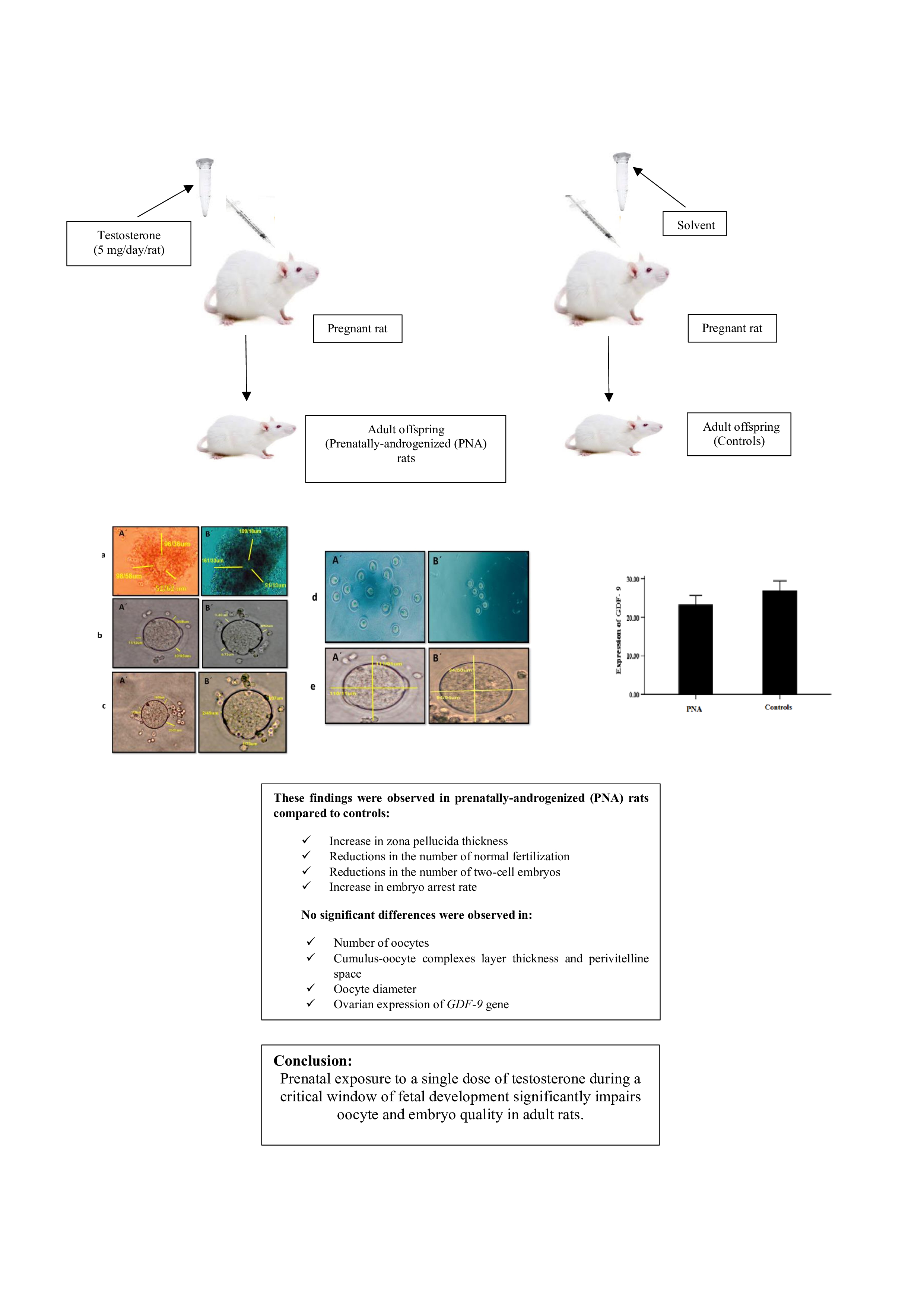
Materials and Methods: In this experimental study, pregnant Wistar rats (12-13 wk, 185 ± 10 gr) in the experimental group (n = 5) subcutaneously received free testosterone (5 mg) on the 20th day of pregnancy, whereas controls received only solvent (n = 5). The offspring were evaluated for oocyte and embryo quality (zona pellucida thickness, number of normal fertilization, number of 2-cell embryos, and embryo arrest rate) and ovarian expression of GDF-9 gene during adulthood.
Results: A significant increase was observed in the zona pellucida thickness (p = 0.02) in prenatally-androgenized (PNA) rats compared to controls. However, reductions in the number of normal fertilization and 2-cell embryos (p = 0.007, p = 0.01, respectively) in PNA rats compared to controls were observed. Furthermore, embryo arrest rate in PNA rats was significantly higher than in controls (p = 0.004). No significant difference was observed in the ovarian expression of GDF-9 gene in PNA rats compared to controls.
Conclusion: Prenatal exposure to a single dose of testosterone during a critical window of fetal development significantly impairs oocyte and embryo quality in adult rats. Further studies are needed to validate these findings and to elucidate the underlying molecular and physiological mechanisms.
Objective: We aimed to investigate whether prenatal exposure to a single dose of testosterone can affect the reproductive system, especially oocyte and embryo quality, and the ovarian expression of growth differentiation factor-9 (GDF-9) gene in rats during adulthood.
Materials and Methods: In this experimental study, pregnant Wistar rats (12-13 wk, 185 ± 10 gr) in the experimental group (n = 5) subcutaneously received free testosterone (5 mg) on the 20th day of pregnancy, whereas controls received only solvent (n = 5). The offspring were evaluated for oocyte and embryo quality (zona pellucida thickness, number of normal fertilization, number of 2-cell embryos, and embryo arrest rate) and ovarian expression of GDF-9 gene during adulthood.
Results: A significant increase was observed in the zona pellucida thickness (p = 0.02) in prenatally-androgenized (PNA) rats compared to controls. However, reductions in the number of normal fertilization and 2-cell embryos (p = 0.007, p = 0.01, respectively) in PNA rats compared to controls were observed. Furthermore, embryo arrest rate in PNA rats was significantly higher than in controls (p = 0.004). No significant difference was observed in the ovarian expression of GDF-9 gene in PNA rats compared to controls.
Conclusion: Prenatal exposure to a single dose of testosterone during a critical window of fetal development significantly impairs oocyte and embryo quality in adult rats. Further studies are needed to validate these findings and to elucidate the underlying molecular and physiological mechanisms.
Type of Study: Original Article |
Subject:
Reproductive Endocrinology
Send email to the article author
| Rights and permissions | |
 |
This work is licensed under a Creative Commons Attribution-NonCommercial 4.0 International License. |
