
International Journal of
Reproductive Biomedicine
Sun, Jul 26, 2026
[Archive]
Volume 24, Issue 1 (January 2026)
IJRM 2026, 24(1): 59-70 |
Back to browse issues page


Ethics code: IR.TBZMED.AEC.1401.087
![]()
![]()
![]()
Download citation:
BibTeX | RIS | EndNote | Medlars | ProCite | Reference Manager | RefWorks
Send citation to:


BibTeX | RIS | EndNote | Medlars | ProCite | Reference Manager | RefWorks
Send citation to:
Bilabari M, Mohammadnejad D, Rahbarghazi R, Shafaei H. Comparative effects of adipose-derived mesenchymal stem cells and their exosomes on interleukin-1 beta-induced testicular inflammation in rats: An experimental study. IJRM 2026; 24 (1) :59-70
URL: http://ijrm.ir/article-1-3690-en.html
URL: http://ijrm.ir/article-1-3690-en.html


1- Stem Cell Research Center, Tabriz University of Medical Sciences, Tabriz, Iran. & Immunology Research Center, Tabriz University of Medical Sciences, Tabriz, Iran., Department of Anatomical Sciences, Faculty of Medicine, Tabriz University of Medical Sciences, Tabriz, Iran.
2- Department of Anatomical Sciences, Faculty of Medicine, Tabriz University of Medical Sciences, Tabriz, Iran.
3- Stem Cell Research Center, Tabriz University of Medical Sciences, Tabriz, Iran.
4- Immunology Research Center, Tabriz University of Medical Sciences, Tabriz, Iran. & Department of Anatomical Sciences, Faculty of Medicine, Tabriz University of Medical Sciences, Tabriz, Iran. ,shafaei49@gmail.com; shafaeih@tbzmed.ac.ir
2- Department of Anatomical Sciences, Faculty of Medicine, Tabriz University of Medical Sciences, Tabriz, Iran.
3- Stem Cell Research Center, Tabriz University of Medical Sciences, Tabriz, Iran.
4- Immunology Research Center, Tabriz University of Medical Sciences, Tabriz, Iran. & Department of Anatomical Sciences, Faculty of Medicine, Tabriz University of Medical Sciences, Tabriz, Iran. ,
Abstract: (465 Views)
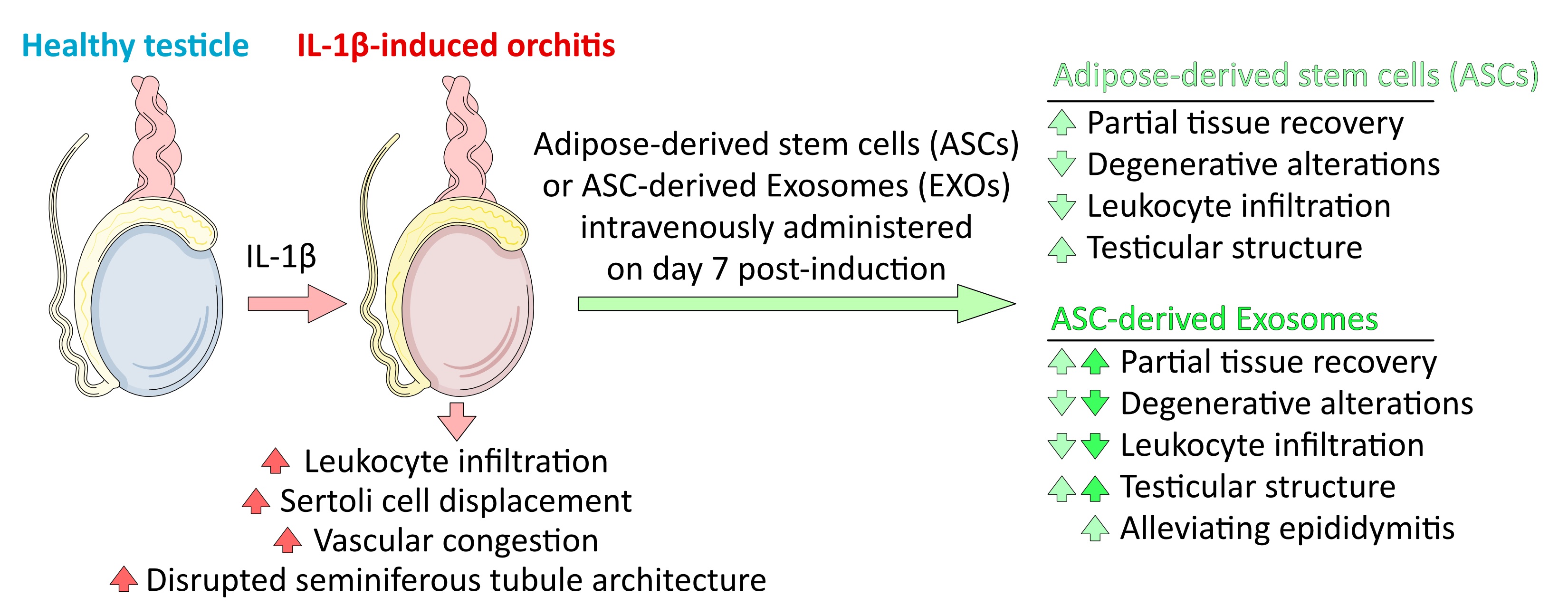
Background: Elevated interleukin-1 beta (IL-1β) in orchitis triggers immune-mediated inflammation, disrupting spermatogenesis and impairing reproductive function.
Objective: This study compared adipose-derived stem cells (ASCs) and their exosomes (EXO) for mitigating inflammation and enhancing repair in IL-1β-induced orchitis.
Materials and Methods: In this experimental study, ASCs were isolated and cultured to passage 3. Flow cytometry and immunocytochemistry confirmed ASCs' surface markers; scanning electron microscopy characterized EXO. 32 male Wistar rats (6-7 wk; 250-300 gr) received daily intraperitoneal IL-1β (10 μg/kg) for 6 days to induce testicular inflammation. Rats were assigned to 4 groups: 1) control; 2) IL-1β only; 3) IL-1β + ASCs (1×10⁶ cells); and 4) IL-1β + EXO (200 μg). After the treatments, testicular histopathology, hematoxylin and eosin staining, and leukocyte infiltration were evaluated.
Results: ASCs displayed spindle morphology and positivity for CD44 and CD105. Isolated EXO averaged 82.6 ± 28.5 nm in diameter. IL-1β induction caused considerable leukocyte infiltration, Sertoli cells displacement, vascular congestion, and disrupted seminiferous tubule structure. ASC administration yielded partial tissue recovery and reduced degenerative alterations. EXO demonstrated superior efficacy, significantly reducing leukocyte infiltration (p = 0.011) and restoring near-normal testicular structure. Exosome treatment also outperformed ASCs in alleviating epididymitis.
Conclusion: Both ASCs and exosome therapies reduced IL-1β-induced inflammation in the rat testis and epididymis. EXO demonstrated superior efficacy, highlighting their significant potential for treating inflammation-induced testicular injury.
Type of Study: Original Article |
Subject:
Stem Cell & Cloning
Send email to the article author
| Rights and permissions | |
 |
This work is licensed under a Creative Commons Attribution-NonCommercial 4.0 International License. |
