
International Journal of
Reproductive Biomedicine
Sat, Nov 7, 2026
[Archive]
Volume 24, Issue 9 (September 2026)
Int J Reprod BioMed. 2026, 24(9): 815-826 |
Back to browse issues page



Ethics code: IR.MAZUMS.RIB.REC.1401.037


Download citation:
BibTeX | RIS | EndNote | Medlars | ProCite | Reference Manager | RefWorks
Send citation to:


BibTeX | RIS | EndNote | Medlars | ProCite | Reference Manager | RefWorks
Send citation to:
Hosseini Gelevardi M H, Daneshfar Z, Farrokhfar S. Maternal morphine exposure during lactation alters hippocampal morphology and Oprm1 expression in rat offspring: An experimental study. Int J Reprod BioMed. 2026; 24 (9) :815-826
URL: http://ijrm.ir/article-1-3772-en.html
URL: http://ijrm.ir/article-1-3772-en.html

1- Student Research Committee, Ramsar Campus, Mazandaran University of Medical Sciences, Ramsar, Iran.
2- Faculty of Medicine, Ramsar Campus, Mazandaran University of Medical Sciences, Ramsar, Iran.
3- Department of Anatomical Sciences, Faculty of Medicine, Ramsar Campus, Mazandaran University of Medical Sciences, Ramsar, Iran. ,s.farrokhfar@mazums.ac.ir
2- Faculty of Medicine, Ramsar Campus, Mazandaran University of Medical Sciences, Ramsar, Iran.
3- Department of Anatomical Sciences, Faculty of Medicine, Ramsar Campus, Mazandaran University of Medical Sciences, Ramsar, Iran. ,
Abstract: (21 Views)
Background: Maternal morphine use during lactation may affect neonatal brain development through drug transfer into breast milk.
Objective: To investigate the effects of short-term maternal morphine exposure during lactation on hippocampal morphology and neurodevelopment-related gene expression in rat offspring.
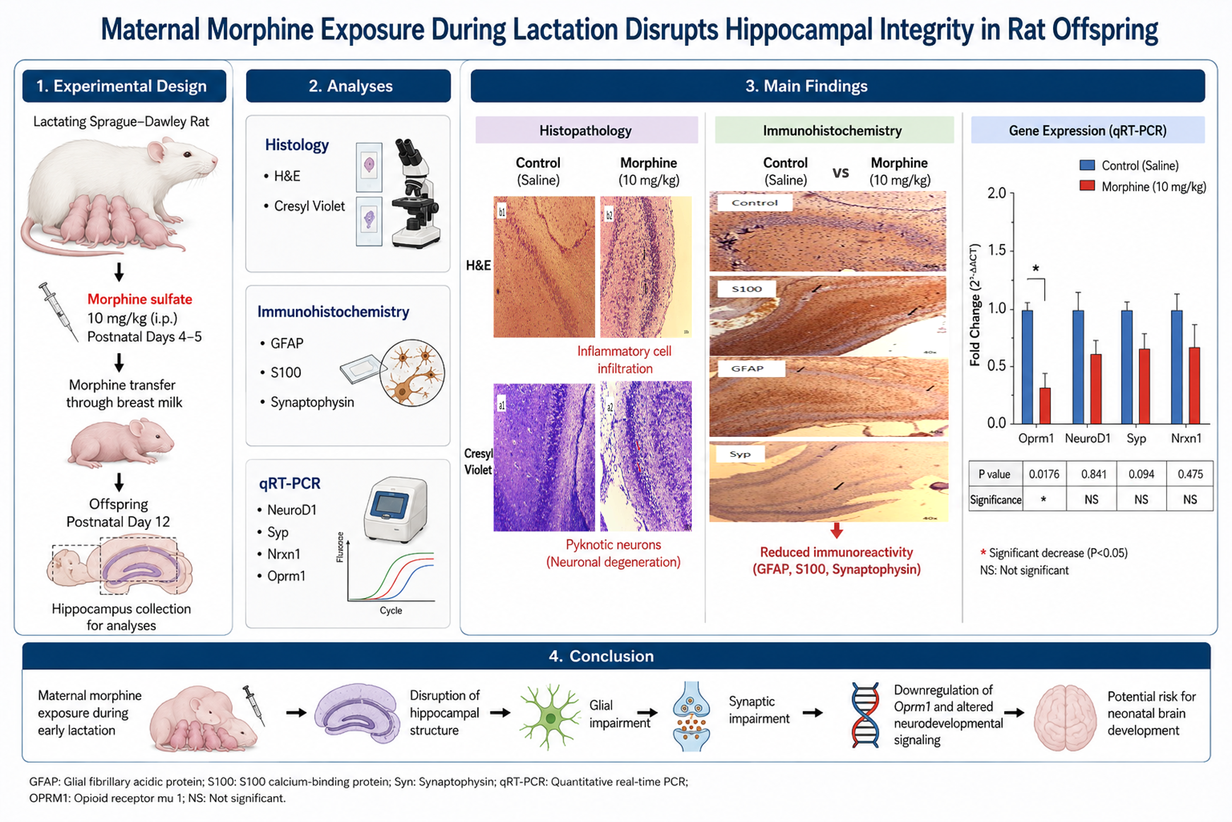
Materials and Methods: This experimental study was conducted at Mazandaran University of Medical Sciences, Ramsar Campus, Ramsar, Iran in 2025. Adult lactating Sprague-Dawley rats (210-240 gr, 2-4 months) were randomly assigned to either the control or morphine-treated group. Lactating female rats in the treatment group received morphine sulfate (10 mg/kg, intraperitoneally) on postnatal days 4 and 5, whereas control animals received normal saline. On postnatal day 12, hippocampal tissues from their offspring (n = 6/group) were collected for histological, immunohistochemical, and quantitative reverse transcription polymerase chain reaction analyses. Immunohistochemical assessment included glial fibrillary acidic protein, S100 calcium-binding protein, and synaptophysin. Gene expression levels of neuronal differentiation-1, synaptophysin, neurexin-1, and opioid receptor Mu 1 were quantified using the ΔΔCt-2 method.
Results: Histological examination revealed qualitative evidence of neuronal degeneration and inflammatory cell accumulation in the hippocampus of morphine-exposed offspring. Analysis demonstrated reduced immunoreactivity for glial fibrillary acidic protein, S100, and synaptophysin immunoreactivity compared with controls. Gene expression analysis showed a significant reduction in Oprm1 expression (p = 0.0176). Although other genes examined exhibited lower mean expression levels in the morphine-exposed group, these differences did not reach statistical significance.
Conclusion: Short-term maternal morphine exposure during lactation was associated with hippocampal histopathological alterations and reduced Oprm1 expression in rat offspring. Further studies are needed to confirm these findings.
Objective: To investigate the effects of short-term maternal morphine exposure during lactation on hippocampal morphology and neurodevelopment-related gene expression in rat offspring.
Materials and Methods: This experimental study was conducted at Mazandaran University of Medical Sciences, Ramsar Campus, Ramsar, Iran in 2025. Adult lactating Sprague-Dawley rats (210-240 gr, 2-4 months) were randomly assigned to either the control or morphine-treated group. Lactating female rats in the treatment group received morphine sulfate (10 mg/kg, intraperitoneally) on postnatal days 4 and 5, whereas control animals received normal saline. On postnatal day 12, hippocampal tissues from their offspring (n = 6/group) were collected for histological, immunohistochemical, and quantitative reverse transcription polymerase chain reaction analyses. Immunohistochemical assessment included glial fibrillary acidic protein, S100 calcium-binding protein, and synaptophysin. Gene expression levels of neuronal differentiation-1, synaptophysin, neurexin-1, and opioid receptor Mu 1 were quantified using the ΔΔCt-2 method.
Results: Histological examination revealed qualitative evidence of neuronal degeneration and inflammatory cell accumulation in the hippocampus of morphine-exposed offspring. Analysis demonstrated reduced immunoreactivity for glial fibrillary acidic protein, S100, and synaptophysin immunoreactivity compared with controls. Gene expression analysis showed a significant reduction in Oprm1 expression (p = 0.0176). Although other genes examined exhibited lower mean expression levels in the morphine-exposed group, these differences did not reach statistical significance.
Conclusion: Short-term maternal morphine exposure during lactation was associated with hippocampal histopathological alterations and reduced Oprm1 expression in rat offspring. Further studies are needed to confirm these findings.
Type of Study: Original Article |
Subject:
Cellular and Molecular Biology of Reproduction
Send email to the article author
| Rights and permissions | |
 |
This work is licensed under a Creative Commons Attribution-NonCommercial 4.0 International License. |