
International Journal of
Reproductive Biomedicine
Tue, May 26, 2026
[Archive]
Volume 23, Issue 11 (November 2025)
IJRM 2025, 23(11): 937-952 |
Back to browse issues page


Ethics code: IR-UU-AEC-32108
![]()
![]()
![]()
Download citation:
BibTeX | RIS | EndNote | Medlars | ProCite | Reference Manager | RefWorks
Send citation to:


BibTeX | RIS | EndNote | Medlars | ProCite | Reference Manager | RefWorks
Send citation to:
Javanmard S, Najdegerami E, Razi M, Nikoo M. An experimental study on shrimp bioactive peptides restoring testicular function in a rat model of fatty liver disease via autophagy, redox balance, and energy transporters. IJRM 2025; 23 (11) :937-952
URL: http://ijrm.ir/article-1-3544-en.html
URL: http://ijrm.ir/article-1-3544-en.html


1- Department of Biology, Faculty of Science, Urmia University, Urmia, Iran.
2- Department of Biology, Faculty of Science, Urmia University, Urmia, Iran. ,e.gerami@urmia.ac.ir
3- Department of Basic Science, Faculty of Veterinary Medicine, Urmia University, Urmia, Iran.
4- Artemia and Aquaculture Research Institute, Urmia University, Urmia, Iran.
2- Department of Biology, Faculty of Science, Urmia University, Urmia, Iran. ,
3- Department of Basic Science, Faculty of Veterinary Medicine, Urmia University, Urmia, Iran.
4- Artemia and Aquaculture Research Institute, Urmia University, Urmia, Iran.
Abstract: (413 Views)
Background: Non-alcoholic fatty liver disease is a widely prevalent condition in the modern era, with potential adverse effects on fertility and the reproductive system.
Objective: This experimental study investigated the adverse effects of non-alcoholic fatty liver disease induced by a high-fat diet (HFD) on testicular structural integrity, with a focus on oxidative stress and metabolic alterations.
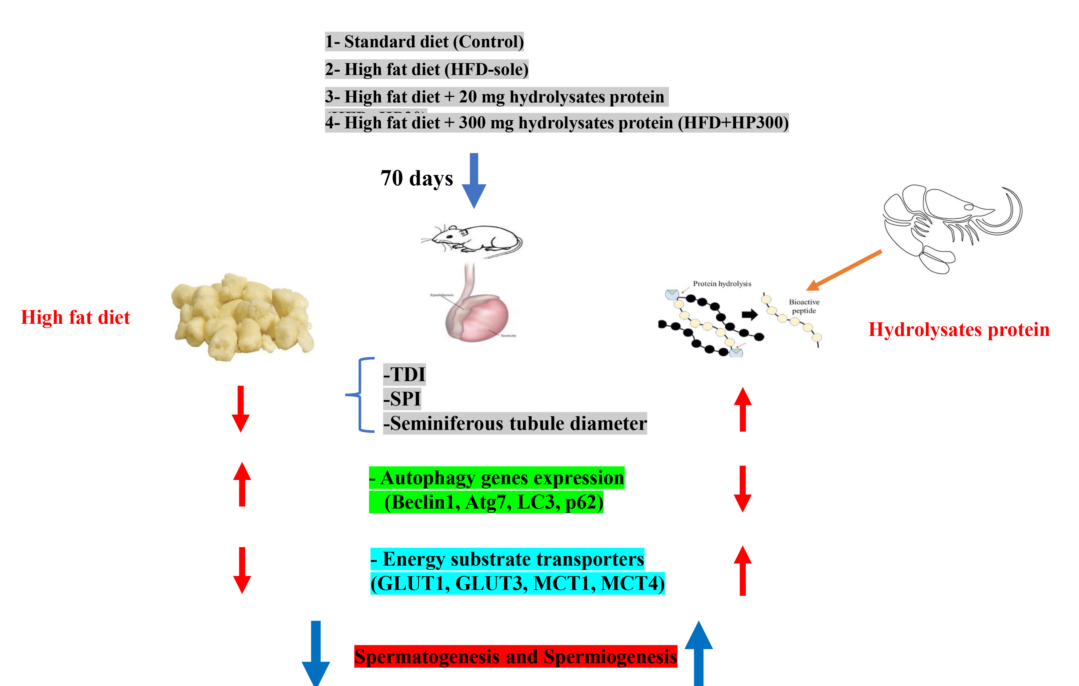
Materials and Methods: To produce bioactive peptides (HPs), whiteleg shrimp by-products were hydrolyzed using alcalase at 50°C for 3 hr. For this experimental study, 24 male Wistar rats (230 ± 23.1 gr, 8 wk) were randomly assigned to 4 groups (n = 6/each): control (standard diet), HFD-sole, HFD+HP20 (HFD supplemented with 20 mg/kg bodyweight of bioactive peptides), and HFD+HP300 (HFD supplemented with 300 mg/kg BW of bioactive peptides). After 10 wk of dietary intervention, testicular tissues were subjected to histological, molecular, and oxidative stress analyses.
Results: After 70 days, the HFD group showed higher malondialdehyde and glutathione, but a lower glutathione/glutathione disulphide ratio (40%, p < 0.001), indicating reductive stress. HPs, especially at higher doses, alleviated stress, improved seminiferous tubule morphology, and increased tubular differential index and spermiogenesis index indices. Autophagy genes (Beclin1, Atg7, LC3-I, p62) rose in HFD but were downregulated by HPs. Glucose transporter 1, 3 (GLUT-1+, GLUT-3+) and monocarboxylate transporter 4 cell distributions decreased in HFD but were restored in HP-received groups.
Conclusion: HPs improved tubular differential index and spermiogenesis index values associated with restored GLUT-1/3 and monocarboxylate transporter 4 expression in Sertoli cells, suggesting that Sertoli cells provided enhanced metabolic support for germ cell development.
Objective: This experimental study investigated the adverse effects of non-alcoholic fatty liver disease induced by a high-fat diet (HFD) on testicular structural integrity, with a focus on oxidative stress and metabolic alterations.
Materials and Methods: To produce bioactive peptides (HPs), whiteleg shrimp by-products were hydrolyzed using alcalase at 50°C for 3 hr. For this experimental study, 24 male Wistar rats (230 ± 23.1 gr, 8 wk) were randomly assigned to 4 groups (n = 6/each): control (standard diet), HFD-sole, HFD+HP20 (HFD supplemented with 20 mg/kg bodyweight of bioactive peptides), and HFD+HP300 (HFD supplemented with 300 mg/kg BW of bioactive peptides). After 10 wk of dietary intervention, testicular tissues were subjected to histological, molecular, and oxidative stress analyses.
Results: After 70 days, the HFD group showed higher malondialdehyde and glutathione, but a lower glutathione/glutathione disulphide ratio (40%, p < 0.001), indicating reductive stress. HPs, especially at higher doses, alleviated stress, improved seminiferous tubule morphology, and increased tubular differential index and spermiogenesis index indices. Autophagy genes (Beclin1, Atg7, LC3-I, p62) rose in HFD but were downregulated by HPs. Glucose transporter 1, 3 (GLUT-1+, GLUT-3+) and monocarboxylate transporter 4 cell distributions decreased in HFD but were restored in HP-received groups.
Conclusion: HPs improved tubular differential index and spermiogenesis index values associated with restored GLUT-1/3 and monocarboxylate transporter 4 expression in Sertoli cells, suggesting that Sertoli cells provided enhanced metabolic support for germ cell development.
Keywords: NAFLD, Bioactive peptides, Oxidative stress, Autophagy, Spermatogenesis, Gene expression, Rat.
Type of Study: Original Article |
Subject:
Cellular and Molecular Biology of Reproduction
Send email to the article author
| Rights and permissions | |
 |
This work is licensed under a Creative Commons Attribution-NonCommercial 4.0 International License. |
