
International Journal of
Reproductive Biomedicine
Thu, Jun 11, 2026
[Archive]
Volume 24, Issue 2 (February 2026)
IJRM 2026, 24(2): 109-116 |
Back to browse issues page

Ethics code: IR.SSU.SPH.REC.1404.040
![]()
![]()
![]()
Download citation:
BibTeX | RIS | EndNote | Medlars | ProCite | Reference Manager | RefWorks
Send citation to:


BibTeX | RIS | EndNote | Medlars | ProCite | Reference Manager | RefWorks
Send citation to:
Fazeli J, Shirmohamadi M, Babakhanzadeh E, Mazaheri-Naeini M, Taatnezhad M, Dadbinpour A. Comparative analysis of DNA methyltransferase 3 alpha and miR-29b expression in eutopic and ectopic endometrial tissues from endometriosis patients compared to healthy controls: A case-control study. IJRM 2026; 24 (2) :109-116
URL: http://ijrm.ir/article-1-3701-en.html
URL: http://ijrm.ir/article-1-3701-en.html
Javad Fazeli1 

 , Maryam Shirmohamadi2 , Emad Babakhanzadeh1 , Mahta Mazaheri-Naeini1 , Milad Taatnezhad3 , Ali Dadbinpour *4
, Maryam Shirmohamadi2 , Emad Babakhanzadeh1 , Mahta Mazaheri-Naeini1 , Milad Taatnezhad3 , Ali Dadbinpour *4
1- Genetic and Environmental Hazards Research Center, Abarkouh School of Medical Sciences, Shahid Sadoughi University of Medical Sciences, Yazd, Iran.
2- Genetic and Environmental Hazards Research Center, Abarkouh School of Medical Sciences, Shahid Sadoughi University of Medical Sciences, Yazd, Iran. & Department of Pharmacognosy, Faculty of Pharmacy, Pharmaceutical Sciences Research Center, Shahid Sadoughi University of Medical Sciences, Yazd, Iran.
3- Department of Medical, Shahid Sadoughi University of Medical Sciences, Yazd, Iran.
4- Department of Medical Genetics, School of Medicine, Shahid Sadoughi University of Medical Sciences, Yazd, Iran. & Genetic and Environmental Hazards Research Center, Abarkouh School of Medical Sciences, Shahid Sadoughi University of Medical Sciences, Yazd, Iran. ,Dadbin11@ssu.ac.ir
2- Genetic and Environmental Hazards Research Center, Abarkouh School of Medical Sciences, Shahid Sadoughi University of Medical Sciences, Yazd, Iran. & Department of Pharmacognosy, Faculty of Pharmacy, Pharmaceutical Sciences Research Center, Shahid Sadoughi University of Medical Sciences, Yazd, Iran.
3- Department of Medical, Shahid Sadoughi University of Medical Sciences, Yazd, Iran.
4- Department of Medical Genetics, School of Medicine, Shahid Sadoughi University of Medical Sciences, Yazd, Iran. & Genetic and Environmental Hazards Research Center, Abarkouh School of Medical Sciences, Shahid Sadoughi University of Medical Sciences, Yazd, Iran. ,
Full-Text [PDF 474 kb]
(242 Downloads)
| Abstract (HTML) (365 Views)
3.2. Mir-29b expression
Full-Text: (25 Views)
1. Introduction
Endometriosis is an often painful uterine disease characterized by the presence of endometrial-like cells outside the uterine cavity, for example, in the tissue lining the pelvic floor and fallopian tubes (1). Although the exact prevalence is not known, on average, one in 10 women of childbearing age is affected. Widely observed factors for the condition include infertility, short periods, certain autoimmune diseases, and early menopause. The final diagnosis of endometriosis is made by laparoscopy and examination of the intra-abdominal lesions with specialized instruments (2). Although the exact pathophysiology of this disease is not yet known, factors such as hormones, oxidative stress, inflammation, and angiogenesis play a significant role. The prominent role of genetics and reversible epigenetic changes in the pathogenesis of endometriosis, including changes in DNA sequences such as methylation, has attracted much attention in recent years. Further research is needed to fully understand and utilize these epigenetic processes for the treatment of this disease.
DNA methyltransferase 3 (DNMT3) alpha and DNMT3b, members of the well-known DNMT family, are responsible for the methylation of unmethylated CpGs and thus regulate DNA methylation patterns (3, 4). Abnormal DNA methylation plays a central role in the development of various diseases, including cancer, by regulating gene expression. However, the exact mechanism of this process is still unknown. Research has shown that increased DNA methylation by DNMT3a in the promoter regions of certain genes leads to the development of various tumors. Tumor suppressor genes, as one of the most critical genes, are among the most important targets of this abnormal methylation, which significantly reduces their expression (5). DNMT3a promotes the progression of breast, colon, and ovarian cancer through signaling pathways and the use of intermediary molecules. Altered expression of DNMT3a has been detected in endometrial tissue from women with endometriosis, although studies have yielded conflicting results and require further studies investigation in this area.
MicroRNAs (miRNAs) are a class of non-coding RNAs that bind to the 3′-UTRs of mRNAs to prevent their degradation or inhibition of translation. The tumor suppressive role of members of the miR-29 family has been observed in various types of cancer, including lung, colon, gastric, and melanoma tumors (6). Abnormal expression of miR-29b has been observed in leukemia, which may play a role in the development and progression of the disease. DNMT3A has been identified as a potential target of miR-29b. In addition, a significant inverse correlation between expression of miR-29 and DNMT3A genes were observed in breast cancer. The interaction between miR-29 and DNMT3A and the roles of these 2 epigenetic regulators in endometriosis are not yet well understood. Understanding this interaction could promise new therapeutic targets and improved diagnostic accuracy in this disease.
The present study aims to compare the expression levels of miR-29 and DNMT3A in eutopic and ectopic tissues of individuals with endometriosis with those of healthy individuals, which could contribute to the development of new diagnostic and therapeutic strategies based on epigenetics.
2. Materials and Methods
This case-control study was conducted on samples taken at Shahid Sadoughi hospital in Yazd, Iran from October 2023 to March 2024. A total of 30 samples were analyzed: 15 endometrial samples from healthy women without clinical signs of endometriosis and 15 histologically confirmed eutopic and ectopic tissue samples from individuals were diagnosed with endometriosis, all from Shahid Sadoughi hospital, Yazd, Iran. According to the laparoscopy results, 28.46% of endometriosis individuals were categorized as stage III and 71.54% as stage IV. Ectopic tissue was obtained from the ovaries, while eutopic samples were obtained using pipelle biopsy techniques. Normal endometrial tissue samples were obtained from women without clinical or histological signs of endometriosis who were admitted to the clinic due to other gynecological complaints, such as pelvic pain or fallopian tube obstruction. Both eutopic and ectopic endometrial samples from individuals diagnosed with endometriosis were collected together with control tissues during surgical procedures.
The collected samples were stored at -80°C in microtubes containing an RNA stabilization solution (RNA later, Yekta Tajhiz Azma, Iran) to ensure the mRNA integrity. Inclusion criteria for selection of participants were: women aged between 20 and 45 yr with regular menstrual cycle and have not been taking hormonal supplements, 6 months prior to the procedure. Exclusion criteria were the presence of malignant uterine or ovarian disease, benign uterine fibroids, and infection with human papillomavirus.
2.1. RNA isolation, cDNA synthesis, quantitative real-time polymerase chain reaction (qRT-PCR)
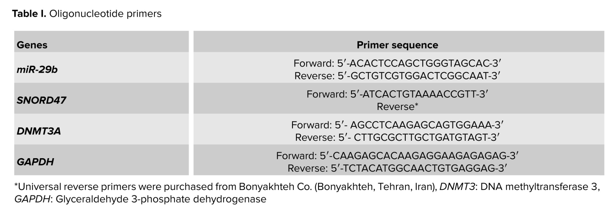
In this study, total RNA was first isolated using the RNX-Plus reagent (Cinna Gen, Iran). The concentration and purity of the extracted RNA were determined spectrophotometrically at 260/280 nm using a NanoDrop instrument (Thermo Fisher Scientific, USA). cDNA synthesis for the DNMT3A gene was performed using a commercially available reverse transcription kit (Yekta Tajhiz Azma, Iran). In contrast, miR-29b cDNA was synthesized by the stem-loop method using the BON-miR miRNA 1st strand cDNA synthesis kit (Bonyakhteh, Tehran, Iran) according to the manufacturer’s protocols. qRT-PCR was performed separately for DNMT3A and miR-29b. For this purpose, 1 µL of synthesized cDNA, 6.5 µL of SYBR Green Master Mix (Bonyakhteh, Tehran, Iran), 0.5 µL of each specific primer (forward and reverse), and 4.5 µL of RNase/DNase-free DEPCK water were used. Amplification reactions were performed on a Rotor-Gene Q instrument (Qiagen, Germany). For normalization, glyceraldehyde 3-phosphate dehydrogenase (GAPDH) was used as an endogenous control for DNMT3A expression and SNORD47 for miR-29b (Table I).


2.2. Ethical Considerations
This research was conducted on samples taken at Shahid Sadoughi hospital in Yazd, Iran. It is worth noting that the project was presented to the ethics committee (Code: IR.SSU.SPH.REC.1404.040) and permission was granted to conduct experiments on the collected samples after receiving the ethics code.

2.3. Statistical Analysis
The relative expression levels of DNMT3A and miR-29b genes in the control, eutopia, and ectopia tissue groups were quantified using the 2^ (-ΔΔCt) method. To evaluate expression differences between the groups and assess a possible correlation between DNMT3A and miR-29b expression levels, a statistical analysis was performed using SPSS Software version 26. A one-way analysis of variance (ANOVA) followed by Tukey’s honestly significant difference post hoc test was conducted to determine statistical significance. A p < 0.05 was considered to indicate a significant difference. The diagrams were created with GraphPad Prism Version 8.

3. Result
3.1. DNMT3A gene expression
2.2. Ethical Considerations
This research was conducted on samples taken at Shahid Sadoughi hospital in Yazd, Iran. It is worth noting that the project was presented to the ethics committee (Code: IR.SSU.SPH.REC.1404.040) and permission was granted to conduct experiments on the collected samples after receiving the ethics code.
2.3. Statistical Analysis
The relative expression levels of DNMT3A and miR-29b genes in the control, eutopia, and ectopia tissue groups were quantified using the 2^ (-ΔΔCt) method. To evaluate expression differences between the groups and assess a possible correlation between DNMT3A and miR-29b expression levels, a statistical analysis was performed using SPSS Software version 26. A one-way analysis of variance (ANOVA) followed by Tukey’s honestly significant difference post hoc test was conducted to determine statistical significance. A p < 0.05 was considered to indicate a significant difference. The diagrams were created with GraphPad Prism Version 8.
3. Result
3.1. DNMT3A gene expression
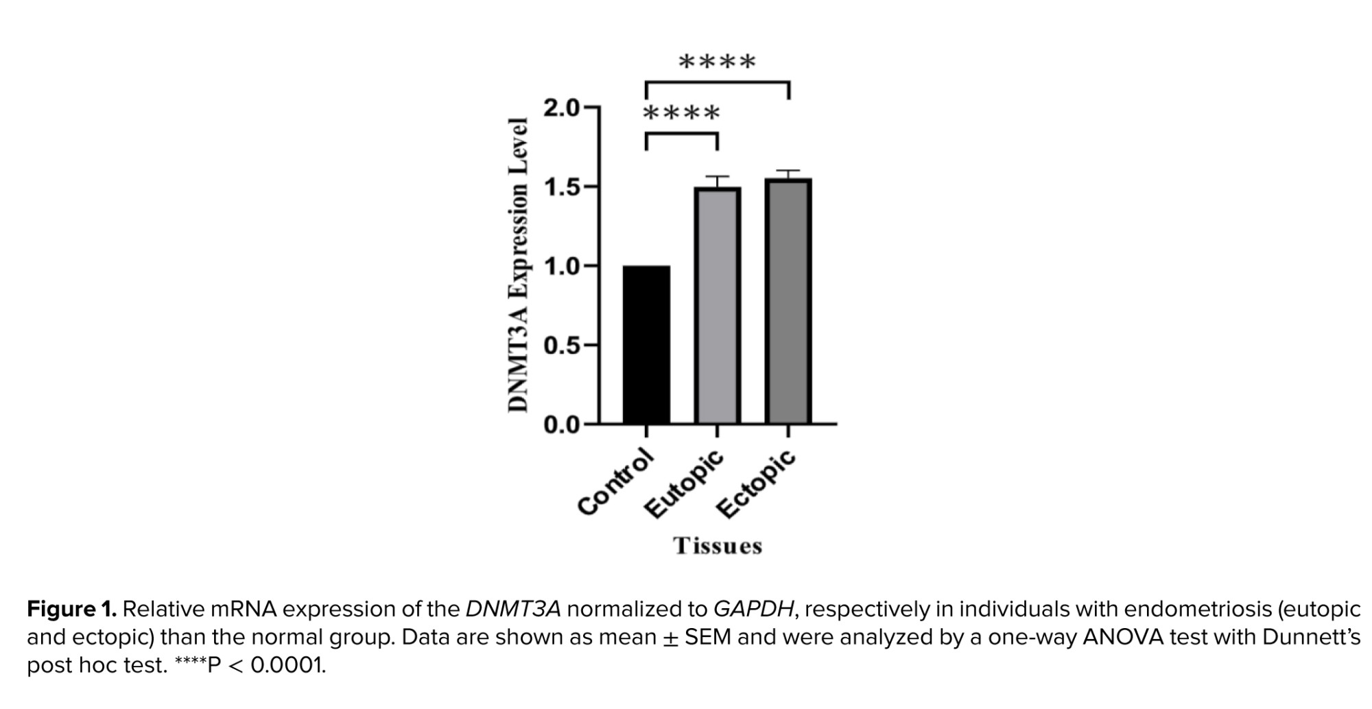
qRT-PCR analysis revealed significant changes in DNMT3A gene expression between endometriosis individuals and healthy controls. In particular, DNMT3A expression levels were significantly increased in both eutopic and ectopic endometrial tissue compared to control samples (p = 0.0001 for both). In addition, a statistically significant upregulation was observed in ectopic tissues compared to eutopic tissues (p = 0.0092) (Figure 1).
3.2. Mir-29b expression
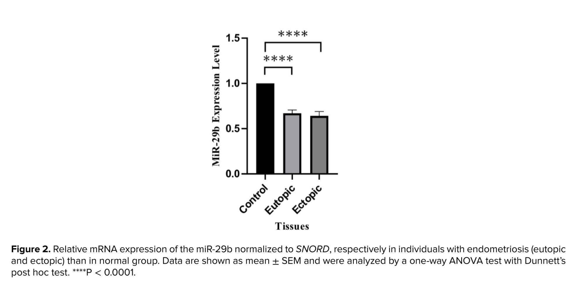
Real-time PCR analysis showed an altered expression of miR-29b in individuals with endometriosis compared to healthy controls. Significant downregulation of miR-29b was observed in both eutopic and ectopic endometrial tissue compared to control samples (p = 0.0001 for both). However, no statistically significant difference was observed between eutopic and ectopic tissue with respect to miR-29b expression (p = 0.640) (Figure 2).
 4. Discussion
DNA methylation is a biochemical process that is important for normal growth and development and cellular differentiation of complex organisms, and alters the pattern of gene expression in cells (7). This process usually involves the addition of a methyl group (-CH3) to the 5th-position of the pyrimidine cytosine (5 mC) ring. It is worth noting that methylation of the C site generally occurs at the CpG sequence. CpG dinucleotides are sometimes scattered throughout the genome, while others occur in dense clusters called CpG islands (8). These sequences are usually located in the gene promoter region and are generally unmethylated. Hypomethylation of CpG leads to activation of gene expression, while hypermethylation leads to translational silencing; thus, methylation genes such as DNMTs play a key role in gene expression. DNMTs have 5 members: DNMT1, DNMT2, DNMT3A, DNMT3B, and DNMT3L, each of which has a different activity (9, 10). DNMT1, for example, is responsible for the maintenance of sequence methylation, while DNMT3A and DNMT3B are mainly responsible for de novo methylation (10). The common feature of DNMT3A and DNMT3B isoforms is that both possess an active and conserved C-terminal domain, responsible for binding the cofactor S-adenosylmethionine (SAM) and targeting to cytosine. Therefore, they can catalyze the transfer of a CH3 group from SAM to the C5 position of cytosine to form 5-methylcytosine (11, 12).
The DNMT3A gene, located on chromosome 2 (13, 14), consists of a protein-coding segment of 2172 bp and produces a protein of approximately 130 kDa. Previous studies suggest that the binding of DNMT3A to DNA is primarily facilitated by a loop of the target recognition domain, the catalytic loop, and the homodimeric interface of DNMT3A, which together form a seamless DNA-binding surface (15). In general, DNMT3A is thought to favor unmethylated DNA, leading to de novo methylation. Among the DNMTs, mutations in DNMT3A have been documented as the most frequently occurring in cancer. In addition, studies from multiple sources have shown that overexpression of DNMT3A correlates with aggressive features in vulvar squamous cell carcinoma, pituitary adenomas, and colorectal cancer, including increased invasion and migration (16-19). Similarly, this study showed that expression of DNMT3A was significantly increased in both ectopic and eutopic tissue compared to normal endometrium. In another study, this increase was only observed in ectopic tissue (3).
miRNAs are a group of short, non-coding RNAs that have attracted the attention of researchers in recent years (20). These small molecules play an important role in regulation of gene expression and control various cellular processes by repressing or degrading mRNAs. Recent studies have shown that changes in expression of miRNAs are critically involved in the occurrence and progression of many diseases, especially cancer. Some of them act as oncogenes and cause tumor growth and invasion, while others suppress tumors and prevent the spread of cancer cells (21-23). According to recent research, miR-29b may play a crucial role in epigenetic regulation, cell proliferation, metastasis, cell differentiation and invasion, and apoptosis. It was found that in AML cells, increased expression of miR-29b can lead to decreased expression of DNMT1, DNMT3A, and DNMT3B at both the protein and RNA levels (24). miR-29b indirectly downregulates DNMT1 by targeting Sp1, resulting in decreased DNA methylation and increased expression of 2 tumor suppressors: ESR1 and INK4b. It was also reported that the introduction of synthetic miR-29b into multiple myeloma cells disrupts cell cycle progression and strongly enhances the growth inhibitory effect of the demethylating agent 5-azacitidine (25).
Another study mentioned that increased miR-29 levels can suppress cell proliferation, invasion, and metastasis in uterine leiomyomas, which may be related to suppression of the STAT3 signaling pathway, introducing miR-29 as a new target for the treatment of uterine leiomyomas (26). In another study, a negative feedback of microRNA on the anti-Mullerian hormone gene was found (27). Considering the role and importance of miR-29b, we were prompted to investigate this microRNA in endometriosis and its relationship with the DNMT3A gene. Our results indicate that expression of miR-29b was significantly reduced in both ectopic and eutopic endometrial tissues compared to controls (p < 0.05). In contrast, DNMT3A expression was significantly increased in these tissues compared to controls (p < 0.05). These observations suggest a possible inverse relationship between miR-29b and DNMT3A. The downregulation of miR-29b likely contributes to the upregulation of DNMT3A, which may subsequently alter DNA methylation patterns. These epigenetic changes may lead to silencing of tumor suppressor genes, such as INK4b and ESR1, and dysregulation of signaling pathways, such as STAT3. These changes can disrupt the balance of cell proliferation, apoptosis, and invasion, potentially contributing to the development and progression of endometriosis. Since the relationship between the DNMT3A gene and miR-29b and their expression in endometriosis has not been fully investigated, miR-29b plays a crucial role in regulating DNA methylation by targeting DNMT3A. This study examined the expression levels of this microRNA and the DNMT3A gene in endometriosis tissues. Although the results of this project were significant, there were limitations, such as the silencing of DNMT3A and miR-29b in endometriosis cell lines, and the evaluation of DNMT3A and miR-29b at the protein level, which was not performed due to financial constraints.
5. Conclusion
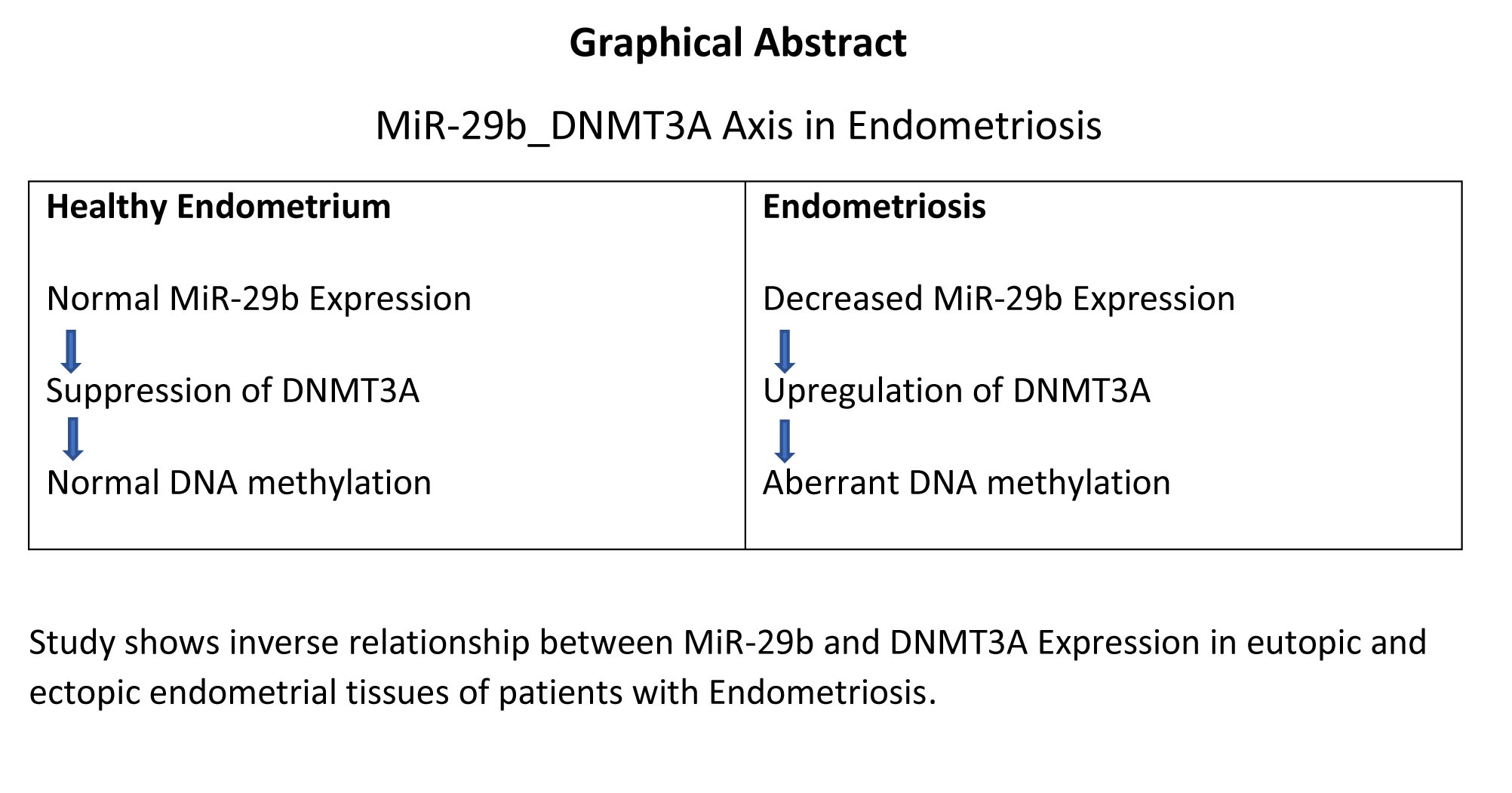
This study reveals that DNMT3A expression is significantly elevated, while miR-29b expression is markedly reduced in both eutopic and ectopic endometrial tissues of women with endometriosis compared to healthy individuals. The observed inverse relationship between these 2 molecules underscores their potential involvement in the epigenetic regulation of endometrial gene expression. These findings contribute to our understanding of endometriosis pathogenesis and support further investigation into the DNMT3A/miR-29b axis as a diagnostic or therapeutic target.
Data Availability
All raw data are available from the corresponding author on reasonable request.
Author Contributions
J. Fazeli and E. Babakhanzadeh designed the study and conducted the research. M. Shirmohamadi, M. Mazaheri-Naeini, and M. Taatnezhad monitored, evaluated, and analyzed the results of the study. Further, A. Dadbinpour reviewed the article. All authors approved the final manuscript and take responsibility for the integrity of the data.
Acknowledgments
This project was funded by Shahid Sadoughi University of Medical Sciences, Yazd, Iran (Code: 11463). The authors would like to express their gratitude to all those who contributed to this work. ChatGPT (OpenAI) has been used to correct spelling and grammatical errors in the article.
Conflict of Interest
All authors declare that there is no conflict of interest.
4. Discussion
DNA methylation is a biochemical process that is important for normal growth and development and cellular differentiation of complex organisms, and alters the pattern of gene expression in cells (7). This process usually involves the addition of a methyl group (-CH3) to the 5th-position of the pyrimidine cytosine (5 mC) ring. It is worth noting that methylation of the C site generally occurs at the CpG sequence. CpG dinucleotides are sometimes scattered throughout the genome, while others occur in dense clusters called CpG islands (8). These sequences are usually located in the gene promoter region and are generally unmethylated. Hypomethylation of CpG leads to activation of gene expression, while hypermethylation leads to translational silencing; thus, methylation genes such as DNMTs play a key role in gene expression. DNMTs have 5 members: DNMT1, DNMT2, DNMT3A, DNMT3B, and DNMT3L, each of which has a different activity (9, 10). DNMT1, for example, is responsible for the maintenance of sequence methylation, while DNMT3A and DNMT3B are mainly responsible for de novo methylation (10). The common feature of DNMT3A and DNMT3B isoforms is that both possess an active and conserved C-terminal domain, responsible for binding the cofactor S-adenosylmethionine (SAM) and targeting to cytosine. Therefore, they can catalyze the transfer of a CH3 group from SAM to the C5 position of cytosine to form 5-methylcytosine (11, 12).
The DNMT3A gene, located on chromosome 2 (13, 14), consists of a protein-coding segment of 2172 bp and produces a protein of approximately 130 kDa. Previous studies suggest that the binding of DNMT3A to DNA is primarily facilitated by a loop of the target recognition domain, the catalytic loop, and the homodimeric interface of DNMT3A, which together form a seamless DNA-binding surface (15). In general, DNMT3A is thought to favor unmethylated DNA, leading to de novo methylation. Among the DNMTs, mutations in DNMT3A have been documented as the most frequently occurring in cancer. In addition, studies from multiple sources have shown that overexpression of DNMT3A correlates with aggressive features in vulvar squamous cell carcinoma, pituitary adenomas, and colorectal cancer, including increased invasion and migration (16-19). Similarly, this study showed that expression of DNMT3A was significantly increased in both ectopic and eutopic tissue compared to normal endometrium. In another study, this increase was only observed in ectopic tissue (3).
miRNAs are a group of short, non-coding RNAs that have attracted the attention of researchers in recent years (20). These small molecules play an important role in regulation of gene expression and control various cellular processes by repressing or degrading mRNAs. Recent studies have shown that changes in expression of miRNAs are critically involved in the occurrence and progression of many diseases, especially cancer. Some of them act as oncogenes and cause tumor growth and invasion, while others suppress tumors and prevent the spread of cancer cells (21-23). According to recent research, miR-29b may play a crucial role in epigenetic regulation, cell proliferation, metastasis, cell differentiation and invasion, and apoptosis. It was found that in AML cells, increased expression of miR-29b can lead to decreased expression of DNMT1, DNMT3A, and DNMT3B at both the protein and RNA levels (24). miR-29b indirectly downregulates DNMT1 by targeting Sp1, resulting in decreased DNA methylation and increased expression of 2 tumor suppressors: ESR1 and INK4b. It was also reported that the introduction of synthetic miR-29b into multiple myeloma cells disrupts cell cycle progression and strongly enhances the growth inhibitory effect of the demethylating agent 5-azacitidine (25).
Another study mentioned that increased miR-29 levels can suppress cell proliferation, invasion, and metastasis in uterine leiomyomas, which may be related to suppression of the STAT3 signaling pathway, introducing miR-29 as a new target for the treatment of uterine leiomyomas (26). In another study, a negative feedback of microRNA on the anti-Mullerian hormone gene was found (27). Considering the role and importance of miR-29b, we were prompted to investigate this microRNA in endometriosis and its relationship with the DNMT3A gene. Our results indicate that expression of miR-29b was significantly reduced in both ectopic and eutopic endometrial tissues compared to controls (p < 0.05). In contrast, DNMT3A expression was significantly increased in these tissues compared to controls (p < 0.05). These observations suggest a possible inverse relationship between miR-29b and DNMT3A. The downregulation of miR-29b likely contributes to the upregulation of DNMT3A, which may subsequently alter DNA methylation patterns. These epigenetic changes may lead to silencing of tumor suppressor genes, such as INK4b and ESR1, and dysregulation of signaling pathways, such as STAT3. These changes can disrupt the balance of cell proliferation, apoptosis, and invasion, potentially contributing to the development and progression of endometriosis. Since the relationship between the DNMT3A gene and miR-29b and their expression in endometriosis has not been fully investigated, miR-29b plays a crucial role in regulating DNA methylation by targeting DNMT3A. This study examined the expression levels of this microRNA and the DNMT3A gene in endometriosis tissues. Although the results of this project were significant, there were limitations, such as the silencing of DNMT3A and miR-29b in endometriosis cell lines, and the evaluation of DNMT3A and miR-29b at the protein level, which was not performed due to financial constraints.
5. Conclusion
This study reveals that DNMT3A expression is significantly elevated, while miR-29b expression is markedly reduced in both eutopic and ectopic endometrial tissues of women with endometriosis compared to healthy individuals. The observed inverse relationship between these 2 molecules underscores their potential involvement in the epigenetic regulation of endometrial gene expression. These findings contribute to our understanding of endometriosis pathogenesis and support further investigation into the DNMT3A/miR-29b axis as a diagnostic or therapeutic target.
Data Availability
All raw data are available from the corresponding author on reasonable request.
Author Contributions
J. Fazeli and E. Babakhanzadeh designed the study and conducted the research. M. Shirmohamadi, M. Mazaheri-Naeini, and M. Taatnezhad monitored, evaluated, and analyzed the results of the study. Further, A. Dadbinpour reviewed the article. All authors approved the final manuscript and take responsibility for the integrity of the data.
Acknowledgments
This project was funded by Shahid Sadoughi University of Medical Sciences, Yazd, Iran (Code: 11463). The authors would like to express their gratitude to all those who contributed to this work. ChatGPT (OpenAI) has been used to correct spelling and grammatical errors in the article.
Conflict of Interest
All authors declare that there is no conflict of interest.
Type of Study: Original Article |
Subject:
Reproductive Genetics
Send email to the article author
| Rights and permissions | |
 |
This work is licensed under a Creative Commons Attribution-NonCommercial 4.0 International License. |
