
International Journal of
Reproductive Biomedicine
Tue, Sep 29, 2026
[Archive]
Volume 24, Issue 5 (May 2026)
IJRM 2026, 24(5): 445-456 |
Back to browse issues page



Ethics code: B/091/UN19.5.1.1.8/UEPKK/2021


Download citation:
BibTeX | RIS | EndNote | Medlars | ProCite | Reference Manager | RefWorks
Send citation to:


BibTeX | RIS | EndNote | Medlars | ProCite | Reference Manager | RefWorks
Send citation to:
Saputra N P K, Samsulhadi S, Hendarto H, Saádi A, Pudjirahardjo W J, Arsana I W, et al . The role of tumor necrosis factor alpha in regulating CYFRA 21-1 as a potential biomarker for endometriosis: A cross-sectional study. IJRM 2026; 24 (5) :445-456
URL: http://ijrm.ir/article-1-3716-en.html
URL: http://ijrm.ir/article-1-3716-en.html
Nicko Pisceski Kusika Saputra *1 

 , Samsulhadi Samsulhadi2 , Hendy Hendarto2 , Ashon Saádi2 , Widodo J Pudjirahardjo3 , I Wayan Arsana4 , Relly Yanuari2 , Sri Ratna Dwiningsih2 , Suri Dwi Lesmana5
, Samsulhadi Samsulhadi2 , Hendy Hendarto2 , Ashon Saádi2 , Widodo J Pudjirahardjo3 , I Wayan Arsana4 , Relly Yanuari2 , Sri Ratna Dwiningsih2 , Suri Dwi Lesmana5
1- Department of Obstetrics and Gynecology, Faculty of Medicine, Universitas Riau, Pekanbaru, Indonesia. , nicko.pisceski@lecturer.unri.ac.id
2- Fertility and Endocrinology Division, Department of Obstetrics and Gynecology, Faculty of Medicine, Universitas Airlangga, Surabaya, Indonesia.
3- Department of Public Health, Faculty of Medicine, Universitas Airlangga, Surabaya, Indonesia.
4- Fertility and Endocrinology Division, Department of Obstetrics and Gynecology, Faculty of Medicine, Universitas Brawijaya, Malang, Indonesia.
5- Department of Parasitology, Faculty of Medicine, Universitas Riau, Pekanbaru, Indonesia. & Lontar Biomedical Laboratory, Faculty of Medicine, Universitas Riau, Pekanbaru, Indonesia.
2- Fertility and Endocrinology Division, Department of Obstetrics and Gynecology, Faculty of Medicine, Universitas Airlangga, Surabaya, Indonesia.
3- Department of Public Health, Faculty of Medicine, Universitas Airlangga, Surabaya, Indonesia.
4- Fertility and Endocrinology Division, Department of Obstetrics and Gynecology, Faculty of Medicine, Universitas Brawijaya, Malang, Indonesia.
5- Department of Parasitology, Faculty of Medicine, Universitas Riau, Pekanbaru, Indonesia. & Lontar Biomedical Laboratory, Faculty of Medicine, Universitas Riau, Pekanbaru, Indonesia.
Full-Text [PDF 406 kb]
(395 Downloads)
| Abstract (HTML) (451 Views)
Full-Text: (57 Views)
1. Introduction
Endometriosis is one of the most common benign gynecological conditions (1) . It is characterized by the presence of endometrium-like tissue comprising glandular cells and stroma outside the uterine cavity, triggering chronic inflammation that is often associated with pelvic pain and infertility (2, 3) . Approximately 30% of women with endometriosis are reported to experience infertility (4) . Endometriosis pathophysiology involving retrograde menstruation, and sexual activity during menstruation may be a possible cause (5) . Endometriosis is often considered an estrogen-dependent lesion model (6) . Progesterone resistance contributes to continuous cellular proliferation (7) .
Currently, laparoscopy is considered the gold standard for diagnosing endometriosis (8) . However, laparoscopy is an invasive procedure associated with potential complications and high costs, often delaying diagnosis and treatment (9) . In fact, diagnosis is typically delayed by 7-10 yr from symptom onset (10). Non-invasive biomarker detection is a promising diagnostic alternative that may expedite the management of infertility and pelvic pain.
In 2011, a urinary protein fragment of cytokeratin-19, identified as cytokeratin fragment 21-1 (CYFRA 21-1), was discovered in endometriosis cases (11) . Urinary CYFRA 21-1 levels show high specificity but very low sensitivity (12) . Previous studies indicate that CYFRA 21-1 levels fluctuate with the menstrual cycle, being significantly higher during the proliferative phase compared to the secretory phase (3) . However, other studies have found no significant differences in CYFRA 21-1 levels between phases (13 ). Research in the Indonesian population demonstrated significantly higher CYFRA 21-1 levels and CYFRA 21-1/creatinine ratios in women with endometriosis (14) .
In the reproductive tract, cytokeratin-19 is expressed in endometrial epithelial cells and is involved in both normal and pathological processes, such as endometriosis and adenomyosis (15) . Cytokeratin-19 is released into the systemic circulation as a fragmented protein, CYFRA 21-1. Ectopic endometrial tissue may increase cellular proliferation, leading to cytokeratin fragmentation and systemic release (3, 13 ). Eventually resulting in urinary excretion. Like other urinary proteins, CYFRA 21-1 levels vary throughout the day due to circadian rhythms, thus urinary creatinine is used to normalize its concentration (3) .
Tumor necrosis factor alpha (TNF-α), a cytokine implicated in the cleavage of cytokeratin-19 into CYFRA 21-1 via caspase-3-mediated epithelial necrosis, may support the role of CYFRA 21-1 in endometriosis pathophysiology. Several studies have shown that CYFRA 21-1 is released through proteolysis during cell necrosis and proliferation, with TNF-α being one of the main stimulants (16) . TNF-α might increase cytokeratin-19 substrate availability through transcriptional upregulation (17) . TNF-α may stimulate endometrial cell proliferation and adhesion and is involved in angiogenesis within endometriotic lesions (18 ). TNF-α demonstrates moderate discriminative ability for diagnosing endometriosis. It can differentiate between cases with and without endometriosis (19 ). TNF-α activates various intracellular signaling pathways that can induce apoptosis, which may indirectly affect cytokeratin-19 levels (20 ).
This study expands on previous findings that merely established the diagnostic potential of urinary CYFRA 21-1 by directly exploring its regulatory relationship with TNF-α. This study aims to analyze the correlation between TNF-α levels and CYFRA 21-1 as an initial step in understanding the role of TNF-α in cytokeratin-19 cleavage.
2. Materials and Methods
2.1. Sample selection
This study was a cross-sectional study. All participants at Fertility Clinic of Arifin Achmad General hospital, Riau province, Indonesia who were indicated for laparoscopic surgery between 2021 and 2022 were included. Examination of CYFRA 21-1 levels in urine and TNF-α were conducted at the Lontar Integrated Biomedical Laboratory, Faculty of Medicine, Universitas Riau, Indonesia. Examination of urine creatinine levels was conducted at the Prodia Laboratory Pekanbaru, Indonesia.
Participants were recruited directly during their visits to the clinic and classified according to predefined inclusion and exclusion criteria. Recruitment was conducted in several stages. Initial screening was performed in participants with suspected endometriosis based on clinical symptoms and ultrasonography findings. The stage of endometriosis was subsequently determined through laparoscopic evaluation using the revised American Society for Reproductive Medicine (r-ASRM) classification and confirmed by histopathological examination of tissue samples. Tissue sampling was lesion-dependent: entire cysts were excised when present, whereas implant lesions were sampled as extensively as possible.
The inclusion criteria consisted of women aged 30-40 yr with a normal body mass index (BMI; 18.5-24.9 kg/m²) and regular menstrual cycles ranging from 26-38 days. Age and BMI were obtained from anamnesis and direct measurement during clinic visits. Menstrual cycle characteristics were assessed through participants interviews regarding the last menstrual period and cycle duration within the preceding 3 months and were reconfirmed during the perioperative period and postoperative follow-up. Menstrual cycle phases (proliferative or secretory) were subsequently determined based on these data. Exclusion criteria included participants receiving hormonal therapy (e.g., contraceptives), anti-inflammatory or antioxidant drugs, as well as those diagnosed with lung or gastrointestinal cancer, urinary tract infections (confirmed through medical records, chest X-ray, urine analysis, or liver function tests), impaired renal function (based on glomerular filtration rate and serum creatinine), or diabetes mellitus.
A total of 73 participants were ultimately enrolled based on laparoscopic findings, comprising 38 participants with endometriosis and 35 without endometriosis. The control group included participants undergoing laparoscopy for non-endometriosis indications, such as female sterilization, uterine septum repair, tubal repair, or intrauterine device translocation. Controls were selected to ensure comparable baseline characteristics with the endometriosis group. Samples collected from the participants included urine, which was obtained prior to surgery, as well as peritoneal fluid collected during the operation.
2.2. Urine CYFRA 21-1 level test
Urine CYFRA 21-1 levels were measured using the Enzyme-Linked Immunosorbent Assay (ELISA) method. Urine samples were collected prior to laparoscopy and stored on ice or refrigerated for no longer than 2 hr before processing. To maintain the cold chain during transport, samples were transported in an icebox. The collected urine was stored at -80°C until analysis. Uncentrifuged urine was used for testing. The Human Cytokeratin 19 ELISA kit (product number RAB1409) was utilized for the assay.
Standard serial dilutions were prepared to generate CYFRA 21-1 concentrations of 0.061 ng/ml, 0.154 ng/ml, 0.384 ng/ml, 0.960 ng/ml, 2.4 ng/ml, 6 ng/ml, and 15 ng/ml, along with a blank. All reagents and samples were equilibrated to room temperature (18-25°C) prior to use. Each standard and sample (100 µl) was added in duplicate to wells, covered, and incubated overnight at 4°C, followed by washing. Subsequently, 100 µl of antibody was added to each well and incubated for 1 hr at room temperature with thorough mixing, followed by washing. Then, 100 µl of streptavidin was added and incubated for 45 min, followed by washing. Finally, 100 µl of 3′-3-5′-5-tetramethylbenzidine substrate was added, followed by 50 µl of stop solution. Absorbance was measured at 450 nm. A standard curve was plotted to derive the equation for sample concentration calculation.
2.3. Urine creatinine and urine CYFRA/creatinine ratio test
Urine creatinine was measured using the Electrochemiluminescence Immunoassay method at Prodia Laboratory, Pekanbaru, Indonesia. The ratio of urine cytokeratin-19 fragments (CYFRA 21-1) to creatinine (CYFRA/Cr) was calculated by comparing their concentrations and expressed in ng/gr units to normalize the variation in urine concentration.
2.4. Intraperitoneal TNF-α
Intraperitoneal TNF-α levels were measured using ELISA. Human TNF-α (RAB0476-1KT merck®) was used for this essay. Peritoneal lavage fluid was obtained by instilling 20 mL of 0.9% NaCl into the peritoneal cavity of the participants and then collected into a sterile tube. The peritoneal fluid was subsequently centrifuged using a refrigerated centrifuge.
2.5. Ethical Considerations
This study was approved by the Medical and Health Research Ethics Unit of the Faculty of Medicine, Universitas Riau, Pekanbaru, Indonesia (Code: B/091/UN19.5.1.1.8/UEPKK/2021) and was deemed to comply with the 7 WHO 2011 ethical standards. The ethical clearance certificate was issued on September 10, 2021. Permission to use participants data was obtained, and all participants provided both written and verbal informed consent after receiving a detailed explanation of the study objectives, sampling procedures, and intended use of their data. Participants were also informed that the results of this research would be published, with the assurance that no personally identifiable information would be disclosed.
2.6. Statistical Analysis
The collected data consisted of both categorical and numerical variables. Categorical data (e.g., menstrual phase, disease status, and r-ASRM stage) were expressed as frequencies and percentages, whereas numerical data (e.g., CYFRA 21-1, TNF-α, and creatinine levels) were presented as mean ± standard deviation or median (minimum-maximum), depending on their distribution. The normality of numerical data was assessed using the Kolmogorov-Smirnov and Shapiro-Wilk tests. For normally distributed data, comparisons between groups were performed using the Independent samples t test; otherwise, the Mann-Whitney U test was applied. Correlations between CYFRA 21-1, CYFRA/Cr ratio, and TNF-α levels were analyzed using the Spearman rank correlation test. A p < 0.05 was considered statistically significant. All statistical analyses were performed using IBM SPSS Statistics version 28.0 (IBM Corp., Armonk, NY, USA).
3. Results
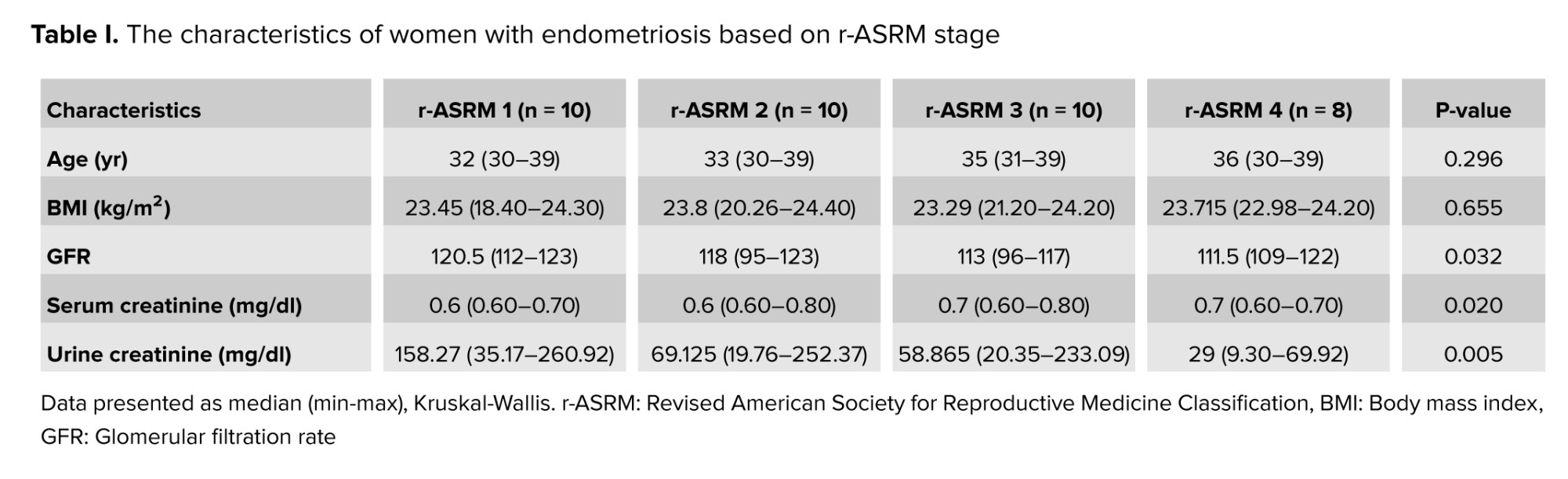
A total of 360 women who presented with indications for laparoscopy were initially enrolled in the study. After screening according to the inclusion and exclusion criteria, 141 participants met the eligibility requirements. Laparoscopic procedures were subsequently performed on all eligible participants. Based on laparoscopic findings and histopathological confirmation, the subjects were classified into 2 groups: 38 women with endometriosis and 103 women without endometriosis. From the non-endometriosis (control) group, 68 participants were later excluded due to the presence of other gynecological disorders, including myoma, pelvic inflammatory disease, malignancy, pelvic tuberculosis, and other types of ovarian cysts. The characteristics of women with endometriosis based on the r-ASRM stage are presented in table I.
Based on table I, it can be observed that all participants included in this study met the predefined inclusion and exclusion criteria. The distribution of participants according to age, BMI, glomerular filtration rate (GFR) and serum creatinine level were homogeneous across the different stages of endometriosis. Table I presents the characteristics of participants according to r-ASRM stage. Age and BMI did not differ significantly among stages (p > 0.05), indicating comparable baseline characteristics. Although GFR, serum creatinine, and urine creatinine showed significant differences (p < 0.05), with declining GFR and urinary creatinine and a slight rise in serum creatinine observed in higher r-ASRM stages. However, both GFR and serum creatinine levels were within normal ranges.
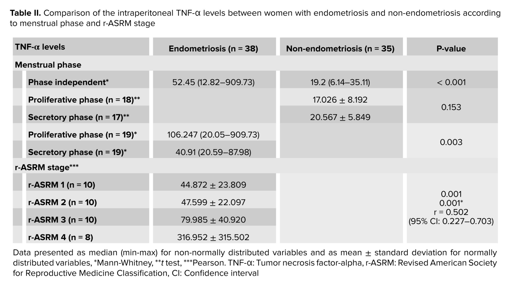
Table II presents the comparison of intraperitoneal TNF-α levels between participants with and without endometriosis, including subgroup analyses based on menstrual phase and disease severity according to the r-ASRM classification. The intraperitoneal TNF-α levels and their relationship with endometriosis severity based on the r-ASRM classification, as well as comparisons with non-endometriosis during both proliferative and secretory phases, are shown in table II.
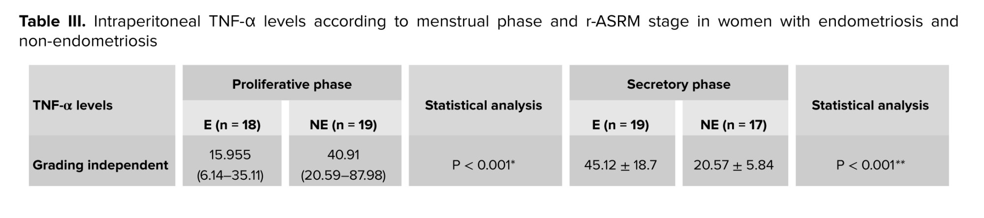
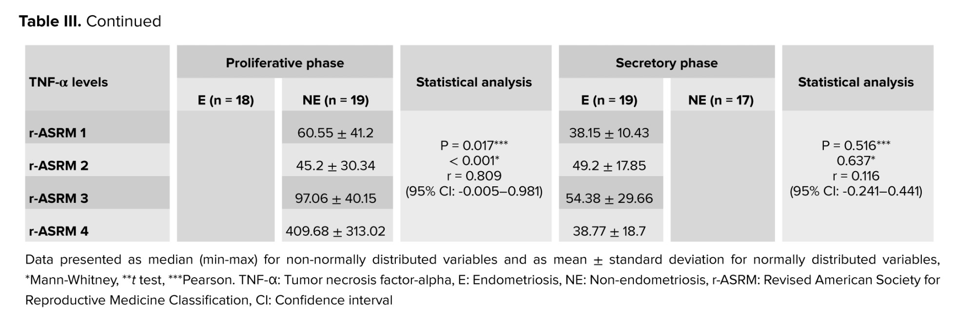
Table III shows the distribution of intraperitoneal TNF-α levels according to menstrual phase and r-ASRM stage, highlighting phase-specific differences in endometriosis and control groups.
Based on tables II, and III, intraperitoneal TNF-α levels were significantly higher in the endometriosis group compared with the non-endometriosis group (p < 0.001). Within the endometriosis group, the mean intraperitoneal TNF-α level was significantly higher during the proliferative phase than in the secretory phase. In contrast, in the non-endometriosis group, no significant difference was observed in mean intraperitoneal TNF-α levels between the proliferative and secretory phases.
A minimally significant difference in intraperitoneal TNF-α levels was observed between groups when stratified by r-ASRM stage, and a moderate positive correlation was found between intraperitoneal TNF-α levels and r-ASRM scores. Higher r-ASRM stages were associated with elevated intraperitoneal TNF-α concentrations.
When analyzed by menstrual cycle phase, intraperitoneal TNF-α levels remained significantly higher in the endometriosis group compared with the non-endometriosis group in both the proliferative and secretory phases. Further analysis of r-ASRM stage in the endometriosis group revealed a strong positive correlation between intraperitoneal TNF-α levels and r-ASRM scores during the proliferative phase (r = 0.809), whereas no significant correlation was observed in the secretory phase.
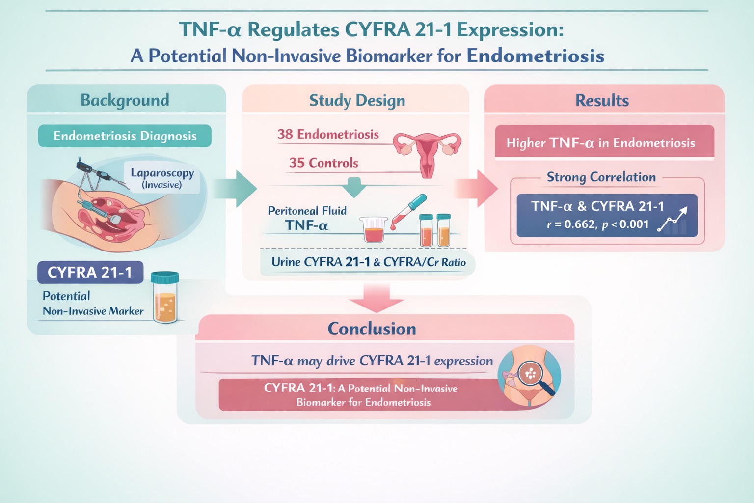
The correlation analysis between CYFRA and TNF-α demonstrated a statistically significant positive association. The median CYFRA concentration was 4.39 ng/ml (range: 0.38-15.78 ng/ml), while the median TNF-α level was 52.45 pg/ml (range: 12.82-909.73 pg/ml). Spearman’s correlation test showed a p < 0.001 with a correlation coefficient (r) of 0.662 and a 95% confidence interval (CI) ranging from 0.426-0.814, indicating a strong positive correlation between CYFRA and TNF-α levels.
Similarly, a significant positive correlation was observed between the CYFRA/Cr ratio and TNF-α. The median CYFRA/Cr ratio was 4,592.47 ng/gr (range: 243.51-58,423.83 ng/gr), while the TNF-α median level remained 52.45 pg/ml (range: 12.82-909.73 pg/ml). Spearman’s correlation analysis yielded a p < 0.001 and a correlation coefficient (r) of 0.626 with a 95% CI of 0.375-0.972, suggesting a strong positive relationship between the CYFRA/Cr ratio and TNF-α concentration.
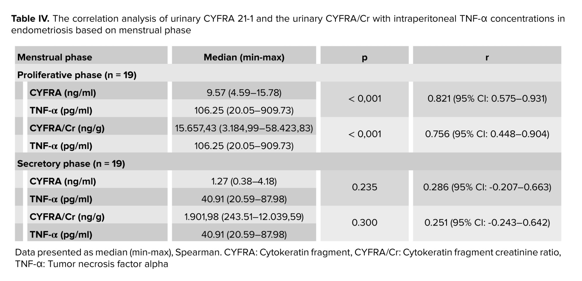
Based on table IV, significant correlations were observed between intraperitoneal TNF-α levels and both CYFRA 21-1 concentrations and the urinary CYFRA/Cr ratio, but only during the proliferative phase. In this phase, the correlations were strong to very strong. In contrast, no significant correlations were found during the secretory phase.
4. Discussion
In this study, there were no significant differences in individual characteristics (age and BMI) among women with endometriosis based on disease stage. Additionally, both GFR and serum creatinine levels were within normal ranges. Therefore, these factors were not considered confounding variables in the assessment of CYFRA 21-1 or TNF-α levels in relation to disease severity.
The findings showed a significant difference in TNF-α levels between women with and without endometriosis, with higher values observed in the endometriosis group. In the context of various pathological processes, TNF-α is an important proinflammatory and angiogenic mediator and, among others, is increasingly secreted by macrophage-1 (21 ). Moreover, a progressive increase in TNF-α concentrations was noted with advancing r-ASRM stages, particularly stage IV, suggesting a direct relationship between disease severity and inflammatory activity. These results are consistent with previous research indicating that TNF-α promotes lesion survival, angiogenesis, and immune evasion, contributing to the progression of endometriosis (22 ). Interestingly, TNF-α levels in women with endometriosis were significantly higher during the proliferative phase compared to the secretory phase. This contrasts with the physiological condition, where TNF-α is typically elevated during the secretory phase due to increased apoptosis and inflammation as the endometrial lining prepares for shedding. In contrast, the proliferative phase, dominated by estrogen, is associated with tissue growth and minimal inflammation (23) . In endometriosis, however, immunological dysregulation occurs (7 ). During the proliferative (follicular) phase, elevated TNF-α levels in the peritoneal fluid may result from retrograde endometrial debris that activates macrophages and dendritic cells. This immune activation triggers the release of proinflammatory cytokines, including TNF-α, in response to residual ectopic tissue. Moreover, rising estrogen levels during this phase may upregulate TNF-α expression via nuclear factor kappa light chain enhancer of activated B cells (NF-κB) signaling in ectopic endometrial cells (24 ). Conversely, during the secretory (luteal) phase, TNF-α levels in the peritoneal fluid decreased. The rise in progesterone during this phase exerts immunomodulatory effects by suppressing TNF-α expression directly on macrophages and indirectly through regulation of the tumor necrosis factor-1 signaling pathway. Although ectopic endometriotic cells exhibit resistance to progesterone, the surrounding peritoneal tissue remains responsive to its anti-inflammatory signals. The reduction in TNF-α also reflects a component of the tolerogenic reprogramming of peritoneal macrophages, characterized by increased secretion of interleukin-10 and transforming growth factor beta (25 ). However, the association between TNF-α levels and menstrual cycle phases in endometriosis remains inconsistent across studies. These discrepancies may be attributed to variations in the timing and determination of the proliferative and secretory phases.
In general, TNF-α demonstrates a strong association and correlation with both CYFRA 21-1 and the CYFRA/Cr ratio. The menstrual phase significantly influenced TNF-α concentrations and CYFRA 21-1 and CYFRA/Cr expression. During the proliferative phase, TNF-α and CYFRA 21-1 or CYFRA/Cr levels were markedly higher compared to the secretory phase, aligning with the hormonal regulation of the endometrium and increased epithelial proliferation during the follicular phase. A strong positive correlation was observed between TNF-α and both CYFRA 21-1 and CYFRA/Cr levels during the proliferative phase, reinforcing the hypothesis that TNF-α mediates cytokeratin-19 cleavage and urinary excretion of CYFRA 21-1. The excessive activity of TNF-α in the pathogenesis of endometriosis is suspected to promote abnormal cellular proliferation. This aberrant proliferation is associated with the cleavage of cytokeratin-19 into CYFRA 21-1. The release of CYFRA 21-1 is linked to proteolytic processes that occur during both cellular necrosis and proliferation, in part stimulated by TNF-α activity. The release of CYFRA 21-1 from cytokeratin-19 under the influence of TNF-α likely involves a complex molecular interplay beyond just caspase-3-mediated cleavage. While caspase-3 has been classically implicated in cytokeratin-19 fragmentation as shown in hepatocarcinoma and lung carcinoma cell models (16 ). In contrast, activation of NF-κB and mitogen-activated protein kinase pathways by TNF-α could indirectly regulate cytokeratin-19 fragmentation by controlling the expression of proteases, inhibitors, or other modulators of cell death. Prosurvival signaling might delay apoptosis long enough to foster necroptosis or other types of regulated cell death, which also involve membrane rupture and release of cytoskeletal fragments. Indeed, the decision between survival and death (apoptosis vs. necroptosis) is modulated by post-translational modifications of receptor-interacting protein kinase 1, the availability of caspase-8, and ubiquitination status. All regulated by TNF-α-driven NF-κB/ mitogen-activated protein kinase signaling (2 6, 27) .
Interestingly, the secretory phase showed weaker correlations, which may reflect reduced epithelial turnover and lower inflammatory stimulation in this phase. These findings underscore the importance of accounting for the menstrual phase when interpreting CYFRA 21-1 levels as a diagnostic biomarker.
This study reinforces the evidence that integrating inflammatory biomarkers such as TNF-α with epithelial-derived markers like CYFRA 21-1 or its creatinine-adjusted ratio (CYFRA 21-1/Cr) may enhance diagnostic accuracy for identifying endometriosis. The use of urine-based biomarkers provides a non-invasive and patient friendly approach, which is particularly valuable given the limitations of laparoscopy. However, before CYFRA 21-1 can be implemented as a reliable diagnostic biomarker, further steps are required. These include validation in larger, multicenter, and external cohorts to confirm reproducibility across diverse populations; standardization of urine collection and assay methodologies to minimize preanalytical and analytical variability; and establishment of diagnostic thresholds and reference ranges across menstrual phases. Additionally, prospective longitudinal studies should evaluate their predictive value for disease progression and response to therapy.
Nonetheless, some limitations must be acknowledged. The relatively small sample size and cross-sectional design limit the generalizability of the findings. Moreover, although urinary CYFRA 21-1 and its ratio to creatinine showed promise, variability due to circadian rhythms and hydration status could affect accuracy. Another limitation is the lack of direct molecular assessment, as this study did not include mRNA or protein expression analysis of KRT19 or TNF-α, nor did it evaluate genetic variants or polymorphisms that might influence their regulation or expression. Future studies incorporating molecular profiling and genetic analyses, alongside longitudinal validation in larger and diverse cohorts, are warranted to strengthen the biological basis and diagnostic reliability of CYFRA 21-1 as a biomarker for endometriosis.
5. Conclusion
In conclusion, the correlation between TNF-α and CYFRA 21-1 supports the potential of this urinary biomarker in reflecting inflammatory processes and disease severity in endometriosis, particularly when interpreted in relation to the menstrual phase.
Data Availability
The data and materials of the current study are available from the corresponding author upon reasonable request.
Author Contributions
NPK. Saputra contributed to the conception of the study, sample collection, interpretation of data and manuscript drafting; S. Samsulhadi contributed to the critical revision and enrichment of the discussion; H. Hendarto contributed to the development of the methodology, enrichment of the discussion, and final approval of the version to be published; A. Saádi contributed to the enrichment of the discussion; WJ. Pudjirahardjo supervised the research methodology and statistical analysis; IW. Arsana contributed to the enrichment of the intellectual content in discussion; R. Yanuari contributed to the enrichment intellectual content of the discussion; SR. Dwiningsih contributed to manuscript editing and enrichment of the discussion; SD. Lesmana conducted laboratory examinations and interpretation of data. All authors reviewed and approved the final version of the manuscript.
Acknowledgments
The authors would like to express their sincere gratitude to all participants and the medical staff at the Fertility Clinic of Arifin Achmad General hospital, Riau Province, Indonesia for their cooperation and support during data and sample collection. The authors also acknowledge the valuable assistance of Lontar Integrated Biomedical Laboratory, Faculty of Medicine, Universitas Riau, Indonesia for providing laboratory facilities used in this study. This research was conducted independently and received no specific grant from any funding agency in the public, commercial, or not-for-profit sectors.
The authors acknowledge that artificial intelligence tool Grammarly (Grammarly Inc., San Francisco, USA) was used solely for language editing and grammar checking under the full supervision of the authors, without influencing the scientific content or interpretation of the results.
Conflict of Interest
The authors declare that there is no conflict of interest.
Endometriosis is one of the most common benign gynecological conditions (
Currently, laparoscopy is considered the gold standard for diagnosing endometriosis (
In 2011, a urinary protein fragment of cytokeratin-19, identified as cytokeratin fragment 21-1 (CYFRA 21-1), was discovered in endometriosis cases (
In the reproductive tract, cytokeratin-19 is expressed in endometrial epithelial cells and is involved in both normal and pathological processes, such as endometriosis and adenomyosis (
Tumor necrosis factor alpha (TNF-α), a cytokine implicated in the cleavage of cytokeratin-19 into CYFRA 21-1 via caspase-3-mediated epithelial necrosis, may support the role of CYFRA 21-1 in endometriosis pathophysiology. Several studies have shown that CYFRA 21-1 is released through proteolysis during cell necrosis and proliferation, with TNF-α being one of the main stimulants (
This study expands on previous findings that merely established the diagnostic potential of urinary CYFRA 21-1 by directly exploring its regulatory relationship with TNF-α. This study aims to analyze the correlation between TNF-α levels and CYFRA 21-1 as an initial step in understanding the role of TNF-α in cytokeratin-19 cleavage.
2. Materials and Methods
2.1. Sample selection
This study was a cross-sectional study. All participants at Fertility Clinic of Arifin Achmad General hospital, Riau province, Indonesia who were indicated for laparoscopic surgery between 2021 and 2022 were included. Examination of CYFRA 21-1 levels in urine and TNF-α were conducted at the Lontar Integrated Biomedical Laboratory, Faculty of Medicine, Universitas Riau, Indonesia. Examination of urine creatinine levels was conducted at the Prodia Laboratory Pekanbaru, Indonesia.
Participants were recruited directly during their visits to the clinic and classified according to predefined inclusion and exclusion criteria. Recruitment was conducted in several stages. Initial screening was performed in participants with suspected endometriosis based on clinical symptoms and ultrasonography findings. The stage of endometriosis was subsequently determined through laparoscopic evaluation using the revised American Society for Reproductive Medicine (r-ASRM) classification and confirmed by histopathological examination of tissue samples. Tissue sampling was lesion-dependent: entire cysts were excised when present, whereas implant lesions were sampled as extensively as possible.
The inclusion criteria consisted of women aged 30-40 yr with a normal body mass index (BMI; 18.5-24.9 kg/m²) and regular menstrual cycles ranging from 26-38 days. Age and BMI were obtained from anamnesis and direct measurement during clinic visits. Menstrual cycle characteristics were assessed through participants interviews regarding the last menstrual period and cycle duration within the preceding 3 months and were reconfirmed during the perioperative period and postoperative follow-up. Menstrual cycle phases (proliferative or secretory) were subsequently determined based on these data. Exclusion criteria included participants receiving hormonal therapy (e.g., contraceptives), anti-inflammatory or antioxidant drugs, as well as those diagnosed with lung or gastrointestinal cancer, urinary tract infections (confirmed through medical records, chest X-ray, urine analysis, or liver function tests), impaired renal function (based on glomerular filtration rate and serum creatinine), or diabetes mellitus.
A total of 73 participants were ultimately enrolled based on laparoscopic findings, comprising 38 participants with endometriosis and 35 without endometriosis. The control group included participants undergoing laparoscopy for non-endometriosis indications, such as female sterilization, uterine septum repair, tubal repair, or intrauterine device translocation. Controls were selected to ensure comparable baseline characteristics with the endometriosis group. Samples collected from the participants included urine, which was obtained prior to surgery, as well as peritoneal fluid collected during the operation.
2.2. Urine CYFRA 21-1 level test
Urine CYFRA 21-1 levels were measured using the Enzyme-Linked Immunosorbent Assay (ELISA) method. Urine samples were collected prior to laparoscopy and stored on ice or refrigerated for no longer than 2 hr before processing. To maintain the cold chain during transport, samples were transported in an icebox. The collected urine was stored at -80°C until analysis. Uncentrifuged urine was used for testing. The Human Cytokeratin 19 ELISA kit (product number RAB1409) was utilized for the assay.
Standard serial dilutions were prepared to generate CYFRA 21-1 concentrations of 0.061 ng/ml, 0.154 ng/ml, 0.384 ng/ml, 0.960 ng/ml, 2.4 ng/ml, 6 ng/ml, and 15 ng/ml, along with a blank. All reagents and samples were equilibrated to room temperature (18-25°C) prior to use. Each standard and sample (100 µl) was added in duplicate to wells, covered, and incubated overnight at 4°C, followed by washing. Subsequently, 100 µl of antibody was added to each well and incubated for 1 hr at room temperature with thorough mixing, followed by washing. Then, 100 µl of streptavidin was added and incubated for 45 min, followed by washing. Finally, 100 µl of 3′-3-5′-5-tetramethylbenzidine substrate was added, followed by 50 µl of stop solution. Absorbance was measured at 450 nm. A standard curve was plotted to derive the equation for sample concentration calculation.
2.3. Urine creatinine and urine CYFRA/creatinine ratio test
Urine creatinine was measured using the Electrochemiluminescence Immunoassay method at Prodia Laboratory, Pekanbaru, Indonesia. The ratio of urine cytokeratin-19 fragments (CYFRA 21-1) to creatinine (CYFRA/Cr) was calculated by comparing their concentrations and expressed in ng/gr units to normalize the variation in urine concentration.
2.4. Intraperitoneal TNF-α
Intraperitoneal TNF-α levels were measured using ELISA. Human TNF-α (RAB0476-1KT merck®) was used for this essay. Peritoneal lavage fluid was obtained by instilling 20 mL of 0.9% NaCl into the peritoneal cavity of the participants and then collected into a sterile tube. The peritoneal fluid was subsequently centrifuged using a refrigerated centrifuge.
2.5. Ethical Considerations
This study was approved by the Medical and Health Research Ethics Unit of the Faculty of Medicine, Universitas Riau, Pekanbaru, Indonesia (Code: B/091/UN19.5.1.1.8/UEPKK/2021) and was deemed to comply with the 7 WHO 2011 ethical standards. The ethical clearance certificate was issued on September 10, 2021. Permission to use participants data was obtained, and all participants provided both written and verbal informed consent after receiving a detailed explanation of the study objectives, sampling procedures, and intended use of their data. Participants were also informed that the results of this research would be published, with the assurance that no personally identifiable information would be disclosed.
2.6. Statistical Analysis
The collected data consisted of both categorical and numerical variables. Categorical data (e.g., menstrual phase, disease status, and r-ASRM stage) were expressed as frequencies and percentages, whereas numerical data (e.g., CYFRA 21-1, TNF-α, and creatinine levels) were presented as mean ± standard deviation or median (minimum-maximum), depending on their distribution. The normality of numerical data was assessed using the Kolmogorov-Smirnov and Shapiro-Wilk tests. For normally distributed data, comparisons between groups were performed using the Independent samples t test; otherwise, the Mann-Whitney U test was applied. Correlations between CYFRA 21-1, CYFRA/Cr ratio, and TNF-α levels were analyzed using the Spearman rank correlation test. A p < 0.05 was considered statistically significant. All statistical analyses were performed using IBM SPSS Statistics version 28.0 (IBM Corp., Armonk, NY, USA).
3. Results
A total of 360 women who presented with indications for laparoscopy were initially enrolled in the study. After screening according to the inclusion and exclusion criteria, 141 participants met the eligibility requirements. Laparoscopic procedures were subsequently performed on all eligible participants. Based on laparoscopic findings and histopathological confirmation, the subjects were classified into 2 groups: 38 women with endometriosis and 103 women without endometriosis. From the non-endometriosis (control) group, 68 participants were later excluded due to the presence of other gynecological disorders, including myoma, pelvic inflammatory disease, malignancy, pelvic tuberculosis, and other types of ovarian cysts. The characteristics of women with endometriosis based on the r-ASRM stage are presented in table I.
Based on table I, it can be observed that all participants included in this study met the predefined inclusion and exclusion criteria. The distribution of participants according to age, BMI, glomerular filtration rate (GFR) and serum creatinine level were homogeneous across the different stages of endometriosis. Table I presents the characteristics of participants according to r-ASRM stage. Age and BMI did not differ significantly among stages (p > 0.05), indicating comparable baseline characteristics. Although GFR, serum creatinine, and urine creatinine showed significant differences (p < 0.05), with declining GFR and urinary creatinine and a slight rise in serum creatinine observed in higher r-ASRM stages. However, both GFR and serum creatinine levels were within normal ranges.
Table II presents the comparison of intraperitoneal TNF-α levels between participants with and without endometriosis, including subgroup analyses based on menstrual phase and disease severity according to the r-ASRM classification. The intraperitoneal TNF-α levels and their relationship with endometriosis severity based on the r-ASRM classification, as well as comparisons with non-endometriosis during both proliferative and secretory phases, are shown in table II.
Table III shows the distribution of intraperitoneal TNF-α levels according to menstrual phase and r-ASRM stage, highlighting phase-specific differences in endometriosis and control groups.
Based on tables II, and III, intraperitoneal TNF-α levels were significantly higher in the endometriosis group compared with the non-endometriosis group (p < 0.001). Within the endometriosis group, the mean intraperitoneal TNF-α level was significantly higher during the proliferative phase than in the secretory phase. In contrast, in the non-endometriosis group, no significant difference was observed in mean intraperitoneal TNF-α levels between the proliferative and secretory phases.
A minimally significant difference in intraperitoneal TNF-α levels was observed between groups when stratified by r-ASRM stage, and a moderate positive correlation was found between intraperitoneal TNF-α levels and r-ASRM scores. Higher r-ASRM stages were associated with elevated intraperitoneal TNF-α concentrations.
When analyzed by menstrual cycle phase, intraperitoneal TNF-α levels remained significantly higher in the endometriosis group compared with the non-endometriosis group in both the proliferative and secretory phases. Further analysis of r-ASRM stage in the endometriosis group revealed a strong positive correlation between intraperitoneal TNF-α levels and r-ASRM scores during the proliferative phase (r = 0.809), whereas no significant correlation was observed in the secretory phase.
The correlation analysis between CYFRA and TNF-α demonstrated a statistically significant positive association. The median CYFRA concentration was 4.39 ng/ml (range: 0.38-15.78 ng/ml), while the median TNF-α level was 52.45 pg/ml (range: 12.82-909.73 pg/ml). Spearman’s correlation test showed a p < 0.001 with a correlation coefficient (r) of 0.662 and a 95% confidence interval (CI) ranging from 0.426-0.814, indicating a strong positive correlation between CYFRA and TNF-α levels.
Similarly, a significant positive correlation was observed between the CYFRA/Cr ratio and TNF-α. The median CYFRA/Cr ratio was 4,592.47 ng/gr (range: 243.51-58,423.83 ng/gr), while the TNF-α median level remained 52.45 pg/ml (range: 12.82-909.73 pg/ml). Spearman’s correlation analysis yielded a p < 0.001 and a correlation coefficient (r) of 0.626 with a 95% CI of 0.375-0.972, suggesting a strong positive relationship between the CYFRA/Cr ratio and TNF-α concentration.
Based on table IV, significant correlations were observed between intraperitoneal TNF-α levels and both CYFRA 21-1 concentrations and the urinary CYFRA/Cr ratio, but only during the proliferative phase. In this phase, the correlations were strong to very strong. In contrast, no significant correlations were found during the secretory phase.
4. Discussion
In this study, there were no significant differences in individual characteristics (age and BMI) among women with endometriosis based on disease stage. Additionally, both GFR and serum creatinine levels were within normal ranges. Therefore, these factors were not considered confounding variables in the assessment of CYFRA 21-1 or TNF-α levels in relation to disease severity.
The findings showed a significant difference in TNF-α levels between women with and without endometriosis, with higher values observed in the endometriosis group. In the context of various pathological processes, TNF-α is an important proinflammatory and angiogenic mediator and, among others, is increasingly secreted by macrophage-1 (
In general, TNF-α demonstrates a strong association and correlation with both CYFRA 21-1 and the CYFRA/Cr ratio. The menstrual phase significantly influenced TNF-α concentrations and CYFRA 21-1 and CYFRA/Cr expression. During the proliferative phase, TNF-α and CYFRA 21-1 or CYFRA/Cr levels were markedly higher compared to the secretory phase, aligning with the hormonal regulation of the endometrium and increased epithelial proliferation during the follicular phase. A strong positive correlation was observed between TNF-α and both CYFRA 21-1 and CYFRA/Cr levels during the proliferative phase, reinforcing the hypothesis that TNF-α mediates cytokeratin-19 cleavage and urinary excretion of CYFRA 21-1. The excessive activity of TNF-α in the pathogenesis of endometriosis is suspected to promote abnormal cellular proliferation. This aberrant proliferation is associated with the cleavage of cytokeratin-19 into CYFRA 21-1. The release of CYFRA 21-1 is linked to proteolytic processes that occur during both cellular necrosis and proliferation, in part stimulated by TNF-α activity. The release of CYFRA 21-1 from cytokeratin-19 under the influence of TNF-α likely involves a complex molecular interplay beyond just caspase-3-mediated cleavage. While caspase-3 has been classically implicated in cytokeratin-19 fragmentation as shown in hepatocarcinoma and lung carcinoma cell models (
Interestingly, the secretory phase showed weaker correlations, which may reflect reduced epithelial turnover and lower inflammatory stimulation in this phase. These findings underscore the importance of accounting for the menstrual phase when interpreting CYFRA 21-1 levels as a diagnostic biomarker.
This study reinforces the evidence that integrating inflammatory biomarkers such as TNF-α with epithelial-derived markers like CYFRA 21-1 or its creatinine-adjusted ratio (CYFRA 21-1/Cr) may enhance diagnostic accuracy for identifying endometriosis. The use of urine-based biomarkers provides a non-invasive and patient friendly approach, which is particularly valuable given the limitations of laparoscopy. However, before CYFRA 21-1 can be implemented as a reliable diagnostic biomarker, further steps are required. These include validation in larger, multicenter, and external cohorts to confirm reproducibility across diverse populations; standardization of urine collection and assay methodologies to minimize preanalytical and analytical variability; and establishment of diagnostic thresholds and reference ranges across menstrual phases. Additionally, prospective longitudinal studies should evaluate their predictive value for disease progression and response to therapy.
Nonetheless, some limitations must be acknowledged. The relatively small sample size and cross-sectional design limit the generalizability of the findings. Moreover, although urinary CYFRA 21-1 and its ratio to creatinine showed promise, variability due to circadian rhythms and hydration status could affect accuracy. Another limitation is the lack of direct molecular assessment, as this study did not include mRNA or protein expression analysis of KRT19 or TNF-α, nor did it evaluate genetic variants or polymorphisms that might influence their regulation or expression. Future studies incorporating molecular profiling and genetic analyses, alongside longitudinal validation in larger and diverse cohorts, are warranted to strengthen the biological basis and diagnostic reliability of CYFRA 21-1 as a biomarker for endometriosis.
5. Conclusion
In conclusion, the correlation between TNF-α and CYFRA 21-1 supports the potential of this urinary biomarker in reflecting inflammatory processes and disease severity in endometriosis, particularly when interpreted in relation to the menstrual phase.
Data Availability
The data and materials of the current study are available from the corresponding author upon reasonable request.
Author Contributions
NPK. Saputra contributed to the conception of the study, sample collection, interpretation of data and manuscript drafting; S. Samsulhadi contributed to the critical revision and enrichment of the discussion; H. Hendarto contributed to the development of the methodology, enrichment of the discussion, and final approval of the version to be published; A. Saádi contributed to the enrichment of the discussion; WJ. Pudjirahardjo supervised the research methodology and statistical analysis; IW. Arsana contributed to the enrichment of the intellectual content in discussion; R. Yanuari contributed to the enrichment intellectual content of the discussion; SR. Dwiningsih contributed to manuscript editing and enrichment of the discussion; SD. Lesmana conducted laboratory examinations and interpretation of data. All authors reviewed and approved the final version of the manuscript.
Acknowledgments
The authors would like to express their sincere gratitude to all participants and the medical staff at the Fertility Clinic of Arifin Achmad General hospital, Riau Province, Indonesia for their cooperation and support during data and sample collection. The authors also acknowledge the valuable assistance of Lontar Integrated Biomedical Laboratory, Faculty of Medicine, Universitas Riau, Indonesia for providing laboratory facilities used in this study. This research was conducted independently and received no specific grant from any funding agency in the public, commercial, or not-for-profit sectors.
The authors acknowledge that artificial intelligence tool Grammarly (Grammarly Inc., San Francisco, USA) was used solely for language editing and grammar checking under the full supervision of the authors, without influencing the scientific content or interpretation of the results.
Conflict of Interest
The authors declare that there is no conflict of interest.
Type of Study: Original Article |
Subject:
Reproductive Endocrinology
Send email to the article author
| Rights and permissions | |
 |
This work is licensed under a Creative Commons Attribution-NonCommercial 4.0 International License. |