
International Journal of
Reproductive Biomedicine
Tue, Oct 13, 2026
[Archive]
Volume 24, Issue 6 (June 2026)
IJRM 2026, 24(6): 483-498 |
Back to browse issues page




Download citation:
BibTeX | RIS | EndNote | Medlars | ProCite | Reference Manager | RefWorks
Send citation to:


BibTeX | RIS | EndNote | Medlars | ProCite | Reference Manager | RefWorks
Send citation to:
Zafari M, Zarei H, Sadeghi E, Lotfi Asrami F, Bahmani F, Jahani S, et al . An overview of the DNA damage response in female reproductive system and breast cancers: A narrative review. IJRM 2026; 24 (6) :483-498
URL: http://ijrm.ir/article-1-3731-en.html
URL: http://ijrm.ir/article-1-3731-en.html
Mandana Zafari1 

 , Hooman Zarei2 , Elham Sadeghi3 , Fatemeh Lotfi Asrami4 , Farima Bahmani5 , Saleheh Jahani6 , Dhifaf Saleem Kareem Maliki7 , Hadis Musavi *8 , Amirsaleh Abdollahi7
, Hooman Zarei2 , Elham Sadeghi3 , Fatemeh Lotfi Asrami4 , Farima Bahmani5 , Saleheh Jahani6 , Dhifaf Saleem Kareem Maliki7 , Hadis Musavi *8 , Amirsaleh Abdollahi7
1- Department of Midwifery, Sar.C., Islamic Azad University, Sari, Iran. & Health Reproductive Research Center, Sari Branch, Islamic Azad University, Sari, Iran.
2- Department of Anatomical Sciences, Faculty of Medicine, Mazandaran University of Medical Sciences, Sari, Iran. & Immunogenetic Research Center, Faculty of Medicine, Mazandaran University of Medical Sciences, Sari, Iran.
3- Department of Anatomical Sciences, School of Medicine, Ilam University of Medical Sciences, Ilam, Iran.
4- Department of Clinical Biochemistry, Faculty of Medicine, Golestan University of Medical Sciences, Golestan, Iran.
5- Department of Biochemistry, Faculty of Medicine, Semnan University of Medical Sciences, Semnan, Iran.
6- Visiting Scholar at the Pathology Department of the University of California, San Dieg, USA.
7- Department of Clinical Biochemistry and Genetics, Faculty of Medicine, Mazandaran University of Medical Sciences, Sari, Iran.
8- Immunogenetic Research Center, Faculty of Medicine, Mazandaran University of Medical Sciences, Sari, Iran. & Department of Clinical Biochemistry and Genetics, Faculty of Medicine, Mazandaran University of Medical Sciences, Sari, Iran. ,msc.musavi66@gmail.com; h.mousavi@mazums.ac.ir
2- Department of Anatomical Sciences, Faculty of Medicine, Mazandaran University of Medical Sciences, Sari, Iran. & Immunogenetic Research Center, Faculty of Medicine, Mazandaran University of Medical Sciences, Sari, Iran.
3- Department of Anatomical Sciences, School of Medicine, Ilam University of Medical Sciences, Ilam, Iran.
4- Department of Clinical Biochemistry, Faculty of Medicine, Golestan University of Medical Sciences, Golestan, Iran.
5- Department of Biochemistry, Faculty of Medicine, Semnan University of Medical Sciences, Semnan, Iran.
6- Visiting Scholar at the Pathology Department of the University of California, San Dieg, USA.
7- Department of Clinical Biochemistry and Genetics, Faculty of Medicine, Mazandaran University of Medical Sciences, Sari, Iran.
8- Immunogenetic Research Center, Faculty of Medicine, Mazandaran University of Medical Sciences, Sari, Iran. & Department of Clinical Biochemistry and Genetics, Faculty of Medicine, Mazandaran University of Medical Sciences, Sari, Iran. ,
Keywords: DNA damage response, Ovarian neoplasms, Endometrial neoplasms, Cervical neoplasms, Breast neoplasms.
Full-Text [PDF 2287 kb]
(347 Downloads)
| Abstract (HTML) (383 Views)
Full-Text: (44 Views)
1. Introduction
The DNA damage response (DDR) is a critical cellular network that detects DNA lesions, coordinates repair, and regulates cell cycle checkpoints to maintain genomic integrity (1). Core DDR components, including Ataxia-telangiectasia mutated (ATM) and Ataxia telangiectasia and Rad3-related (ATR) kinases, respond to double-strand breaks (DSBs) and replication stress, initiating signaling cascades that halt cell cycle progression and recruit repair machinery (2, 3). Specialized pathways such as homologous recombination (HR) repair, mismatch repair (MMR), nucleotide excision repair (NER), and base excision repair (BER) work together to resolve diverse DNA lesions (4). In gynecologic and breast cancers, including ovarian, endometrial, cervical, and breast malignancies, DDR defects are prevalent and influence tumor initiation, progression, therapy response, and resistance (5, 6). These cancers exhibit distinct DDR alteration patterns reflecting differences in tissue origin, hormonal regulation, and oncogenic drivers. High-grade serous ovarian carcinoma often exhibits HR repair deficiencies, exploited therapeutically with PARP inhibitors (7). Endometrial cancer displays heterogeneous DDR landscapes, with HR deficiency particularly common in high-grade, tumor protein p53 (TP53)-mutated subtypes and serous carcinomas, while Fanconi anemia (FA) pathway alterations characterize aggressive subtypes. In breast cancer, interactions between DDR and hormone signaling highlight mechanisms of genomic instability and potential combinatorial treatments (8). Cervical cancer pathogenesis is driven by human papillomavirus (HPV) oncoproteins that compromise DDR through viral integration and replication stress (9, 10). DDR alterations also impact tumor aggressiveness, metastasis, and immune interactions through impaired checkpoint activation and sustained replication stress (11). This review integrates molecular insights with clinical relevance, focusing on DDR dysregulation across gynecologic and breast cancers, and the therapeutic opportunities arising from these vulnerabilities. Literature was systematically collected from PubMed/MEDLINE, Scopus, and Web of Science, prioritizing original research, meta-analyses, and key reviews from the past decade, with seminal earlier studies included for context.
2. Molecular basis of DDR
DDR is a highly coordinated network of sensors, transducers, and effectors that detects DNA lesions, signals their presence, and orchestrates appropriate repair or cell cycle control to preserve genomic integrity. DDR maintains cellular homeostasis in response to both endogenous insults, such as reactive oxygen species (ROS) and replication stress, as well as exogenous factors, including radiation and chemical agents (1). The initial recognition of DNA damage is mediated by specialized sensor proteins. DSBs are detected by the meiotic recombination 11 homolog 1, RAD50 recombinase (RAD50), and Nijmegen breakage syndrome 1 (MRN) complex, which recruits and activates the ATM kinase, whereas stalled replication forks and single-stranded DNA regions primarily trigger the ATR kinase (1, 12). Single-strand breaks (SSBs) are sensed by PARP1/2/3, which facilitates recruitment of repair factors through poly (ADP-ribosyl) ation (13). Once activated, DDR engages multiple DNA repair pathways according to the type of lesion. HR represents a high-fidelity DSB repair mechanism that utilizes a sister chromatid as a template. HR requires the coordinated activity of BRCA1/2 genes, RAD51 recombinase, PALB2, and the MRN complex, and is primarily active during the S and G2 phases of the cell cycle (14). Non-homologous end joining (NHEJ), in contrast, is an error-prone pathway that directly ligates DNA ends with minimal sequence homology (1). Its core components include the Ku70/Ku80 heterodimer and DNA-dependent protein kinase catalytic subunit, and it operates throughout the cell cycle, predominantly in G0/G1 (1, 15). Microhomology-mediated end joining (MMEJ) represents an alternative, mutagenic DSB repair mechanism that uses short homologous sequences at break termini and relies on DNA polymerase theta (POLQ) (16). BER addresses SSBs and small base modifications, involving glycosylases, APE endonucleases such as apurinic/APE1, polymerases, and ligases, with PARP1/2 functioning as critical sensors and recruiters (17). NER removes bulky lesions, including thymine dimers and chemical adducts, through protein complexes such as xeroderma pigmentosum group C-RAD23 homolog B and xeroderma pigmentosum group A and ERCC1/xeroderma pigmentosum group F (XPF) (18). MMR corrects replication errors, such as base mispairings or small insertions/deletions, via MutL homolog (MLH1), MutS homolog 2 (MSH2), MutS homolog 6 (MSH6), and postmeiotic segregation increased 2 (PMS2) (19). The FA pathway repairs interstrand crosslinks (ICLs) by monoubiquitination of FANCD2/FA complementation group-I through the FA core complex, often coordinating with BRCA1/2 and HR-mediated repair (20). Checkpoint signaling is an integral component of DDR, ensuring repair occurs before cell cycle progression. ATM and ATR activate downstream kinases, checkpoint kinase 1 and 2, which induce transient arrest at G1/S, intra-S, or G2/M phases. This checkpoint regulation allows time for DNA repair and prevents propagation of damaged DNA (21). TP53 and cyclin-dependent kinase inhibitor 1A are key effectors in this process, mediating cell cycle arrest or apoptosis if lesions are irreparable (21, 22). Deficiencies in DDR create exploitable vulnerabilities, exemplified by the concept of synthetic lethality. HR deficiency renders cells particularly sensitive to PARP inhibitors, as they are unable to repair accumulated DNA damage effectively (23). Beyond PARP inhibitors, other DDR-targeting agents, including ATM, ATR, checkpoint kinase 1 and 2, and WEE1 G2 checkpoint kinase (WEE1) inhibitors, are under investigation across various cancer types (24). In addition to direct repair functions, DDR maintains chromatin integrity, telomere stability, and crosstalk with innate immunity. Cytosolic DNA fragments arising from defective repair activate the cyclic GMP-AMP synthase (cGAS)-stimulator of interferon genes (STING) pathway, inducing type I interferon responses and influencing the tumor microenvironment (TME) (25). Telomere protection is mediated by shelterin proteins (telomeric repeat-binding factor 1 and 2 [TERF1, TERF2], protection of telomeres 1 [POT1]) and chromatin remodelers such as alpha thalassemia/mental retardation syndrome X-linked (ATRX), and death domain-associated protein, preventing chromosome ends from being recognized as DSBs and maintaining chromosomal stability (26). Collectively, DDR functions as the central guardian of genomic integrity, and its impairment underlies mutagenesis, cancer susceptibility, and differential therapeutic responses, forming the foundation for the subsequent cancer-specific analyses.
3. DDR in endometriosis-associated cancer
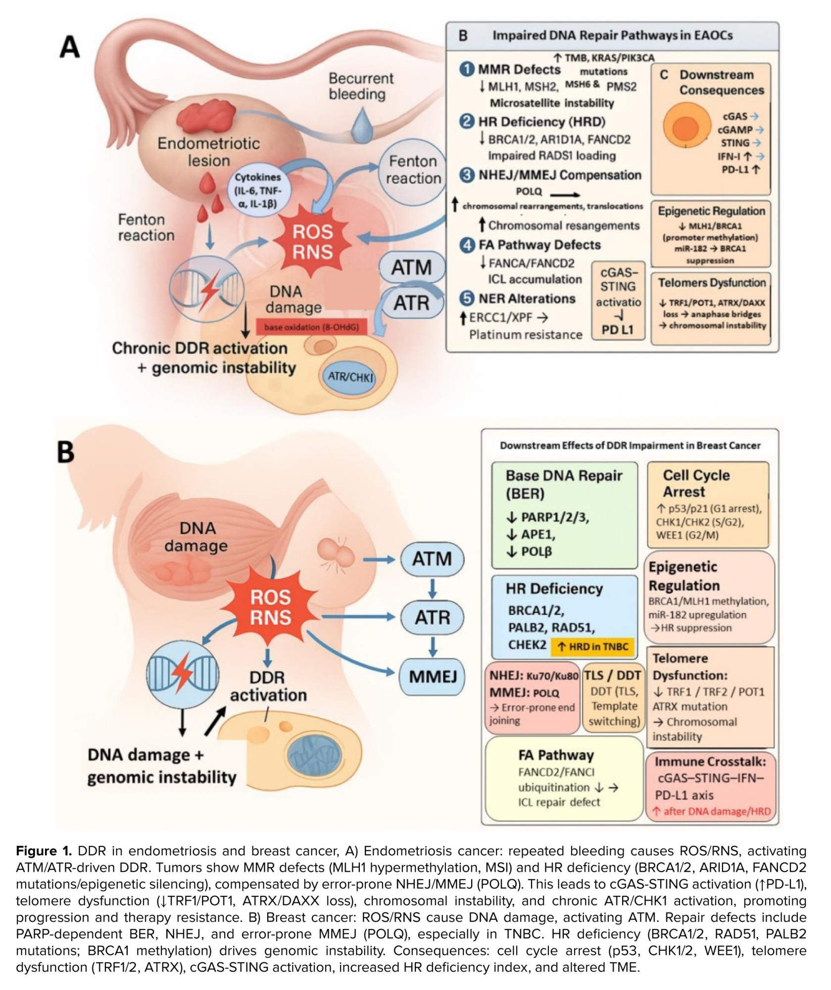
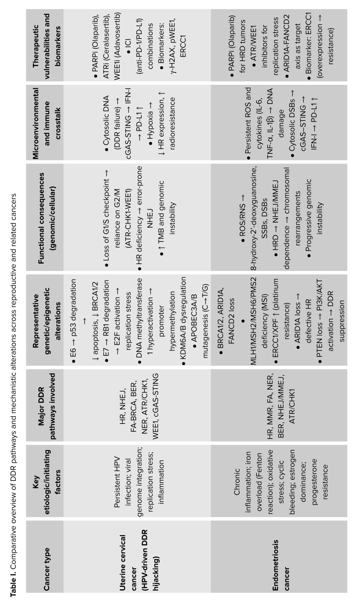
Endometriosis is a chronic inflammatory and estrogen-dependent disorder characterized by ectopic endometrial-like tissue growth outside the uterine cavity, most frequently affecting the ovaries, fallopian tubes, and peritoneal surfaces (27). Although histologically benign, endometriosis exhibits cancer-associated features, including invasive growth, resistance to apoptosis, increased angiogenesis, and an established risk of malignant transformation (28). These characteristics have positioned genomic instability and dysregulated DDR signaling as central contributors to disease progression under chronic inflammatory and oxidative stress (6). Persistent cyclic bleeding of ectopic lesions, combined with impaired clearance of blood and cellular debris, sustains a pro-inflammatory microenvironment. This milieu generates ROS and reactive nitrogen species (RNS), promoting oxidative DNA lesions such as 8-hydroxy-2′-deoxyguanosine, SSBs, and DSBs, particularly in ovarian endometriomas where iron-mediated fenton reactions amplify oxidative stress (29, 30). Chronic inflammation and hormonal dysregulation jointly impose sustained replication stress and progressive impairment of DDR fidelity in endometriotic lesions (29, 31). Among DDR alterations, MMR deficiency represents an early and prominent event in endometriosis-associated endometrioid carcinoma. Reduced expression of MLH1, MSH2, MSH6, and PMS2 results in microsatellite instability (MSI) and elevated tumor mutational burden (TMB), facilitating acquisition of oncogenic mutations such as KRAS proto-oncogene, GTPase, and phosphatidylinositol-4,5-bisphosphate 3-kinase catalytic subunit alpha (PIK3CA) (31). Notably, partial loss of MMR proteins has also been detected in atypical endometriosis lesions, suggesting that DDR dysfunction precedes malignant transformation (29, 31). HR deficiency is a defining feature of endometriosis-associated ovarian cancers (EAOCs), particularly clear cell carcinoma and endometrioid carcinoma (32). In many cases, this phenotype arises through "BRCA-like" mechanisms rather than germline BRCA gene mutations, including epigenetic silencing and loss of HR-associated regulators such as AT-rich interaction domain 1A (ARID1A) and FANCD2 (33). The resulting chromosomal instability promotes reliance on error-prone DNA repair pathways, accelerating malignant progression (32, 33). Additional DDR-related pathways are frequently perturbed in EAOCs. Dysregulation of the FA pathway compromises repair of ICLs generated by oxidative and iron-mediated damage, particularly in tumors with ARID1A loss (34, 35). Altered NER activity has also been reported, with ERCC1 overexpression correlating with platinum resistance (36). Loss of phosphatase and tensin homolog (PTEN), a common feature in endometrial cancer, further suppresses DDR signaling through the activation of the phosphoinositide 3-kinase (PI3K)/protein kinase B(AKT)/mammalian target of rapamycin (mTOR) pathway (34, 37). Epigenetic mechanisms represent an additional layer of DDR impairment. Promoter hypermethylation of MLH1 and BRCA1, histone modifications, and non-coding RNAs such as microRNA-182 targeting BRCA1 contribute to a "BRCAness" phenotype in EAOCs (33, 34). Chronic replication stress further disrupts replication fork protection mediated by RAD51C, FA complementation group M, and BRCA2, thereby enhancing genomic instability (35). Telomere dysfunction, driven by oxidative damage and loss of shelterin components and chromatin remodelers, promotes chromosome fusions and is frequently observed in advanced EAOCs (38). DDR alterations in endometriosis-associated cancers have direct clinical relevance. Mismatch repair-deficient and MSI-high (MSI-H) tumors exhibit high TMB and distinct molecular profiles, supporting their classification as a biologically and therapeutically relevant subgroup (31, 39). HR-deficient EAOCs demonstrate increased sensitivity to platinum-based chemotherapy and DDR-targeted therapies, consistent with their underlying genomic instability. Beyond DNA repair, defective DDR promotes cytosolic accumulation of unrepaired DNA fragments, activating Cgas-STING signaling and programmed death-ligand 1 (PD-L1) upregulation (40, 41). This immunogenic reprogramming suggests potential responsiveness to immunotherapeutic strategies in selected EAOCs (11). Despite therapeutic opportunities, several challenges remain. Intratumoral heterogeneity of DDR defects complicates patient stratification, while secondary resistance mechanisms may limit the durability of DDR-targeted therapies. Identification of robust biomarkers integrating genomic, epigenetic, and replication stress signatures is critical for optimizing treatment selection. Future research should focus on longitudinal mapping of DDR alterations during the transition from benign endometriosis to malignancy, as well as rational combination strategies integrating DDR-targeted agents with immunotherapy or epigenetic modulators. A comprehensive understanding of DDR dysregulation across repair pathways may enable earlier detection and personalized therapeutic approaches for endometriosis-associated cancers (Figure 1A; Table I) (6, 39).
4. DDR in breast cancer
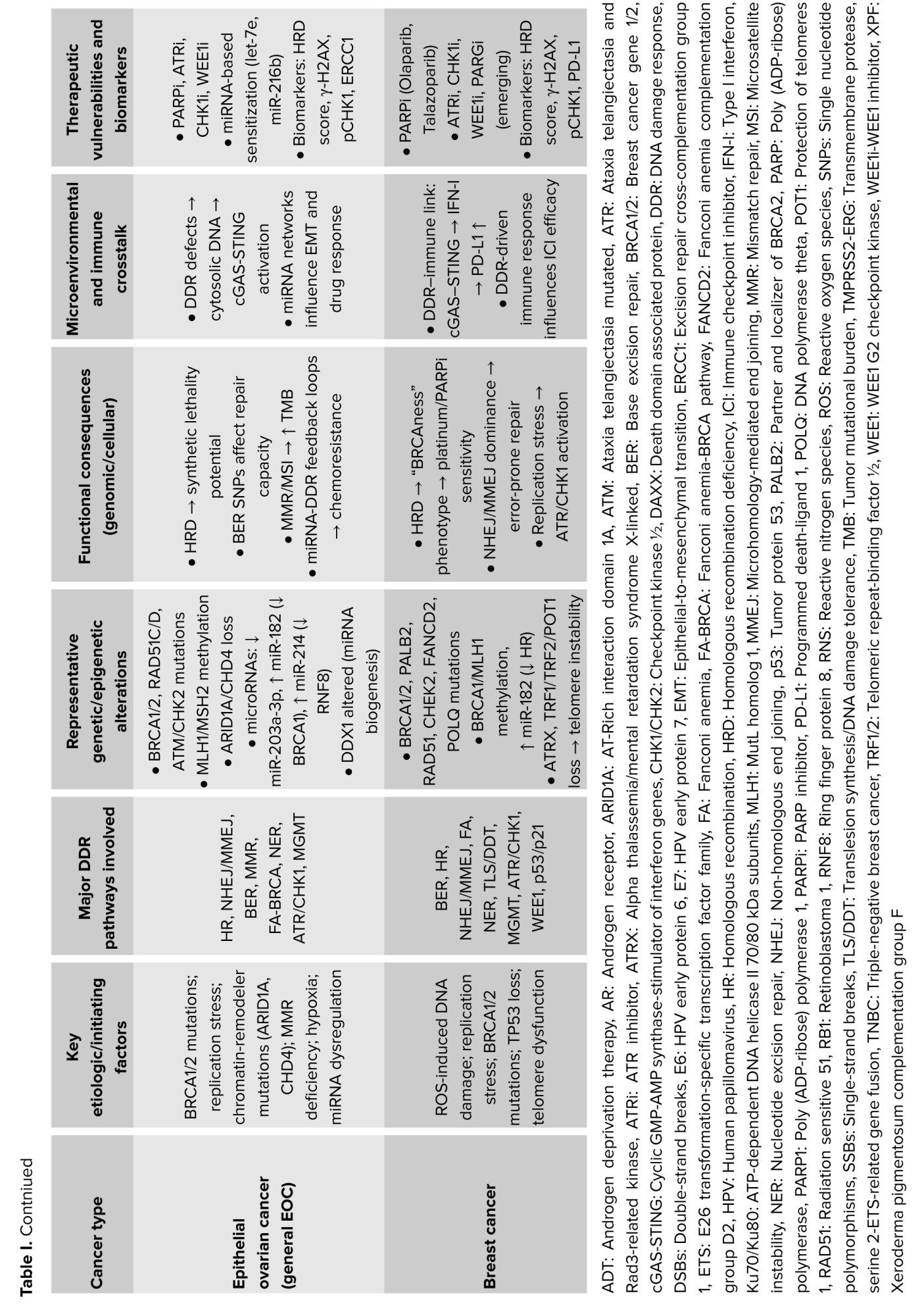
Breast cancer cells are continuously exposed to endogenous and exogenous sources of DNA damage, including oxidative lesions, strand breaks, insertions/deletions, and DNA crosslinks. Accumulation of such damage, if not accurately repaired, promotes genomic instability and breast tumorigenesis. Dysregulation of the DDR is therefore a fundamental driver of mutational burden, tumor heterogeneity, and therapeutic vulnerability in breast cancer. Oxidative stress-induced SSBs are particularly frequent in breast cancer cells, increasing reliance on BER for genome maintenance (42). Elevated expression and activity of PARP family members and other BER-associated factors have been observed in multiple breast cancer subtypes, reflecting adaptation to chronic oxidative stress (43). Defects in DSBs repair pathways are central to breast cancer biology. HR deficiency arises in up to 30% of breast cancers, either through germline or somatic mutations in BRCA1/2 or via "BRCAness" mechanisms involving alterations in RAD51, PALB2, CHEK2, epigenetic silencing of BRCA1, or microRNA dysregulation such as miR-182 (44). HR deficiency prevalence varies by subtype, reaching approximately 50% in triple-negative breast cancer (TNBC), while being less frequent in luminal tumors. Human epidermal growth factor receptor 2 (HER2)-positive tumors commonly exhibit HR deficiency linked to BRCA2 loss or BRCA1 promoter methylation (45). In HR-deficient tumors, reliance on alternative error-prone repair pathways contributes to genomic instability. Nuclear overexpression of NHEJ-associated Ku70 and Ku80 DNA repair proteins (Ku70/Ku80) correlates with aggressive disease and poor prognosis, while Ku80 is also associated with increased breast cancer risk (46, 47). MMEJ, driven by POLQ, is frequently upregulated in HR-deficient tumors and promotes mutagenic repair (48). Additional DDR alterations include dysregulation of the FA pathway, which compromises repair of interstrand cross-links induced by platinum agents. Mutations in the FA complementation group A increase BRCA and functionally overlap with BRCA1/2 defects (49). Deficient MMR, although rare in breast cancer, is associated with elevated TMB, PD-L1 expression, and enhanced chemotherapy sensitivity, particularly in HR-negative tumors (50). Germline mutations in MSH6 and PMS2 have been reported in Lynch syndrome-associated breast cancers (51). Beyond repair defects, replication stress tolerance mechanisms are frequently engaged. DNA damage tolerance pathways, including translesion synthesis and template switching, enable lesion bypass during replication but contribute to mutagenesis when dysregulated. Telomere maintenance is also perturbed, with altered telomerase activity and shelterin complex dysfunction promoting chromosome instability; mutations in ATRX are associated with aggressive breast cancer phenotypes (26). DDR alterations have major therapeutic relevance in breast cancer. HR deficiency tumors show increased sensitivity to platinum-based chemotherapy and DDR-targeted strategies, particularly in TNBC and BRCA-mutated disease (52). Elevated BER activity has positioned BER-associated proteins as actionable targets, while MMR-deficient tumors may benefit from immunotherapeutic approaches due to increased TMB and immune activation (53). Pharmacologic inhibition of DDR signaling has expanded beyond PARP inhibitors. Targeting upstream kinases and cell-cycle regulators has demonstrated efficacy in selected settings, especially in advanced TNBC and HER2-negative disease (54). Moreover, DDR-induced cytosolic DNA accumulation activates (cGAS-STING) signaling, linking genomic instability to antitumor immune responses, and enhancing therapeutic synergy with immunotherapy (55). Despite significant progress, therapeutic resistance remains a major challenge, driven by restoration of HR, activation of compensatory repair pathways, and tumor heterogeneity. Identification of robust biomarkers integrating HR deficiency scores, replication stress signatures, and immune markers is critical for patient stratification. Future strategies will likely focus on rational combination therapies targeting multiple DDR nodes, integration with immunotherapy, and exploitation of emerging vulnerabilities such as DNA damage tolerance and chromatin remodeling defects. A deeper understanding of DDR pathway interplay across breast cancer subtypes will continue to refine precision medicine approaches, particularly in therapy-resistant and high-risk disease (Figure 1B; Table I) (42).
5. DDR in ovarian cancer
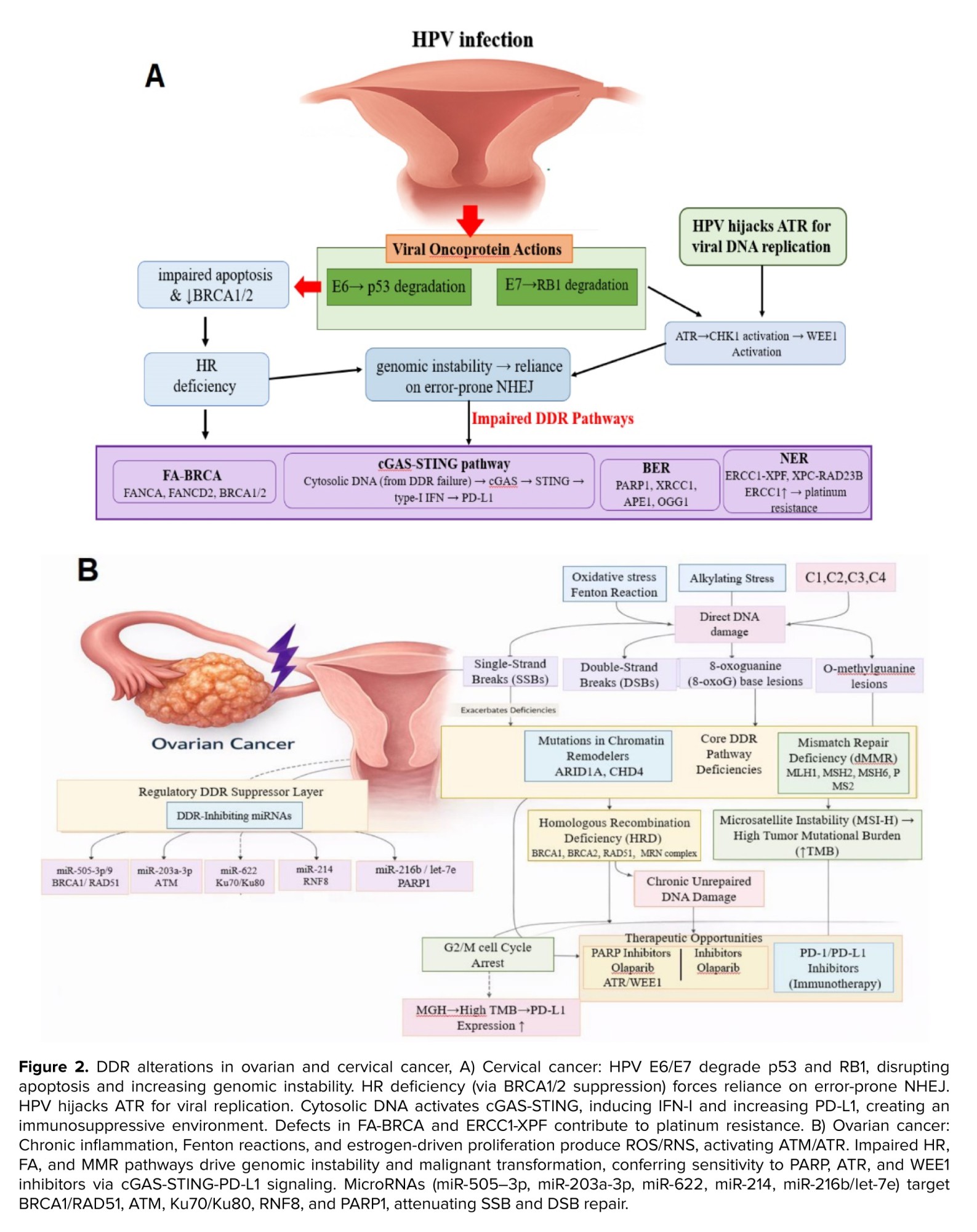
Epithelial ovarian cancer is a biologically heterogeneous malignancy comprising 5 major subtypes: high-grade serous carcinoma (HGSC), low-grade serous carcinoma, endometrioid carcinoma, clear cell carcinoma, and mucinous carcinoma (56). Among these, HGSC represents the most prevalent and genomically unstable subtype, with widespread DDR alterations contributing to disease initiation, progression, and therapeutic response. Both genetic and epigenetic disruptions of DDR pathways critically influence sensitivity or resistance to platinum-based chemotherapy and targeted therapies in ovarian cancer (56, 57). Defects associated with HR deficiency are observed in up to 50% of epithelial ovarian cancers. Mutations in BRCA1/2 remain the strongest predictors of platinum responsiveness and therapeutic vulnerability. Additional alterations involve RAD51 paralogs (RAD51C, RAD51D), components of the MRN complex, reduced RAD50 expression, and promoter hypermethylation of RAD51, collectively driving a "BRCAness" phenotype (57). Aberrant engagement of error-prone repair pathways contributes to disease progression and treatment resistance. Alterations in NHEJ-associated factors, including overexpression or mutation of DNA-dependent protein kinase, POLQ, and X-ray repair cross-complementing protein 4 (XRCC4), have been linked to genomic instability and reduced therapeutic efficacy. Single-nucleotide polymorphisms (SNPs) in DNA ligase IV and XRCC1 further compromise repair fidelity and promote resistance, independent of HR status (58). BER alterations primarily involve SNPs in 8-oxoguanine DNA glycosylase, apurinic/APE1, and XRCC1 (59). Notably, apurinic/APE1 overexpression in HGSC correlates with enhanced repair capacity and platinum resistance (60). MMR deficiency arises through germline mutations in Lynch syndrome-associated genes (MLH1, MSH2, MSH6, and PMS2) or epigenetic silencing of MLH1, resulting in MSI, particularly in endometrioid and clear cell ovarian carcinomas (61). HGSCs exhibit near-universal TP53 mutations and loss of heterozygosity, impairing apoptotic responses to DNA damage. Additional alterations in checkpoint regulators and chromatin remodelers-including ARID1A, chromodomain helicase DNA-binding proteins (chromodomain helicase DNA-binding protein4/5/8 [CHD4, CHD5, CHD8]), and actin-like protein 6A-further disrupt DDR signaling by altering chromatin accessibility (62, 63). MicroRNAs represent a major epigenetic layer modulating DDR in ovarian cancer. Dysregulation of tumor-suppressive microRNAs targeting DDR components promotes genomic instability and tumor progression (64). For instance, reduced miR-203a-3p expression correlates with poor prognosis, while miR-191, miR-214-5p, and miR-647 indirectly impair p53 regulation via mouse double minute 4 and 2 homolog targeting. Other microRNAs, including miR-4324, miR-134-3p, and miR-328-3p, influence proliferation, migration, and tumor aggressiveness through modulation of flap endonuclease 1, DNA damage-binding protein 2, and aldehyde dehydrogenase 1 family member A1 (64). DDR alterations have direct clinical relevance in ovarian cancer. HR-deficient tumors show enhanced sensitivity to platinum-based chemotherapy and DDR-targeted therapies. In contrast, aberrant activation of alternative repair pathways contributes to intrinsic or acquired resistance, including reduced efficacy of targeted agents (65). MicroRNAs-mediated regulation significantly influences therapeutic response. Downregulation of miR-519a-3p elevates PARP1 expression and promotes resistance, whereas let-7e and miR-216b restore platinum sensitivity by suppressing PARP1. Other microRNAs, such as miR-9, miR-506-3p, miR-379-5p, and miR-770-5p, modulate sensitivity to cisplatin and targeted therapies by influencing key DDR and NER components. Despite advances in DDR-targeted strategies, ovarian cancer remains characterized by high relapse rates and therapy resistance. Tumor heterogeneity, restoration of repair capacity, and dynamic epigenetic regulation complicate durable treatment responses. Comprehensive profiling of DDR defects, including miRNA signatures and chromatin remodeling alterations, is essential for improving patient stratification (64). Future therapeutic approaches will likely integrate DDR-targeted agents with epigenetic modulators and immunotherapy, aiming to overcome resistance and exploit context-specific vulnerabilities. Understanding the complex regulatory network linking DDR proteins, microRNAs, and chromatin architecture will be critical for advancing precision medicine in ovarian cancer (Figure 2; Table I) (64, 66).
6. DDR in uterine cervical cancer
Uterine cervical cancer is etiologically driven by persistent infection with HPV, which profoundly reshapes genomic stability and treatment response through disruption of DDR regulation. Viral-mediated genomic instability represents a defining molecular feature of cervical cancer and underlies both radiosensitivity and chemotherapeutic outcomes. HPV oncoproteins E6 and E7 inactivate key tumor suppressors, including TP53 and retinoblastoma 1 (RB1), abolishing G1/S checkpoint control and enforcing uncontrolled proliferation. This unchecked cell cycle progression induces severe replication stress and dependence on post-replication checkpoints, creating a distinctive DDR dependency profile in cervical cancer cells (67). HPV infection also drives extensive epigenetic remodeling. E7-mediated activation of DNA methyltransferase-1 promotes promoter hypermethylation and transcriptional silencing of regulatory and immune-related genes, while modulation of lysine demethylase 6A and 6B (KDM6A/B) alters chromatin accessibility and facilitates malignant progression. Viral genome integration into host DNA further accelerates genomic instability, producing characteristic mutational signatures dominated by apolipoprotein B mRNA editing catalytic polypeptide-like (APOBEC)-driven cytidine deamination (68). HPV-associated DDR impairment extends to multiple repair pathways. E6 disrupts the function of BRCA1/2, reducing HR efficiency and shifting repair toward error-prone mechanisms. Altered NHEJ capacity has prognostic significance, as reduced XRCC6 expression correlates with improved radiotherapy outcomes (69). In addition, dysregulation of the FA-BRCA axis compromises repair of cisplatin-induced ICLs, while NER capacity, particularly ERCC1 expression, modulates platinum sensitivity and overall survival (70). Accumulation of unrepaired DNA damage leads to cytosolic DNA fragments that activate cGAS-STING signaling, linking DDR failure to innate immune activation within the TME. Hypoxia further modulates DDR gene expression and therapeutic response, adding another layer of context-dependent regulation (71). DDR deficiencies render HPV-positive cervical cancers particularly sensitive to DNA-damaging modalities, including radiotherapy and platinum-based chemotherapy. These vulnerabilities have prompted clinical evaluation of DDR-targeted strategies. Inhibition of PARP exploits HR impairment, enhancing tumor cell death while potentially mitigating cisplatin-induced nephrotoxicity in normal tissues (70). Targeting cell-cycle checkpoint kinases has also shown promise. WEE1 inhibition (e.g., adavosertib) induces checkpoint abrogation and mitotic catastrophe in DDR-compromised cells (72). DDR-associated biomarkers, including phosphorylated histone H2AX (γ-H2AX), phosphorylated WEE1, and ERCC1, provide prognostic and predictive value, while dynamic γ-H2AX foci resolution may forecast response to chemoradiotherapy (73). Despite intrinsic radiosensitivity, therapeutic resistance and disease recurrence remain significant challenges in cervical cancer. Tumor heterogeneity, hypoxia-driven DDR modulation, and adaptive repair responses limit the long-term efficacy of single-agent strategies. High TMBI generated by HPV-driven DDR failure supports a rational combination of DDR-targeted therapies with immune checkpoint inhibitors targeting programmed cell death protein-1 (Figure 2; Table I) (74). Future directions include integrated biomarker-driven patient stratification, combinatorial DDR-immunotherapy regimens, and exploitation of virus-specific DDR dependencies. A refined understanding of HPV-mediated DDR reprogramming will be critical for advancing precision therapeutic approaches in cervical cancer.
7. Conclusion
DDR pathways are central to the maintenance of genomic integrity and represent key determinants of tumor initiation, progression, and therapeutic vulnerability in reproductive cancers. While the specific DDR alterations differ across ovarian, breast, and cervical cancers (driven by tissue-specific factors, hormonal influences, viral oncogenes, and epigenetic regulation) these pathways consistently influence genomic stability, immune activation, and treatment response. Narrative synthesis of the literature highlights that HR deficiency, FA pathway defects, MMR deficiency, and replication stress converge on shared vulnerabilities can be therapeutically exploited using PARP inhibitors, ATR and WEE1 inhibitors, radiotherapy, and immune checkpoint inhibitors. DDR profiling, including assessment of HR deficiency status, miRNA networks, and biomarkers such as γ-H2AX, RAD51, and ERCC1, provides a framework for patient stratification and personalized treatment strategies. Furthermore, integrating mechanistic insights with clinical observations underscores opportunities for combination therapies and guides rational selection of DDR-targeted agents. While this review does not provide quantitative meta-analyses or detailed epidemiologic data, it offers a comprehensive qualitative synthesis, identifying key mechanistic and therapeutic trends and highlighting emerging directions, including novel DDR inhibitors, biomarker-guided interventions, and strategies to overcome resistance. Overall, a precise understanding of DDR dynamics in reproductive malignancies can inform translational research and optimize clinical management, supporting the development of tailored, mechanism-driven therapies.
Author Contributions
M. Zafari: Conceptualization, writing, visualization and supervision. H. Zarei: Conceptualization, writing, and supervision. E. Sadeghi: Writing and visualization. F. Lotfi Asrami, F. Bahmani, and DSK. Maliki: Writing. S. Jahani and A. Abdollahi: Review, validation, and supervision. H. Musavi: Conceptualization, review, project administration, supervision, and is also the corresponding author.
Acknowledgements
The authors gratefully acknowledge the support and assistance of the staff at the Immunogenetic Research Center and the Departments of Anatomical Sciences and Clinical Biochemistry, Mazandaran University of Medical Sciences, Mazandaran, Iran. The authors also acknowledge the use of artificial intelligence tools (Perplexity) for language polishing and grammar checking during the preparation of this manuscript. This research did not receive any specific grant from funding agencies in the public, commercial, or not-for-profit sectors.
Conflict of Interest
The authors declare that there is no conflict of interest.
The DNA damage response (DDR) is a critical cellular network that detects DNA lesions, coordinates repair, and regulates cell cycle checkpoints to maintain genomic integrity (1). Core DDR components, including Ataxia-telangiectasia mutated (ATM) and Ataxia telangiectasia and Rad3-related (ATR) kinases, respond to double-strand breaks (DSBs) and replication stress, initiating signaling cascades that halt cell cycle progression and recruit repair machinery (2, 3). Specialized pathways such as homologous recombination (HR) repair, mismatch repair (MMR), nucleotide excision repair (NER), and base excision repair (BER) work together to resolve diverse DNA lesions (4). In gynecologic and breast cancers, including ovarian, endometrial, cervical, and breast malignancies, DDR defects are prevalent and influence tumor initiation, progression, therapy response, and resistance (5, 6). These cancers exhibit distinct DDR alteration patterns reflecting differences in tissue origin, hormonal regulation, and oncogenic drivers. High-grade serous ovarian carcinoma often exhibits HR repair deficiencies, exploited therapeutically with PARP inhibitors (7). Endometrial cancer displays heterogeneous DDR landscapes, with HR deficiency particularly common in high-grade, tumor protein p53 (TP53)-mutated subtypes and serous carcinomas, while Fanconi anemia (FA) pathway alterations characterize aggressive subtypes. In breast cancer, interactions between DDR and hormone signaling highlight mechanisms of genomic instability and potential combinatorial treatments (8). Cervical cancer pathogenesis is driven by human papillomavirus (HPV) oncoproteins that compromise DDR through viral integration and replication stress (9, 10). DDR alterations also impact tumor aggressiveness, metastasis, and immune interactions through impaired checkpoint activation and sustained replication stress (11). This review integrates molecular insights with clinical relevance, focusing on DDR dysregulation across gynecologic and breast cancers, and the therapeutic opportunities arising from these vulnerabilities. Literature was systematically collected from PubMed/MEDLINE, Scopus, and Web of Science, prioritizing original research, meta-analyses, and key reviews from the past decade, with seminal earlier studies included for context.
2. Molecular basis of DDR
DDR is a highly coordinated network of sensors, transducers, and effectors that detects DNA lesions, signals their presence, and orchestrates appropriate repair or cell cycle control to preserve genomic integrity. DDR maintains cellular homeostasis in response to both endogenous insults, such as reactive oxygen species (ROS) and replication stress, as well as exogenous factors, including radiation and chemical agents (1). The initial recognition of DNA damage is mediated by specialized sensor proteins. DSBs are detected by the meiotic recombination 11 homolog 1, RAD50 recombinase (RAD50), and Nijmegen breakage syndrome 1 (MRN) complex, which recruits and activates the ATM kinase, whereas stalled replication forks and single-stranded DNA regions primarily trigger the ATR kinase (1, 12). Single-strand breaks (SSBs) are sensed by PARP1/2/3, which facilitates recruitment of repair factors through poly (ADP-ribosyl) ation (13). Once activated, DDR engages multiple DNA repair pathways according to the type of lesion. HR represents a high-fidelity DSB repair mechanism that utilizes a sister chromatid as a template. HR requires the coordinated activity of BRCA1/2 genes, RAD51 recombinase, PALB2, and the MRN complex, and is primarily active during the S and G2 phases of the cell cycle (14). Non-homologous end joining (NHEJ), in contrast, is an error-prone pathway that directly ligates DNA ends with minimal sequence homology (1). Its core components include the Ku70/Ku80 heterodimer and DNA-dependent protein kinase catalytic subunit, and it operates throughout the cell cycle, predominantly in G0/G1 (1, 15). Microhomology-mediated end joining (MMEJ) represents an alternative, mutagenic DSB repair mechanism that uses short homologous sequences at break termini and relies on DNA polymerase theta (POLQ) (16). BER addresses SSBs and small base modifications, involving glycosylases, APE endonucleases such as apurinic/APE1, polymerases, and ligases, with PARP1/2 functioning as critical sensors and recruiters (17). NER removes bulky lesions, including thymine dimers and chemical adducts, through protein complexes such as xeroderma pigmentosum group C-RAD23 homolog B and xeroderma pigmentosum group A and ERCC1/xeroderma pigmentosum group F (XPF) (18). MMR corrects replication errors, such as base mispairings or small insertions/deletions, via MutL homolog (MLH1), MutS homolog 2 (MSH2), MutS homolog 6 (MSH6), and postmeiotic segregation increased 2 (PMS2) (19). The FA pathway repairs interstrand crosslinks (ICLs) by monoubiquitination of FANCD2/FA complementation group-I through the FA core complex, often coordinating with BRCA1/2 and HR-mediated repair (20). Checkpoint signaling is an integral component of DDR, ensuring repair occurs before cell cycle progression. ATM and ATR activate downstream kinases, checkpoint kinase 1 and 2, which induce transient arrest at G1/S, intra-S, or G2/M phases. This checkpoint regulation allows time for DNA repair and prevents propagation of damaged DNA (21). TP53 and cyclin-dependent kinase inhibitor 1A are key effectors in this process, mediating cell cycle arrest or apoptosis if lesions are irreparable (21, 22). Deficiencies in DDR create exploitable vulnerabilities, exemplified by the concept of synthetic lethality. HR deficiency renders cells particularly sensitive to PARP inhibitors, as they are unable to repair accumulated DNA damage effectively (23). Beyond PARP inhibitors, other DDR-targeting agents, including ATM, ATR, checkpoint kinase 1 and 2, and WEE1 G2 checkpoint kinase (WEE1) inhibitors, are under investigation across various cancer types (24). In addition to direct repair functions, DDR maintains chromatin integrity, telomere stability, and crosstalk with innate immunity. Cytosolic DNA fragments arising from defective repair activate the cyclic GMP-AMP synthase (cGAS)-stimulator of interferon genes (STING) pathway, inducing type I interferon responses and influencing the tumor microenvironment (TME) (25). Telomere protection is mediated by shelterin proteins (telomeric repeat-binding factor 1 and 2 [TERF1, TERF2], protection of telomeres 1 [POT1]) and chromatin remodelers such as alpha thalassemia/mental retardation syndrome X-linked (ATRX), and death domain-associated protein, preventing chromosome ends from being recognized as DSBs and maintaining chromosomal stability (26). Collectively, DDR functions as the central guardian of genomic integrity, and its impairment underlies mutagenesis, cancer susceptibility, and differential therapeutic responses, forming the foundation for the subsequent cancer-specific analyses.
3. DDR in endometriosis-associated cancer
Endometriosis is a chronic inflammatory and estrogen-dependent disorder characterized by ectopic endometrial-like tissue growth outside the uterine cavity, most frequently affecting the ovaries, fallopian tubes, and peritoneal surfaces (27). Although histologically benign, endometriosis exhibits cancer-associated features, including invasive growth, resistance to apoptosis, increased angiogenesis, and an established risk of malignant transformation (28). These characteristics have positioned genomic instability and dysregulated DDR signaling as central contributors to disease progression under chronic inflammatory and oxidative stress (6). Persistent cyclic bleeding of ectopic lesions, combined with impaired clearance of blood and cellular debris, sustains a pro-inflammatory microenvironment. This milieu generates ROS and reactive nitrogen species (RNS), promoting oxidative DNA lesions such as 8-hydroxy-2′-deoxyguanosine, SSBs, and DSBs, particularly in ovarian endometriomas where iron-mediated fenton reactions amplify oxidative stress (29, 30). Chronic inflammation and hormonal dysregulation jointly impose sustained replication stress and progressive impairment of DDR fidelity in endometriotic lesions (29, 31). Among DDR alterations, MMR deficiency represents an early and prominent event in endometriosis-associated endometrioid carcinoma. Reduced expression of MLH1, MSH2, MSH6, and PMS2 results in microsatellite instability (MSI) and elevated tumor mutational burden (TMB), facilitating acquisition of oncogenic mutations such as KRAS proto-oncogene, GTPase, and phosphatidylinositol-4,5-bisphosphate 3-kinase catalytic subunit alpha (PIK3CA) (31). Notably, partial loss of MMR proteins has also been detected in atypical endometriosis lesions, suggesting that DDR dysfunction precedes malignant transformation (29, 31). HR deficiency is a defining feature of endometriosis-associated ovarian cancers (EAOCs), particularly clear cell carcinoma and endometrioid carcinoma (32). In many cases, this phenotype arises through "BRCA-like" mechanisms rather than germline BRCA gene mutations, including epigenetic silencing and loss of HR-associated regulators such as AT-rich interaction domain 1A (ARID1A) and FANCD2 (33). The resulting chromosomal instability promotes reliance on error-prone DNA repair pathways, accelerating malignant progression (32, 33). Additional DDR-related pathways are frequently perturbed in EAOCs. Dysregulation of the FA pathway compromises repair of ICLs generated by oxidative and iron-mediated damage, particularly in tumors with ARID1A loss (34, 35). Altered NER activity has also been reported, with ERCC1 overexpression correlating with platinum resistance (36). Loss of phosphatase and tensin homolog (PTEN), a common feature in endometrial cancer, further suppresses DDR signaling through the activation of the phosphoinositide 3-kinase (PI3K)/protein kinase B(AKT)/mammalian target of rapamycin (mTOR) pathway (34, 37). Epigenetic mechanisms represent an additional layer of DDR impairment. Promoter hypermethylation of MLH1 and BRCA1, histone modifications, and non-coding RNAs such as microRNA-182 targeting BRCA1 contribute to a "BRCAness" phenotype in EAOCs (33, 34). Chronic replication stress further disrupts replication fork protection mediated by RAD51C, FA complementation group M, and BRCA2, thereby enhancing genomic instability (35). Telomere dysfunction, driven by oxidative damage and loss of shelterin components and chromatin remodelers, promotes chromosome fusions and is frequently observed in advanced EAOCs (38). DDR alterations in endometriosis-associated cancers have direct clinical relevance. Mismatch repair-deficient and MSI-high (MSI-H) tumors exhibit high TMB and distinct molecular profiles, supporting their classification as a biologically and therapeutically relevant subgroup (31, 39). HR-deficient EAOCs demonstrate increased sensitivity to platinum-based chemotherapy and DDR-targeted therapies, consistent with their underlying genomic instability. Beyond DNA repair, defective DDR promotes cytosolic accumulation of unrepaired DNA fragments, activating Cgas-STING signaling and programmed death-ligand 1 (PD-L1) upregulation (40, 41). This immunogenic reprogramming suggests potential responsiveness to immunotherapeutic strategies in selected EAOCs (11). Despite therapeutic opportunities, several challenges remain. Intratumoral heterogeneity of DDR defects complicates patient stratification, while secondary resistance mechanisms may limit the durability of DDR-targeted therapies. Identification of robust biomarkers integrating genomic, epigenetic, and replication stress signatures is critical for optimizing treatment selection. Future research should focus on longitudinal mapping of DDR alterations during the transition from benign endometriosis to malignancy, as well as rational combination strategies integrating DDR-targeted agents with immunotherapy or epigenetic modulators. A comprehensive understanding of DDR dysregulation across repair pathways may enable earlier detection and personalized therapeutic approaches for endometriosis-associated cancers (Figure 1A; Table I) (6, 39).
4. DDR in breast cancer
Breast cancer cells are continuously exposed to endogenous and exogenous sources of DNA damage, including oxidative lesions, strand breaks, insertions/deletions, and DNA crosslinks. Accumulation of such damage, if not accurately repaired, promotes genomic instability and breast tumorigenesis. Dysregulation of the DDR is therefore a fundamental driver of mutational burden, tumor heterogeneity, and therapeutic vulnerability in breast cancer. Oxidative stress-induced SSBs are particularly frequent in breast cancer cells, increasing reliance on BER for genome maintenance (42). Elevated expression and activity of PARP family members and other BER-associated factors have been observed in multiple breast cancer subtypes, reflecting adaptation to chronic oxidative stress (43). Defects in DSBs repair pathways are central to breast cancer biology. HR deficiency arises in up to 30% of breast cancers, either through germline or somatic mutations in BRCA1/2 or via "BRCAness" mechanisms involving alterations in RAD51, PALB2, CHEK2, epigenetic silencing of BRCA1, or microRNA dysregulation such as miR-182 (44). HR deficiency prevalence varies by subtype, reaching approximately 50% in triple-negative breast cancer (TNBC), while being less frequent in luminal tumors. Human epidermal growth factor receptor 2 (HER2)-positive tumors commonly exhibit HR deficiency linked to BRCA2 loss or BRCA1 promoter methylation (45). In HR-deficient tumors, reliance on alternative error-prone repair pathways contributes to genomic instability. Nuclear overexpression of NHEJ-associated Ku70 and Ku80 DNA repair proteins (Ku70/Ku80) correlates with aggressive disease and poor prognosis, while Ku80 is also associated with increased breast cancer risk (46, 47). MMEJ, driven by POLQ, is frequently upregulated in HR-deficient tumors and promotes mutagenic repair (48). Additional DDR alterations include dysregulation of the FA pathway, which compromises repair of interstrand cross-links induced by platinum agents. Mutations in the FA complementation group A increase BRCA and functionally overlap with BRCA1/2 defects (49). Deficient MMR, although rare in breast cancer, is associated with elevated TMB, PD-L1 expression, and enhanced chemotherapy sensitivity, particularly in HR-negative tumors (50). Germline mutations in MSH6 and PMS2 have been reported in Lynch syndrome-associated breast cancers (51). Beyond repair defects, replication stress tolerance mechanisms are frequently engaged. DNA damage tolerance pathways, including translesion synthesis and template switching, enable lesion bypass during replication but contribute to mutagenesis when dysregulated. Telomere maintenance is also perturbed, with altered telomerase activity and shelterin complex dysfunction promoting chromosome instability; mutations in ATRX are associated with aggressive breast cancer phenotypes (26). DDR alterations have major therapeutic relevance in breast cancer. HR deficiency tumors show increased sensitivity to platinum-based chemotherapy and DDR-targeted strategies, particularly in TNBC and BRCA-mutated disease (52). Elevated BER activity has positioned BER-associated proteins as actionable targets, while MMR-deficient tumors may benefit from immunotherapeutic approaches due to increased TMB and immune activation (53). Pharmacologic inhibition of DDR signaling has expanded beyond PARP inhibitors. Targeting upstream kinases and cell-cycle regulators has demonstrated efficacy in selected settings, especially in advanced TNBC and HER2-negative disease (54). Moreover, DDR-induced cytosolic DNA accumulation activates (cGAS-STING) signaling, linking genomic instability to antitumor immune responses, and enhancing therapeutic synergy with immunotherapy (55). Despite significant progress, therapeutic resistance remains a major challenge, driven by restoration of HR, activation of compensatory repair pathways, and tumor heterogeneity. Identification of robust biomarkers integrating HR deficiency scores, replication stress signatures, and immune markers is critical for patient stratification. Future strategies will likely focus on rational combination therapies targeting multiple DDR nodes, integration with immunotherapy, and exploitation of emerging vulnerabilities such as DNA damage tolerance and chromatin remodeling defects. A deeper understanding of DDR pathway interplay across breast cancer subtypes will continue to refine precision medicine approaches, particularly in therapy-resistant and high-risk disease (Figure 1B; Table I) (42).
5. DDR in ovarian cancer
Epithelial ovarian cancer is a biologically heterogeneous malignancy comprising 5 major subtypes: high-grade serous carcinoma (HGSC), low-grade serous carcinoma, endometrioid carcinoma, clear cell carcinoma, and mucinous carcinoma (56). Among these, HGSC represents the most prevalent and genomically unstable subtype, with widespread DDR alterations contributing to disease initiation, progression, and therapeutic response. Both genetic and epigenetic disruptions of DDR pathways critically influence sensitivity or resistance to platinum-based chemotherapy and targeted therapies in ovarian cancer (56, 57). Defects associated with HR deficiency are observed in up to 50% of epithelial ovarian cancers. Mutations in BRCA1/2 remain the strongest predictors of platinum responsiveness and therapeutic vulnerability. Additional alterations involve RAD51 paralogs (RAD51C, RAD51D), components of the MRN complex, reduced RAD50 expression, and promoter hypermethylation of RAD51, collectively driving a "BRCAness" phenotype (57). Aberrant engagement of error-prone repair pathways contributes to disease progression and treatment resistance. Alterations in NHEJ-associated factors, including overexpression or mutation of DNA-dependent protein kinase, POLQ, and X-ray repair cross-complementing protein 4 (XRCC4), have been linked to genomic instability and reduced therapeutic efficacy. Single-nucleotide polymorphisms (SNPs) in DNA ligase IV and XRCC1 further compromise repair fidelity and promote resistance, independent of HR status (58). BER alterations primarily involve SNPs in 8-oxoguanine DNA glycosylase, apurinic/APE1, and XRCC1 (59). Notably, apurinic/APE1 overexpression in HGSC correlates with enhanced repair capacity and platinum resistance (60). MMR deficiency arises through germline mutations in Lynch syndrome-associated genes (MLH1, MSH2, MSH6, and PMS2) or epigenetic silencing of MLH1, resulting in MSI, particularly in endometrioid and clear cell ovarian carcinomas (61). HGSCs exhibit near-universal TP53 mutations and loss of heterozygosity, impairing apoptotic responses to DNA damage. Additional alterations in checkpoint regulators and chromatin remodelers-including ARID1A, chromodomain helicase DNA-binding proteins (chromodomain helicase DNA-binding protein4/5/8 [CHD4, CHD5, CHD8]), and actin-like protein 6A-further disrupt DDR signaling by altering chromatin accessibility (62, 63). MicroRNAs represent a major epigenetic layer modulating DDR in ovarian cancer. Dysregulation of tumor-suppressive microRNAs targeting DDR components promotes genomic instability and tumor progression (64). For instance, reduced miR-203a-3p expression correlates with poor prognosis, while miR-191, miR-214-5p, and miR-647 indirectly impair p53 regulation via mouse double minute 4 and 2 homolog targeting. Other microRNAs, including miR-4324, miR-134-3p, and miR-328-3p, influence proliferation, migration, and tumor aggressiveness through modulation of flap endonuclease 1, DNA damage-binding protein 2, and aldehyde dehydrogenase 1 family member A1 (64). DDR alterations have direct clinical relevance in ovarian cancer. HR-deficient tumors show enhanced sensitivity to platinum-based chemotherapy and DDR-targeted therapies. In contrast, aberrant activation of alternative repair pathways contributes to intrinsic or acquired resistance, including reduced efficacy of targeted agents (65). MicroRNAs-mediated regulation significantly influences therapeutic response. Downregulation of miR-519a-3p elevates PARP1 expression and promotes resistance, whereas let-7e and miR-216b restore platinum sensitivity by suppressing PARP1. Other microRNAs, such as miR-9, miR-506-3p, miR-379-5p, and miR-770-5p, modulate sensitivity to cisplatin and targeted therapies by influencing key DDR and NER components. Despite advances in DDR-targeted strategies, ovarian cancer remains characterized by high relapse rates and therapy resistance. Tumor heterogeneity, restoration of repair capacity, and dynamic epigenetic regulation complicate durable treatment responses. Comprehensive profiling of DDR defects, including miRNA signatures and chromatin remodeling alterations, is essential for improving patient stratification (64). Future therapeutic approaches will likely integrate DDR-targeted agents with epigenetic modulators and immunotherapy, aiming to overcome resistance and exploit context-specific vulnerabilities. Understanding the complex regulatory network linking DDR proteins, microRNAs, and chromatin architecture will be critical for advancing precision medicine in ovarian cancer (Figure 2; Table I) (64, 66).
6. DDR in uterine cervical cancer
Uterine cervical cancer is etiologically driven by persistent infection with HPV, which profoundly reshapes genomic stability and treatment response through disruption of DDR regulation. Viral-mediated genomic instability represents a defining molecular feature of cervical cancer and underlies both radiosensitivity and chemotherapeutic outcomes. HPV oncoproteins E6 and E7 inactivate key tumor suppressors, including TP53 and retinoblastoma 1 (RB1), abolishing G1/S checkpoint control and enforcing uncontrolled proliferation. This unchecked cell cycle progression induces severe replication stress and dependence on post-replication checkpoints, creating a distinctive DDR dependency profile in cervical cancer cells (67). HPV infection also drives extensive epigenetic remodeling. E7-mediated activation of DNA methyltransferase-1 promotes promoter hypermethylation and transcriptional silencing of regulatory and immune-related genes, while modulation of lysine demethylase 6A and 6B (KDM6A/B) alters chromatin accessibility and facilitates malignant progression. Viral genome integration into host DNA further accelerates genomic instability, producing characteristic mutational signatures dominated by apolipoprotein B mRNA editing catalytic polypeptide-like (APOBEC)-driven cytidine deamination (68). HPV-associated DDR impairment extends to multiple repair pathways. E6 disrupts the function of BRCA1/2, reducing HR efficiency and shifting repair toward error-prone mechanisms. Altered NHEJ capacity has prognostic significance, as reduced XRCC6 expression correlates with improved radiotherapy outcomes (69). In addition, dysregulation of the FA-BRCA axis compromises repair of cisplatin-induced ICLs, while NER capacity, particularly ERCC1 expression, modulates platinum sensitivity and overall survival (70). Accumulation of unrepaired DNA damage leads to cytosolic DNA fragments that activate cGAS-STING signaling, linking DDR failure to innate immune activation within the TME. Hypoxia further modulates DDR gene expression and therapeutic response, adding another layer of context-dependent regulation (71). DDR deficiencies render HPV-positive cervical cancers particularly sensitive to DNA-damaging modalities, including radiotherapy and platinum-based chemotherapy. These vulnerabilities have prompted clinical evaluation of DDR-targeted strategies. Inhibition of PARP exploits HR impairment, enhancing tumor cell death while potentially mitigating cisplatin-induced nephrotoxicity in normal tissues (70). Targeting cell-cycle checkpoint kinases has also shown promise. WEE1 inhibition (e.g., adavosertib) induces checkpoint abrogation and mitotic catastrophe in DDR-compromised cells (72). DDR-associated biomarkers, including phosphorylated histone H2AX (γ-H2AX), phosphorylated WEE1, and ERCC1, provide prognostic and predictive value, while dynamic γ-H2AX foci resolution may forecast response to chemoradiotherapy (73). Despite intrinsic radiosensitivity, therapeutic resistance and disease recurrence remain significant challenges in cervical cancer. Tumor heterogeneity, hypoxia-driven DDR modulation, and adaptive repair responses limit the long-term efficacy of single-agent strategies. High TMBI generated by HPV-driven DDR failure supports a rational combination of DDR-targeted therapies with immune checkpoint inhibitors targeting programmed cell death protein-1 (Figure 2; Table I) (74). Future directions include integrated biomarker-driven patient stratification, combinatorial DDR-immunotherapy regimens, and exploitation of virus-specific DDR dependencies. A refined understanding of HPV-mediated DDR reprogramming will be critical for advancing precision therapeutic approaches in cervical cancer.
7. Conclusion
DDR pathways are central to the maintenance of genomic integrity and represent key determinants of tumor initiation, progression, and therapeutic vulnerability in reproductive cancers. While the specific DDR alterations differ across ovarian, breast, and cervical cancers (driven by tissue-specific factors, hormonal influences, viral oncogenes, and epigenetic regulation) these pathways consistently influence genomic stability, immune activation, and treatment response. Narrative synthesis of the literature highlights that HR deficiency, FA pathway defects, MMR deficiency, and replication stress converge on shared vulnerabilities can be therapeutically exploited using PARP inhibitors, ATR and WEE1 inhibitors, radiotherapy, and immune checkpoint inhibitors. DDR profiling, including assessment of HR deficiency status, miRNA networks, and biomarkers such as γ-H2AX, RAD51, and ERCC1, provides a framework for patient stratification and personalized treatment strategies. Furthermore, integrating mechanistic insights with clinical observations underscores opportunities for combination therapies and guides rational selection of DDR-targeted agents. While this review does not provide quantitative meta-analyses or detailed epidemiologic data, it offers a comprehensive qualitative synthesis, identifying key mechanistic and therapeutic trends and highlighting emerging directions, including novel DDR inhibitors, biomarker-guided interventions, and strategies to overcome resistance. Overall, a precise understanding of DDR dynamics in reproductive malignancies can inform translational research and optimize clinical management, supporting the development of tailored, mechanism-driven therapies.
Author Contributions
M. Zafari: Conceptualization, writing, visualization and supervision. H. Zarei: Conceptualization, writing, and supervision. E. Sadeghi: Writing and visualization. F. Lotfi Asrami, F. Bahmani, and DSK. Maliki: Writing. S. Jahani and A. Abdollahi: Review, validation, and supervision. H. Musavi: Conceptualization, review, project administration, supervision, and is also the corresponding author.
Acknowledgements
The authors gratefully acknowledge the support and assistance of the staff at the Immunogenetic Research Center and the Departments of Anatomical Sciences and Clinical Biochemistry, Mazandaran University of Medical Sciences, Mazandaran, Iran. The authors also acknowledge the use of artificial intelligence tools (Perplexity) for language polishing and grammar checking during the preparation of this manuscript. This research did not receive any specific grant from funding agencies in the public, commercial, or not-for-profit sectors.
Conflict of Interest
The authors declare that there is no conflict of interest.
Type of Study: Review Article |
Subject:
Reproductive Oncology
Send email to the article author
| Rights and permissions | |
 |
This work is licensed under a Creative Commons Attribution-NonCommercial 4.0 International License. |