
International Journal of
Reproductive Biomedicine
Tue, Oct 13, 2026
[Archive]
Volume 24, Issue 6 (June 2026)
IJRM 2026, 24(6): 465-482 |
Back to browse issues page




Download citation:
BibTeX | RIS | EndNote | Medlars | ProCite | Reference Manager | RefWorks
Send citation to:


BibTeX | RIS | EndNote | Medlars | ProCite | Reference Manager | RefWorks
Send citation to:
Ghaedrahmati A, Zandi M. Advanced three-dimensional culture technologies in reproductive medicine: A narrative review. IJRM 2026; 24 (6) :465-482
URL: http://ijrm.ir/article-1-3768-en.html
URL: http://ijrm.ir/article-1-3768-en.html


1- Department of Animal Science, Agricultural Sciences and Natural Resources, University of Khuzestan, Ahvaz, Iran.
2- Department of Agriculture, Iranian Research Organization for Science and Technology (IROST), Tehran, Iran. ,mz1075@yahoo.com; mz1075@irost.ir
2- Department of Agriculture, Iranian Research Organization for Science and Technology (IROST), Tehran, Iran. ,
Full-Text [PDF 850 kb]
(327 Downloads)
| Abstract (HTML) (389 Views)
Full-Text: (62 Views)
1. Introduction
In vitro embryo production (IVEP), as one of the most advanced assisted reproductive technologies (ART), has brought remarkable advancements in livestock breeding and reproductive medicine. In the livestock industry, this technique plays a key role in enhancing bovine embryo production efficiency by optimizing assisted reproductive methods (1). In medicine, it has achieved significant success, with over 8 million babies worldwide born using in vitro production-related technologies to date (2). It is noteworthy that the quality and quantity of embryos obtained through IVEP are significantly influenced by in vitro culture conditions (3). However, despite substantial progress, there are challenges regarding the quality and efficiency of in vitro produced embryos, necessitating further research (1). One of the most significant limitations of produced in vitro is the low efficiency of embryo development to the blastocyst stage, with only about 30% in vitro-matured oocytes reaching this stage (4). Recent studies have revealed that these embryos harbor notable epigenetic differences, potentially stemming from alterations in DNA methylation, histone modifications, or regulation by non-coding RNAs (e.g., miRNAs). These modifications may impact not only embryonic development but also the offspring’s long-term health (5).
In this context, the development of 3-dimensional (3D) culture systems has emerged as an innovative strategy. By better mimicking physiological conditions, these systems can enhance the quality of in vitro-produced embryos. Reproductive technologies, particularly IVEP, have revolutionized the livestock industry by overcoming the limitations of traditional methods. With the growing demand for high-yield livestock, optimizing oocyte and embryo culture techniques have become both a scientific and economic imperative (6, 7). One of the main challenges in this field involves improving the quality of in vitro-matured oocytes, where significant progress has been made through the development of 3D culture systems that better simulate physiological conditions. These systems overcome the limitations of 2-dimensional (2D) cultures, such as the flattening of cumulus-oocyte complexes on the culture dish surface and reduced cell-to-medium contact (7). 3D follicular culture systems demonstrate superior performance compared to 2D systems by increasing follicular survival and growth rates, maintaining natural morphology, and enhancing the expression of oocyte maturation genes. These systems are particularly suitable for large mammals and humans as they effectively preserve critical cell-cell and cell-matrix interactions (8, 9).
An examination of embryo culture strategies would be incomplete without considering the physical culture substrate. Under in vitro conditions, the 2 main culture medium approaches (sequential-step systems “back to nature” and single-step systems “let the embryo choose”) were developed primarily to address the limitations of the conventional 2D culture system. In 2D culture, where embryos grow on a flat surface, these strategies aim to compensate for the nutritional and environmental shortcomings of this simplistic system. In contrast, 3D culture, by more accurately simulating the natural architecture of the uterus, provides more optimal physical substrate that enables more complex interactions with nutritional strategies (10).
Although 2D culture is widely used, it has significant limitations, including its failure to simulate the in vivo environment (11), adhesion of oocytes to the culture dish surface and disruption of natural morphology (12), and inability to study cell-cell and cell-matrix interactions (13). In contrast, 3D culture offers multiple advantages: preservation of spherical follicular structure and cellular connections (14), better simulation of physiological conditions with improved oocyte and embryo development (7), and the ability to investigate complex cellular interactions impossible in 2D models (13).
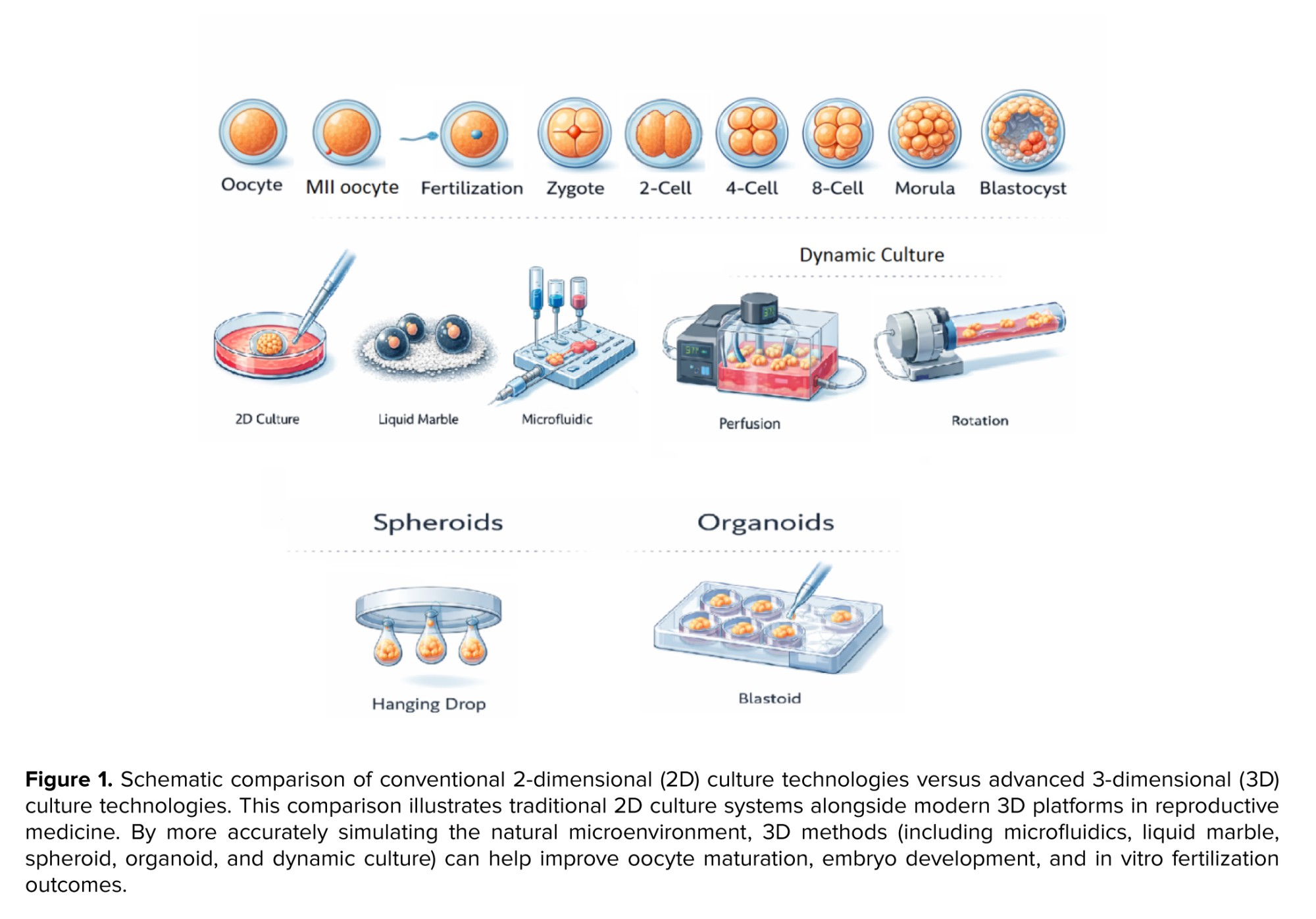
This review article aims to examine modern 3D culture systems for follicles, oocytes, and embryos, analyze their advantages and limitations compared to traditional methods, and propose practical solutions to enhance produced in vitro efficiency in both livestock production and medical applications. For this narrative review, a comprehensive literature search was conducted across major scientific databases, including PubMed, Scopus, and Google Scholar. The main keywords used were "Three-Dimensional Culture", "Microfluidics", "Spheroid", "Organoid", "Liquid Marble", "Dynamic Culture", "Oocyte Maturation", and "Embryo Development". The search focused on seminal and recent studies published until 2025 in order to cover the latest advancements in 3D culture technologies in reproductive medicine. Figure 1 shows a schematic comparison of conventional 2D culture technologies versus advanced 3D culture technologies.
2. 3D embryonic culture systems
In this context, the development of 3-dimensional (3D) culture systems has emerged as an innovative strategy. By better mimicking physiological conditions, these systems can enhance the quality of in vitro-produced embryos. Reproductive technologies, particularly IVEP, have revolutionized the livestock industry by overcoming the limitations of traditional methods. With the growing demand for high-yield livestock, optimizing oocyte and embryo culture techniques have become both a scientific and economic imperative (6, 7). One of the main challenges in this field involves improving the quality of in vitro-matured oocytes, where significant progress has been made through the development of 3D culture systems that better simulate physiological conditions. These systems overcome the limitations of 2-dimensional (2D) cultures, such as the flattening of cumulus-oocyte complexes on the culture dish surface and reduced cell-to-medium contact (7). 3D follicular culture systems demonstrate superior performance compared to 2D systems by increasing follicular survival and growth rates, maintaining natural morphology, and enhancing the expression of oocyte maturation genes. These systems are particularly suitable for large mammals and humans as they effectively preserve critical cell-cell and cell-matrix interactions (8, 9).
An examination of embryo culture strategies would be incomplete without considering the physical culture substrate. Under in vitro conditions, the 2 main culture medium approaches (sequential-step systems “back to nature” and single-step systems “let the embryo choose”) were developed primarily to address the limitations of the conventional 2D culture system. In 2D culture, where embryos grow on a flat surface, these strategies aim to compensate for the nutritional and environmental shortcomings of this simplistic system. In contrast, 3D culture, by more accurately simulating the natural architecture of the uterus, provides more optimal physical substrate that enables more complex interactions with nutritional strategies (10).
Although 2D culture is widely used, it has significant limitations, including its failure to simulate the in vivo environment (11), adhesion of oocytes to the culture dish surface and disruption of natural morphology (12), and inability to study cell-cell and cell-matrix interactions (13). In contrast, 3D culture offers multiple advantages: preservation of spherical follicular structure and cellular connections (14), better simulation of physiological conditions with improved oocyte and embryo development (7), and the ability to investigate complex cellular interactions impossible in 2D models (13).
This review article aims to examine modern 3D culture systems for follicles, oocytes, and embryos, analyze their advantages and limitations compared to traditional methods, and propose practical solutions to enhance produced in vitro efficiency in both livestock production and medical applications. For this narrative review, a comprehensive literature search was conducted across major scientific databases, including PubMed, Scopus, and Google Scholar. The main keywords used were "Three-Dimensional Culture", "Microfluidics", "Spheroid", "Organoid", "Liquid Marble", "Dynamic Culture", "Oocyte Maturation", and "Embryo Development". The search focused on seminal and recent studies published until 2025 in order to cover the latest advancements in 3D culture technologies in reproductive medicine. Figure 1 shows a schematic comparison of conventional 2D culture technologies versus advanced 3D culture technologies.
2. 3D embryonic culture systems
2.1. Liquid marbles (LMs)
The cultivation system using hydrophobic particles (or LMs) is considered a novel method in 3D culture. In this system, highly hydrophobic particles adhere to a droplet of liquid, forming stable and adjustable spheres that enable simultaneous group or individual cell culture. Key advantages of this method include the need for small culture volumes, optimal gas permeability, and cost-effectiveness (1). This system has broad applications in culturing various cell types, including embryonic stem cells, fibroblasts, and red blood cells (15). Studies have shown that LMs can be a suitable option for the in vitro maturation (IVM) of oocytes, as they create a stable micro-environment, provide optimal gas exchange, and minimize cell exposure to harmful chemicals. Empirical evidence suggests that LM micro-bioreactors are a practical method for establishing a suitable micro-environment for in vitro oocyte maturation in different species. Results across various species indicate that the response to the LM system may be species-dependent, requiring further studies to optimize this method (7).
Numerous studies have examined the impact of 3D culture systems, particularly LMs, on in vitro oocyte maturation and embryonic development in different species. This system can enhance the developmental competence of oocytes, particularly in low-quality gametes, by creating a physiological microenvironment. In this section, the results of applying the LM system in various species, including cattle, sheep, and goats, are reviewed, and its effects on key parameters such as oocyte maturation, blastocyst formation, epigenetic status, and mitochondrial function are analyzed.
The 3D LM culture system has emerged as a promising technology in ARTs for farm animals. This system exhibits species-specific effects on oocyte maturation and embryonic development. In cattle, while the LM system supported blastocyst formation, it resulted in a significant reduction in blastocyst cell numbers. It altered DNA methylation patterns, indicating the need for further optimization of this system for this particular species (1). In contrast, in sheep, the LM system improved blastocyst formation rates, likely due to better preservation of cumulus-oocyte complexes in the 3D environment (16). Studies in goats revealed that although the system did not significantly affect nuclear maturation rates, it led to increased mitochondrial activity and changes in mitochondrial distribution patterns, which may indicate enhanced cytoplasmic maturation (7).
The main advantages of the LM system include creating a stable microenvironment, eliminating the need for mineral oil, and reducing contact with plastic surfaces, all of which can contribute to improving gamete and embryo quality (15, 17). This system has been particularly effective in maintaining oxidative-antioxidant balance and the physiological organization of cumulus cells. However, challenges such as droplet fragility and species-specific differences in response to this system indicate the need for technical and species-specific modifications to achieve optimal results. One of the key findings of these studies was the impact of the LM system on the epigenetic profile and gene expression. The observed changes in DNA methylation patterns and reduced expression of miR-615 in cattle highlight the importance of further investigating the long-term effects of this system on the epigenetic programming of embryos. In contrast, the improved mitochondrial activity observed in goats may indicate the system's potential to enhance oocyte energy metabolism, which could be particularly significant for oocytes with low developmental competence (1, 7).
Another advantage of the LM system is its simplicity, speed, and cost-effectiveness. Unlike complex 3D culture methods that require hydrogels or scaffolds, this system can be implemented with simple coatings. Additionally, the LM system enables individual monitoring of oocytes and embryos, allowing researchers to easily perform sampling and assess the metabolic needs of the cells. This method is particularly beneficial for oocytes with low developmental competence, such as immature oocytes, as it preserves the 3D structure, prevents cumulus cell spreading, and enhances the oocyte’s molecular and metabolic quality (7, 15, 16).
To broaden the applications of this technology, further studies are essential in several areas, optimizing the physicochemical conditions of the system to reduce oocyte and embryo loss, investigating the molecular mechanisms that influence gamete and embryo quality in this system, and assessing the long-term health of animals born through this method. Additionally, comparing the LM with other 3D culture systems could provide a better understanding of its advantages and limitations (7). Overall, current findings suggest that the LM system, despite its technical challenges, holds significant potential as an alternative method in ARTs. So far, research on LM for oocyte maturation and embryo culture has primarily focused on sheep, cattle, and goat models, with no published data on its applications in humans or pigs. Although LM systems offer unique advantages, such as better micro-environmental control and reduced oxidative stress, their direct impact on human or porcine reproductive cells remains unknown.
2.2. Spheroids/organoids
The 3D culture systems using spheroids and organoids have revolutionized reproductive research. These complex structures, formed by the aggregation of stem and somatic cells, are capable of mimicking the architecture and function of ovarian follicles and early-stage embryos (18). There are 2 main approaches to generating these structures: scaffold-based systems that utilize natural matrices such as Matrigel or agarose, and scaffold-free systems that rely on cellular self-organization (19, 20).
The hanging drop method, as a scaffold-free system, enables high-efficiency production of spheroids by creating optimal conditions for cell aggregation at the liquid-air interface. This approach is considered a favorable option due to its simplicity, consistent results, and lack of requirement for an external matrix. In contrast, scaffold-based systems provide a suitable environment for cell adhesion, proliferation, and differentiation (21). Conversely, in scaffold-based systems, human endometrial mesenchymal stem cells (hEnMSCs) have been widely used for endometrial regeneration and organoid formation (22, 23). Additionally, numerous efforts have been made to develop ovarian organoids using epithelial, granulosa, theca, and even cancer cells in 3D culture systems such as Matrigel and agarose (20, 24).
Recent advances in ovarian tissue engineering have led to the development of more complex models. For example, by isolating theca and granulosa cells and culturing them in gel micro-molds, 3D ovary-like spheroids capable of supporting oocyte maturation have been successfully created (25). Similarly, by encapsulating granulosa and theca cells in a collagen gel, steroid hormone-secreting spheroids (such as estradiol and progesterone) have been produced, which maintain their endocrine function for up to 30 days (26). A groundbreaking achievement in this field was the generation of ovarian organoids from female germline stem cells, which not only produced functional oocytes but also gave rise to live offspring (27). Additionally, it has been demonstrated that even spermatogonial stem cells could be reprogrammed into functional oocytes in an organoid culture system (28).
A study investigated the co-culture of hEnMSCs with mouse germinal vesicle (GV) oocytes in 2 3D culture systems (hanging drop and alginate hydrogel). The results demonstrated that hEnMSCs, under the influence of paracrine factors secreted by oocytes, differentiated into germ cell-like cells. Some of these cells exhibited morphological features resembling oocytes (large cytoplasm), while others resembled nurse cells (smaller size). These findings represent a significant step in understanding the differentiation of stem cells toward germline cells and hold high potential for research and therapeutic applications in reproductive medicine (18).
3D reproductive organoids are self-organizing structures derived from stem cells that recapitulate the architecture and function of native tissues (29, 30). Blastoids are formed through self-organization of human pluripotent stem cells and, by possessing 3 main cell lineages, recapitulate the morphology of human blastocysts and are capable of implantation into the mouse uterus. These models are used to study embryogenesis, implantation, and drug screening (31). In the ovarian field, organoids have enhanced follicle survival using decellularized scaffolds, and ovarian cancer models have been developed for drug response evaluation (32, 33). Endometrial organoids recapitulate the glandular structure of the endometrium, respond to hormonal treatment, and are used to study implantation and embryo-maternal interactions (34).
A common challenge across all these models is the lack of cellular complexity, which can be addressed through strategies such as integration with microfluidics, replacement of Matrigel with stable hydrogels or decellularized matrices, and co-culture with stromal cells (33, 34). Overall, research indicates that 3D culture systems (such as ovarian spheroids and organoids) mimic the structure and function of natural follicles, offering a novel approach to improving oocyte maturation and ARTs efficiency. These systems demonstrate that ovarian organoids are not only valuable tools for studying follicular maturation and cell-cell interactions but also hold significant promise for clinical applications in reproductive medicine. However, further studies are needed to optimize culture conditions and evaluate the potential of these structures to mature into fully functional oocytes.
3. Co-culture
The cultivation system using hydrophobic particles (or LMs) is considered a novel method in 3D culture. In this system, highly hydrophobic particles adhere to a droplet of liquid, forming stable and adjustable spheres that enable simultaneous group or individual cell culture. Key advantages of this method include the need for small culture volumes, optimal gas permeability, and cost-effectiveness (1). This system has broad applications in culturing various cell types, including embryonic stem cells, fibroblasts, and red blood cells (15). Studies have shown that LMs can be a suitable option for the in vitro maturation (IVM) of oocytes, as they create a stable micro-environment, provide optimal gas exchange, and minimize cell exposure to harmful chemicals. Empirical evidence suggests that LM micro-bioreactors are a practical method for establishing a suitable micro-environment for in vitro oocyte maturation in different species. Results across various species indicate that the response to the LM system may be species-dependent, requiring further studies to optimize this method (7).
Numerous studies have examined the impact of 3D culture systems, particularly LMs, on in vitro oocyte maturation and embryonic development in different species. This system can enhance the developmental competence of oocytes, particularly in low-quality gametes, by creating a physiological microenvironment. In this section, the results of applying the LM system in various species, including cattle, sheep, and goats, are reviewed, and its effects on key parameters such as oocyte maturation, blastocyst formation, epigenetic status, and mitochondrial function are analyzed.
The 3D LM culture system has emerged as a promising technology in ARTs for farm animals. This system exhibits species-specific effects on oocyte maturation and embryonic development. In cattle, while the LM system supported blastocyst formation, it resulted in a significant reduction in blastocyst cell numbers. It altered DNA methylation patterns, indicating the need for further optimization of this system for this particular species (1). In contrast, in sheep, the LM system improved blastocyst formation rates, likely due to better preservation of cumulus-oocyte complexes in the 3D environment (16). Studies in goats revealed that although the system did not significantly affect nuclear maturation rates, it led to increased mitochondrial activity and changes in mitochondrial distribution patterns, which may indicate enhanced cytoplasmic maturation (7).
The main advantages of the LM system include creating a stable microenvironment, eliminating the need for mineral oil, and reducing contact with plastic surfaces, all of which can contribute to improving gamete and embryo quality (15, 17). This system has been particularly effective in maintaining oxidative-antioxidant balance and the physiological organization of cumulus cells. However, challenges such as droplet fragility and species-specific differences in response to this system indicate the need for technical and species-specific modifications to achieve optimal results. One of the key findings of these studies was the impact of the LM system on the epigenetic profile and gene expression. The observed changes in DNA methylation patterns and reduced expression of miR-615 in cattle highlight the importance of further investigating the long-term effects of this system on the epigenetic programming of embryos. In contrast, the improved mitochondrial activity observed in goats may indicate the system's potential to enhance oocyte energy metabolism, which could be particularly significant for oocytes with low developmental competence (1, 7).
Another advantage of the LM system is its simplicity, speed, and cost-effectiveness. Unlike complex 3D culture methods that require hydrogels or scaffolds, this system can be implemented with simple coatings. Additionally, the LM system enables individual monitoring of oocytes and embryos, allowing researchers to easily perform sampling and assess the metabolic needs of the cells. This method is particularly beneficial for oocytes with low developmental competence, such as immature oocytes, as it preserves the 3D structure, prevents cumulus cell spreading, and enhances the oocyte’s molecular and metabolic quality (7, 15, 16).
To broaden the applications of this technology, further studies are essential in several areas, optimizing the physicochemical conditions of the system to reduce oocyte and embryo loss, investigating the molecular mechanisms that influence gamete and embryo quality in this system, and assessing the long-term health of animals born through this method. Additionally, comparing the LM with other 3D culture systems could provide a better understanding of its advantages and limitations (7). Overall, current findings suggest that the LM system, despite its technical challenges, holds significant potential as an alternative method in ARTs. So far, research on LM for oocyte maturation and embryo culture has primarily focused on sheep, cattle, and goat models, with no published data on its applications in humans or pigs. Although LM systems offer unique advantages, such as better micro-environmental control and reduced oxidative stress, their direct impact on human or porcine reproductive cells remains unknown.
2.2. Spheroids/organoids
The 3D culture systems using spheroids and organoids have revolutionized reproductive research. These complex structures, formed by the aggregation of stem and somatic cells, are capable of mimicking the architecture and function of ovarian follicles and early-stage embryos (18). There are 2 main approaches to generating these structures: scaffold-based systems that utilize natural matrices such as Matrigel or agarose, and scaffold-free systems that rely on cellular self-organization (19, 20).
The hanging drop method, as a scaffold-free system, enables high-efficiency production of spheroids by creating optimal conditions for cell aggregation at the liquid-air interface. This approach is considered a favorable option due to its simplicity, consistent results, and lack of requirement for an external matrix. In contrast, scaffold-based systems provide a suitable environment for cell adhesion, proliferation, and differentiation (21). Conversely, in scaffold-based systems, human endometrial mesenchymal stem cells (hEnMSCs) have been widely used for endometrial regeneration and organoid formation (22, 23). Additionally, numerous efforts have been made to develop ovarian organoids using epithelial, granulosa, theca, and even cancer cells in 3D culture systems such as Matrigel and agarose (20, 24).
Recent advances in ovarian tissue engineering have led to the development of more complex models. For example, by isolating theca and granulosa cells and culturing them in gel micro-molds, 3D ovary-like spheroids capable of supporting oocyte maturation have been successfully created (25). Similarly, by encapsulating granulosa and theca cells in a collagen gel, steroid hormone-secreting spheroids (such as estradiol and progesterone) have been produced, which maintain their endocrine function for up to 30 days (26). A groundbreaking achievement in this field was the generation of ovarian organoids from female germline stem cells, which not only produced functional oocytes but also gave rise to live offspring (27). Additionally, it has been demonstrated that even spermatogonial stem cells could be reprogrammed into functional oocytes in an organoid culture system (28).
A study investigated the co-culture of hEnMSCs with mouse germinal vesicle (GV) oocytes in 2 3D culture systems (hanging drop and alginate hydrogel). The results demonstrated that hEnMSCs, under the influence of paracrine factors secreted by oocytes, differentiated into germ cell-like cells. Some of these cells exhibited morphological features resembling oocytes (large cytoplasm), while others resembled nurse cells (smaller size). These findings represent a significant step in understanding the differentiation of stem cells toward germline cells and hold high potential for research and therapeutic applications in reproductive medicine (18).
3D reproductive organoids are self-organizing structures derived from stem cells that recapitulate the architecture and function of native tissues (29, 30). Blastoids are formed through self-organization of human pluripotent stem cells and, by possessing 3 main cell lineages, recapitulate the morphology of human blastocysts and are capable of implantation into the mouse uterus. These models are used to study embryogenesis, implantation, and drug screening (31). In the ovarian field, organoids have enhanced follicle survival using decellularized scaffolds, and ovarian cancer models have been developed for drug response evaluation (32, 33). Endometrial organoids recapitulate the glandular structure of the endometrium, respond to hormonal treatment, and are used to study implantation and embryo-maternal interactions (34).
A common challenge across all these models is the lack of cellular complexity, which can be addressed through strategies such as integration with microfluidics, replacement of Matrigel with stable hydrogels or decellularized matrices, and co-culture with stromal cells (33, 34). Overall, research indicates that 3D culture systems (such as ovarian spheroids and organoids) mimic the structure and function of natural follicles, offering a novel approach to improving oocyte maturation and ARTs efficiency. These systems demonstrate that ovarian organoids are not only valuable tools for studying follicular maturation and cell-cell interactions but also hold significant promise for clinical applications in reproductive medicine. However, further studies are needed to optimize culture conditions and evaluate the potential of these structures to mature into fully functional oocytes.
3. Co-culture
3.1. Co-culture with ovarian supportive cells
The 3D co-culture systems that simulate the physiological environment of the ovarian follicle offer a novel approach in research on IVM of human oocytes. Studies indicate that presence of extracellular matrix and the establishment of a 3D space significantly enhance the functionality of granulosa and cumulus cells, thereby providing optimal conditions for oocyte maturation. Research suggests that these systems can effectively maintain the rate of progression to metaphase II (MII), cytoskeletal organization, and chromatin configuration at desirable levels. Among the key advantages of this method are the preservation of 3D cell structure, the simplicity of protocol implementation, and the ability to investigate critical molecular mechanisms such as M-phase promoting factor and mitogen-activated protein kinase enzyme activity. The advancement of this technology could play a significant role in improving our understanding of oocyte maturation processes in ART (35).
A study investigated the role of oviductal epithelial cells and their conditioned medium in the in vitro fertilization (IVF) process of sheep. Contrary to expectations, the results revealed that the presence of oviductal epithelial cells and conditioned medium not only failed to enhance embryo development but also significantly reduced cleavage rates, blastocyst formation, and hatching. These findings highlight important species-specific differences in the response to environmental factors affecting embryo development, suggesting that the optimal culture conditions for sheep embryos may differ from those of other species (36).
3.2. Co-culture with stem cells
Stem cells, particularly mesenchymal types, can play a significant role in enhancing embryo development in in vitro systems. For instance, it has been reported that mesenchymal stem cells derived from follicular fluid and other sources can differentiate into germ-like cells or oocyte-like cells under the influence of oocyte-secreted factors such as bone morphogenetic protein 15 (37, 38). These findings have laid the foundation for developing novel co-culture systems, including a method that utilizes ovarian stem cells (OSCs) derived from hiPSCs for IVM of human oocytes. This system offers key advantages such as compatibility with standard ART protocols, scalability, and effective performance under varying ovarian stimulation conditions, representing a significant step toward optimizing embryo culture (39).
In line with this, findings have demonstrated that co-culture with adipose tissue-derived mesenchymal stem cells can significantly enhance zygote cleavage rate, blastocyst formation, and implantation potential, particularly in embryos derived from aged mice. These effects are likely mediated through paracrine factor secretion, improved energy metabolism (via increased glucose 6-phosphate dehydrogenase activity), and maintenance of pluripotency (via upregulated OCT4), without inducing cellular stress, as heat shock protein 70 remains unchanged (40). These results align with a study that reported the successful maintenance of human embryonic stem cell characteristics in fully defined culture media, further underscoring the critical role of controlled culture conditions in clinical applications (41).
The development of defined culture media (41) and paracrine systems such as ovarian support cells (OSCs)-IVM (39) could bridge the gap between basic research and therapeutic applications. Although these results are promising, significant challenges remain. The precise mechanisms by which adipose tissue-derived mesenchymal stem cells influence embryo development and the identification of key molecules involved require further investigation. Additionally, evaluating implantation and post-implantation development outcomes under in vivo conditions are a major limitation of current studies. It appears that translating these findings into clinical applications will require longer-term studies with human samples (40). Overall, 3D co-culture systems, by mimicking natural cell-cell and cell-extracellular matrix interactions, have significantly improved oocyte maturation and embryo quality. However, further research is needed to fully understand the underlying mechanisms and facilitate the clinical application of these technologies.
4. Microfluidic technologies
The 3D co-culture systems that simulate the physiological environment of the ovarian follicle offer a novel approach in research on IVM of human oocytes. Studies indicate that presence of extracellular matrix and the establishment of a 3D space significantly enhance the functionality of granulosa and cumulus cells, thereby providing optimal conditions for oocyte maturation. Research suggests that these systems can effectively maintain the rate of progression to metaphase II (MII), cytoskeletal organization, and chromatin configuration at desirable levels. Among the key advantages of this method are the preservation of 3D cell structure, the simplicity of protocol implementation, and the ability to investigate critical molecular mechanisms such as M-phase promoting factor and mitogen-activated protein kinase enzyme activity. The advancement of this technology could play a significant role in improving our understanding of oocyte maturation processes in ART (35).
A study investigated the role of oviductal epithelial cells and their conditioned medium in the in vitro fertilization (IVF) process of sheep. Contrary to expectations, the results revealed that the presence of oviductal epithelial cells and conditioned medium not only failed to enhance embryo development but also significantly reduced cleavage rates, blastocyst formation, and hatching. These findings highlight important species-specific differences in the response to environmental factors affecting embryo development, suggesting that the optimal culture conditions for sheep embryos may differ from those of other species (36).
3.2. Co-culture with stem cells
Stem cells, particularly mesenchymal types, can play a significant role in enhancing embryo development in in vitro systems. For instance, it has been reported that mesenchymal stem cells derived from follicular fluid and other sources can differentiate into germ-like cells or oocyte-like cells under the influence of oocyte-secreted factors such as bone morphogenetic protein 15 (37, 38). These findings have laid the foundation for developing novel co-culture systems, including a method that utilizes ovarian stem cells (OSCs) derived from hiPSCs for IVM of human oocytes. This system offers key advantages such as compatibility with standard ART protocols, scalability, and effective performance under varying ovarian stimulation conditions, representing a significant step toward optimizing embryo culture (39).
In line with this, findings have demonstrated that co-culture with adipose tissue-derived mesenchymal stem cells can significantly enhance zygote cleavage rate, blastocyst formation, and implantation potential, particularly in embryos derived from aged mice. These effects are likely mediated through paracrine factor secretion, improved energy metabolism (via increased glucose 6-phosphate dehydrogenase activity), and maintenance of pluripotency (via upregulated OCT4), without inducing cellular stress, as heat shock protein 70 remains unchanged (40). These results align with a study that reported the successful maintenance of human embryonic stem cell characteristics in fully defined culture media, further underscoring the critical role of controlled culture conditions in clinical applications (41).
The development of defined culture media (41) and paracrine systems such as ovarian support cells (OSCs)-IVM (39) could bridge the gap between basic research and therapeutic applications. Although these results are promising, significant challenges remain. The precise mechanisms by which adipose tissue-derived mesenchymal stem cells influence embryo development and the identification of key molecules involved require further investigation. Additionally, evaluating implantation and post-implantation development outcomes under in vivo conditions are a major limitation of current studies. It appears that translating these findings into clinical applications will require longer-term studies with human samples (40). Overall, 3D co-culture systems, by mimicking natural cell-cell and cell-extracellular matrix interactions, have significantly improved oocyte maturation and embryo quality. However, further research is needed to fully understand the underlying mechanisms and facilitate the clinical application of these technologies.
4. Microfluidic technologies
Numerous studies have explored in vitro folliculogenesis improvement through dynamic culture systems designed to more accurately mimic natural follicular activation and growth processes. These systems enable efficient exchange of nutrients, oxygen, and metabolic byproducts, representing a significant advancement in reproductive biology and fertility preservation. Microfluidic technologies, combined with perfusion bioreactors, can culture cells in oxygen-rich environments. In vivo, organs are continuously exposed to various stimuli (such as mechanical, electrical, and biochemical factors) essential for natural physiology, growth, and function. The primary goal of microfluidic follicular culture is to precisely replicate the natural ovarian environment to enhance in-follicle oocyte maturation (9).
Microfluidic technology has broad applications in ARTs, including oocyte and sperm manipulation, IVF, and embryo culture (42). Additionally, this technology has revolutionized cell cryopreservation by enabling precise control of fluid dynamics, allowing the gradual addition and removal of cryoprotective agents (CPAs). This significantly reduces osmotic damage to oocytes. Key advantages of microfluidics include high integration, automation, operational precision, and low cost. The unique characteristics of micro- and nanoscale systems, such as high surface-to-volume ratio, efficient heat transfer, and optimized shear stress, make this technology ideal for vitrification-based cryopreservation (43). Although microfluidics holds great potential as an alternative to conventional culture methods, further studies are needed to validate the outcomes. Nevertheless, microfluidic systems are expected to establish new standards in reproductive medicine in the near future, potentially revolutionizing infertility treatment (44).
In one study, researchers developed an advanced microfluidic system for single-cell analysis of oocytes and embryos. This system utilizes intelligent hydrodynamic traps to enable long-term observation and high-resolution examination of samples under controlled conditions (temperature, precise culture medium perfusion, and live imaging). Their results demonstrated that this technology can accurately load and analyze individual oocytes and embryos without sample loss. Key advantages include reduced human error, the ability to conduct stable longitudinal studies, preservation of sample integrity, and parallel processing of multiple samples. This system has broad applications in reproductive research, infertility drug screening, and IVF centers, representing a significant advancement in ARTs and embryology (45).
Microfluidic devices based on electrowetting-on-dielectric (EWOD) have found wide applications in chemistry and life sciences due to their advantages, such as handling extremely small liquid volumes, the ability to integrate sensors on-chip, and portability. They also have great potential for developing point-of-care diagnostic systems. These microdevices can control environmental parameters like pH and temperature and can be equipped with electrochemical, optical, and electrical sensors. The integration of this technology with magnetic beads has been successfully used for analyzing embryo culture media and isolating DNA and biomarkers, a feature particularly valuable in assisted reproduction. This approach enables the simultaneous detection of multiple molecules from a very small volume of single-embryo culture media. By simulating dynamic culture conditions and allowing targeted delivery of growth factors and miRNAs at different developmental stages, these systems can closely mimic embryo-maternal interactions in vitro. However, the application of this technology in assisted reproduction is still in its early stages, with only successful on-chip manipulation and culture of mouse embryos reported so far (46).
In 2017, researchers developed an “oviduct-on-a-chip” system using 3D printing. While this system improved sperm penetration and reduced abnormal fertilization in bovines, it faced limitations due to the toxicity of the materials used for fertilized oocytes (47). In another study, a disposable oil-free polydimethylsiloxane (PDMS) microfluidic device was developed, enabling the culture of mouse zygotes from the single-cell stage to blastocyst. A key advantage of this system is the reduced need for manual embryo pipetting, as embryo loading and washing (whether in groups or individually) are performed via controlled culture medium flow. This feature allows for medium exchange without directly handling the embryos (48).
Numerous studies have focused on developing microfluidic systems for separating cumulus cells from oocytes, in one of which a device was designed that gradually removes cumulus cells from bovine zygotes using narrow channels and suction ports; however, this system was only capable of processing one zygote at a time and required manual flow control (49). Another study successfully separated cumulus cells from mouse oocytes by implementing microchannels with uneven and constricting geometries (50). Additionally, researchers successfully isolated high-quality bovine oocytes by applying mechanical stress in microchannels, thereby enhancing IVF rates (51). In a more recent study, a novel microfluidic chip was designed with a hybrid structure featuring serrated channels and expansion units, enabling oocyte denudation without requiring hyaluronidase. The chip was fabricated cost-effectively using laser engraving on polymethyl methacrylate (PMMA), and computational fluid dynamics simulations optimized the serration angles and flow rates. Laboratory results demonstrated 96.7% denudation efficiency and a 90% oocyte recovery rate. By combining structural simplicity with high performance, this system represents a significant advancement in replacing enzymatic methods with mechanical approaches for oocyte denudation (52).
An intelligent microfluidic system has been developed that combines deep learning and differential pressure sensors to enable non-invasive, automated measurement of the elastic modulus of zebrafish embryos at various developmental stages. By employing a power-law rheology model, the system dynamically measures changes in the mechanical properties of embryos from cleavage to segmentation, which can serve as a novel indicator for assessing embryonic developmental quality (53). In another study, researchers developed an automated microfluidic device for denuding bovine and human oocytes. This system features an intelligent open-chamber design and can process 16 samples simultaneously, addressing the limitations of manual methods, such as operator-dependent variability and high workload. Results demonstrate that this method does not affect zona pellucida permeability and yields developmental outcomes comparable to those obtained by manual techniques performed by experts, while also improving blastocyst formation rates and post-hatching stages. This technology could help standardize IVF procedures and enhance the reliability of results in infertility clinics (54). Both studies highlight significant advancements in microfluidic technologies for embryological research and clinical applications.
The use of dynamic microfluidic culture has the potential to enhance in vitro embryo development in mice, cattle, and possibly humans. This method, by controlling culture conditions (such as temperature, environment, and gases), enhances embryo quality, reduces apoptosis, increases blastocyst formation rates, and strengthens the inner cell mass (ICM). Studies on mouse embryos have shown that dynamic culture, compared to static culture, normalizes the expression of placental imprinted genes more effectively and may influence offspring health. In human embryos, this technique has led to better early-stage development and increased the likelihood of live births. Overall, the programmable and dynamic microfluidic system is safe, efficient, and capable of supporting pre-implantation embryo growth (55).
Generally, microfluidic technology has brought about a fundamental transformation in ARTs by accurately simulating physiological conditions in the body, overcoming the major challenges of traditional culture systems. With its unique advantages, this technology has achieved remarkable success in improving oocyte quality, increasing fertilization rates, and enhancing blastocyst formation. The development of advanced systems such as organ-on-a-chip and automated microfluidic devices has enabled more precise studies of reproductive mechanisms and the provision of more targeted therapeutic solutions. Although challenges remain in standardizing and clinically applying these technologies, their immense potential to revolutionize the fields of assisted reproduction and fertility preservation is undeniable, promising a bright future for infertility treatment and embryological research. However, it should be noted that the majority of these findings are derived from animal models or proof-of-concept studies; therefore, comprehensive clinical validation in humans is essential before widespread adoption in reproductive medicine.
4.1. Microfluidic systems for oocyte vitrification
Oocyte vitrification techniques play a vital role in assisted reproduction and fertility preservation. However, challenges such as operator dependency, variability in outcomes, and the toxicity of CPAs have driven the need for more automated and precise technologies. In this context, microfluidic systems have emerged as a promising solution, enabling precise control over CPA concentration and minimizing human error. One study introduced the microfluidic-based automated vitrification device designed for oocyte vitrification. This system compared 3 CPA loading protocols (linear, stepwise) against the conventional 2-step method. The results showed that the highest CPA concentration (VS1-TS1 group) achieved an 86.18% survival rate, while the VS3-TS3 group (with half the concentration) exhibited a higher blastocyst formation rate (56). Another study further confirmed the impact of CPA concentration, demonstrating that a 7.5% dimethylsulfoxide and ethylene glycol mixture yielded the highest blastocyst formation rate (57). The microfluidic-based automated vitrification system, by standardizing the process and reducing human error, shows great potential for improving assisted reproductive outcomes. This technology could be particularly impactful in fertility preservation for cancer patients and embryological research, though further studies are needed to optimize its clinical application (56).
In one study, an advanced automated microfluidic system was introduced that is fully compatible with the conventional Cryotop method. This system consists of an open microfluidic chip and automated devices capable of precisely adjusting CPA concentration and transferring oocytes with minimal solution volume to the Cryotop. Experiments on mouse oocytes at the GV and MII stages demonstrated that this system successfully achieves 100% vitrification pretreatment and oocyte transfer within a specified time frame. A comparison with manual methods revealed that the new system performs better, with a survival rate of 97.6% (compared to 94% for the manual method) and maintains optimal mitochondrial membrane potential levels while reducing oxidative stress. The most significant advantage of this technology is the elimination of operator dependency and the establishment of unprecedented standardization in the vitrification process, which is crucial for high-value human oocytes (58).
Numerous studies have shown that microfluidic systems provide more precise control over CPA concentration compared to manual methods. For example, one study designed a syringe-pump-based microfluidic system that generated a linear concentration profile, reducing osmotic stress; however, it was unable to extract oocytes (59). In contrast, another study developed a device capable of linearly loading equilibration solution, dehydration solution, and extracting oocytes, leading to better preservation of cytoplasmic lipids and improved developmental competence in bovine and murine models (60). In a subsequent study, a low-cost and user-friendly microfluidic system was introduced, automating the CPA loading process. This technology utilizes a fluidic capacitor and requires no complex electronic equipment, enabling linear ES loading and potentially improving access to IVF treatment in remote areas. Further development of this system by integrating a microfluidic transistor could represent the next step toward clinical application (61).
A study introduced a transparent alumina ceramics-based microfluidic chip that addresses the challenges of conventional methods (such as process complexity and toxicity from prolonged CPA exposure). Results demonstrated that oocytes vitrified using this method exhibited higher quality, fewer genetic alterations, and better developmental competence compared to traditional techniques. This chip provides an integrated semi-automated oocyte cryopreservation platform, which could facilitate access to fertility preservation methods. However, challenges such as standardization and achieving a fully automated process still require further research. Automated microfluidic systems (with their precise CPA concentration control, reduced osmotic stress, and elimination of operator dependency) represent a transformative advancement in oocyte vitrification. These technologies not only improve oocyte survival rates and quality but could also expand access to assisted reproductive treatments in diverse regions. Nevertheless, further research is needed to optimize these systems for clinical applications (43).
Organ-on-a-chip technology has transformed biomedical research by enabling precise simulation of human organs using microfluidic systems. These platforms accurately replicate tissue functions and paracrine signaling, with particular promise for infertility research. The ovary-on-a-chip model specifically allows controlled study of reproductive toxicology and folliculogenesis while reducing animal testing. Integration with other organ-on-a-chip systems and 3D modeling could further enhance its applications in assisted reproduction and fertility preservation (9).
5. Dynamic culture
Microfluidic technology has broad applications in ARTs, including oocyte and sperm manipulation, IVF, and embryo culture (42). Additionally, this technology has revolutionized cell cryopreservation by enabling precise control of fluid dynamics, allowing the gradual addition and removal of cryoprotective agents (CPAs). This significantly reduces osmotic damage to oocytes. Key advantages of microfluidics include high integration, automation, operational precision, and low cost. The unique characteristics of micro- and nanoscale systems, such as high surface-to-volume ratio, efficient heat transfer, and optimized shear stress, make this technology ideal for vitrification-based cryopreservation (43). Although microfluidics holds great potential as an alternative to conventional culture methods, further studies are needed to validate the outcomes. Nevertheless, microfluidic systems are expected to establish new standards in reproductive medicine in the near future, potentially revolutionizing infertility treatment (44).
In one study, researchers developed an advanced microfluidic system for single-cell analysis of oocytes and embryos. This system utilizes intelligent hydrodynamic traps to enable long-term observation and high-resolution examination of samples under controlled conditions (temperature, precise culture medium perfusion, and live imaging). Their results demonstrated that this technology can accurately load and analyze individual oocytes and embryos without sample loss. Key advantages include reduced human error, the ability to conduct stable longitudinal studies, preservation of sample integrity, and parallel processing of multiple samples. This system has broad applications in reproductive research, infertility drug screening, and IVF centers, representing a significant advancement in ARTs and embryology (45).
Microfluidic devices based on electrowetting-on-dielectric (EWOD) have found wide applications in chemistry and life sciences due to their advantages, such as handling extremely small liquid volumes, the ability to integrate sensors on-chip, and portability. They also have great potential for developing point-of-care diagnostic systems. These microdevices can control environmental parameters like pH and temperature and can be equipped with electrochemical, optical, and electrical sensors. The integration of this technology with magnetic beads has been successfully used for analyzing embryo culture media and isolating DNA and biomarkers, a feature particularly valuable in assisted reproduction. This approach enables the simultaneous detection of multiple molecules from a very small volume of single-embryo culture media. By simulating dynamic culture conditions and allowing targeted delivery of growth factors and miRNAs at different developmental stages, these systems can closely mimic embryo-maternal interactions in vitro. However, the application of this technology in assisted reproduction is still in its early stages, with only successful on-chip manipulation and culture of mouse embryos reported so far (46).
In 2017, researchers developed an “oviduct-on-a-chip” system using 3D printing. While this system improved sperm penetration and reduced abnormal fertilization in bovines, it faced limitations due to the toxicity of the materials used for fertilized oocytes (47). In another study, a disposable oil-free polydimethylsiloxane (PDMS) microfluidic device was developed, enabling the culture of mouse zygotes from the single-cell stage to blastocyst. A key advantage of this system is the reduced need for manual embryo pipetting, as embryo loading and washing (whether in groups or individually) are performed via controlled culture medium flow. This feature allows for medium exchange without directly handling the embryos (48).
Numerous studies have focused on developing microfluidic systems for separating cumulus cells from oocytes, in one of which a device was designed that gradually removes cumulus cells from bovine zygotes using narrow channels and suction ports; however, this system was only capable of processing one zygote at a time and required manual flow control (49). Another study successfully separated cumulus cells from mouse oocytes by implementing microchannels with uneven and constricting geometries (50). Additionally, researchers successfully isolated high-quality bovine oocytes by applying mechanical stress in microchannels, thereby enhancing IVF rates (51). In a more recent study, a novel microfluidic chip was designed with a hybrid structure featuring serrated channels and expansion units, enabling oocyte denudation without requiring hyaluronidase. The chip was fabricated cost-effectively using laser engraving on polymethyl methacrylate (PMMA), and computational fluid dynamics simulations optimized the serration angles and flow rates. Laboratory results demonstrated 96.7% denudation efficiency and a 90% oocyte recovery rate. By combining structural simplicity with high performance, this system represents a significant advancement in replacing enzymatic methods with mechanical approaches for oocyte denudation (52).
An intelligent microfluidic system has been developed that combines deep learning and differential pressure sensors to enable non-invasive, automated measurement of the elastic modulus of zebrafish embryos at various developmental stages. By employing a power-law rheology model, the system dynamically measures changes in the mechanical properties of embryos from cleavage to segmentation, which can serve as a novel indicator for assessing embryonic developmental quality (53). In another study, researchers developed an automated microfluidic device for denuding bovine and human oocytes. This system features an intelligent open-chamber design and can process 16 samples simultaneously, addressing the limitations of manual methods, such as operator-dependent variability and high workload. Results demonstrate that this method does not affect zona pellucida permeability and yields developmental outcomes comparable to those obtained by manual techniques performed by experts, while also improving blastocyst formation rates and post-hatching stages. This technology could help standardize IVF procedures and enhance the reliability of results in infertility clinics (54). Both studies highlight significant advancements in microfluidic technologies for embryological research and clinical applications.
The use of dynamic microfluidic culture has the potential to enhance in vitro embryo development in mice, cattle, and possibly humans. This method, by controlling culture conditions (such as temperature, environment, and gases), enhances embryo quality, reduces apoptosis, increases blastocyst formation rates, and strengthens the inner cell mass (ICM). Studies on mouse embryos have shown that dynamic culture, compared to static culture, normalizes the expression of placental imprinted genes more effectively and may influence offspring health. In human embryos, this technique has led to better early-stage development and increased the likelihood of live births. Overall, the programmable and dynamic microfluidic system is safe, efficient, and capable of supporting pre-implantation embryo growth (55).
Generally, microfluidic technology has brought about a fundamental transformation in ARTs by accurately simulating physiological conditions in the body, overcoming the major challenges of traditional culture systems. With its unique advantages, this technology has achieved remarkable success in improving oocyte quality, increasing fertilization rates, and enhancing blastocyst formation. The development of advanced systems such as organ-on-a-chip and automated microfluidic devices has enabled more precise studies of reproductive mechanisms and the provision of more targeted therapeutic solutions. Although challenges remain in standardizing and clinically applying these technologies, their immense potential to revolutionize the fields of assisted reproduction and fertility preservation is undeniable, promising a bright future for infertility treatment and embryological research. However, it should be noted that the majority of these findings are derived from animal models or proof-of-concept studies; therefore, comprehensive clinical validation in humans is essential before widespread adoption in reproductive medicine.
4.1. Microfluidic systems for oocyte vitrification
Oocyte vitrification techniques play a vital role in assisted reproduction and fertility preservation. However, challenges such as operator dependency, variability in outcomes, and the toxicity of CPAs have driven the need for more automated and precise technologies. In this context, microfluidic systems have emerged as a promising solution, enabling precise control over CPA concentration and minimizing human error. One study introduced the microfluidic-based automated vitrification device designed for oocyte vitrification. This system compared 3 CPA loading protocols (linear, stepwise) against the conventional 2-step method. The results showed that the highest CPA concentration (VS1-TS1 group) achieved an 86.18% survival rate, while the VS3-TS3 group (with half the concentration) exhibited a higher blastocyst formation rate (56). Another study further confirmed the impact of CPA concentration, demonstrating that a 7.5% dimethylsulfoxide and ethylene glycol mixture yielded the highest blastocyst formation rate (57). The microfluidic-based automated vitrification system, by standardizing the process and reducing human error, shows great potential for improving assisted reproductive outcomes. This technology could be particularly impactful in fertility preservation for cancer patients and embryological research, though further studies are needed to optimize its clinical application (56).
In one study, an advanced automated microfluidic system was introduced that is fully compatible with the conventional Cryotop method. This system consists of an open microfluidic chip and automated devices capable of precisely adjusting CPA concentration and transferring oocytes with minimal solution volume to the Cryotop. Experiments on mouse oocytes at the GV and MII stages demonstrated that this system successfully achieves 100% vitrification pretreatment and oocyte transfer within a specified time frame. A comparison with manual methods revealed that the new system performs better, with a survival rate of 97.6% (compared to 94% for the manual method) and maintains optimal mitochondrial membrane potential levels while reducing oxidative stress. The most significant advantage of this technology is the elimination of operator dependency and the establishment of unprecedented standardization in the vitrification process, which is crucial for high-value human oocytes (58).
Numerous studies have shown that microfluidic systems provide more precise control over CPA concentration compared to manual methods. For example, one study designed a syringe-pump-based microfluidic system that generated a linear concentration profile, reducing osmotic stress; however, it was unable to extract oocytes (59). In contrast, another study developed a device capable of linearly loading equilibration solution, dehydration solution, and extracting oocytes, leading to better preservation of cytoplasmic lipids and improved developmental competence in bovine and murine models (60). In a subsequent study, a low-cost and user-friendly microfluidic system was introduced, automating the CPA loading process. This technology utilizes a fluidic capacitor and requires no complex electronic equipment, enabling linear ES loading and potentially improving access to IVF treatment in remote areas. Further development of this system by integrating a microfluidic transistor could represent the next step toward clinical application (61).
A study introduced a transparent alumina ceramics-based microfluidic chip that addresses the challenges of conventional methods (such as process complexity and toxicity from prolonged CPA exposure). Results demonstrated that oocytes vitrified using this method exhibited higher quality, fewer genetic alterations, and better developmental competence compared to traditional techniques. This chip provides an integrated semi-automated oocyte cryopreservation platform, which could facilitate access to fertility preservation methods. However, challenges such as standardization and achieving a fully automated process still require further research. Automated microfluidic systems (with their precise CPA concentration control, reduced osmotic stress, and elimination of operator dependency) represent a transformative advancement in oocyte vitrification. These technologies not only improve oocyte survival rates and quality but could also expand access to assisted reproductive treatments in diverse regions. Nevertheless, further research is needed to optimize these systems for clinical applications (43).
Organ-on-a-chip technology has transformed biomedical research by enabling precise simulation of human organs using microfluidic systems. These platforms accurately replicate tissue functions and paracrine signaling, with particular promise for infertility research. The ovary-on-a-chip model specifically allows controlled study of reproductive toxicology and folliculogenesis while reducing animal testing. Integration with other organ-on-a-chip systems and 3D modeling could further enhance its applications in assisted reproduction and fertility preservation (9).
5. Dynamic culture
The currently used static ovarian culture systems face significant challenges, including limited nutrient exchange and accumulation of waste products in the stagnant layer surrounding the tissue. These limitations can substantially impair ovarian tissue survival and function under in vitro conditions. In contrast, dynamic culture systems address these issues by establishing continuous medium perfusion around the tissue, enabling optimal nutrient exchange and efficient removal of metabolic waste products. This approach not only overcomes the constraints of static methods but also provides a more physiologically accurate simulation of early folliculogenesis in vitro. Such advancements represent a critical step toward improving ovarian tissue culture techniques and enhancing follicular development studies (62). The fallopian tube is responsible for transporting gametes, timely transfer of the embryo to the uterus, and providing a suitable environment for fertilization and pre-implantation development. These processes are facilitated by motile cilia, smooth muscles, and secretory currents. Although the precise role of each of these factors is not fully understood, the cilia of the fallopian tube are generally recognized as the primary factor in the movement of eggs and embryos toward the uterus for implantation (63).
A dynamic bioreactor system with perfused flow and mechanical stimulation provides optimal conditions for culturing ovarian tissue slices. Compared to static culture methods, this system significantly improves the number and quality of secondary follicles, tissue viability, and hormone secretion. These benefits result from better oxygenation, nutrient exchange, and effective waste removal. This dynamic system can address the limitations of static methods, although further research is still needed (62). An ex vivo ovarian perfusion system for fertility preservation in cancer patients, particularly prepubertal girls, has been investigated. Testing on sheep ovaries showed that the non-stimulated group produced mature oocytes (MII), whereas the stimulated group primarily yielded immature oocytes (GV-MI). By maintaining physiological conditions, this system enables 8-day culture and production of mature oocytes while reducing the risk of cancer cell transfer (64).
A study introduces the innovative "OoTrap" device, which provides optimal conditions for oocyte maturation through both static and perfusion modes. The perfusion mode has demonstrated remarkable results: increased IVM rates, production of oocytes with fewer chromosomal abnormalities, and preservation of spindle structure. Key advantages of OoTrap include its portable design with a specialized heating system, transport case suitable for environments without incubators, and use of 3D-printed pumps-making it a cost-effective and practical solution for resource-limited facilities. This technology represents a significant advancement in ARTs (65).
Microfluidic dynamic culture systems have demonstrated significant improvements in embryo development quality across various species, including mice, cattle, and humans. By creating more physiological conditions, this method enhances blastomere count, reduces cellular apoptosis and fragmentation, improves blastocyst conversion rates, and induces favorable changes in placental gene expression. Notably, these superior results (achieved independently of culture medium composition, temperature, or gas conditions) have even led to live human births in clinical studies. These advancements not only enhance the clinical efficacy of ARTs but also hold remarkable potential for biodiversity preservation and fertility enhancement across species, potentially revolutionizing in vitro embryology practices (55).
The results of a study demonstrate that an advanced dynamic bioreactor system for ovarian tissue culture, which combines optimized oxygen transfer with controlled mechanical stimulation, effectively simulates the physiological ovarian environment. Developed in response to the limitations of static culture methods, this dynamic system has shown significant improvements in follicle survival and growth to secondary stages. The researchers found that using biocompatible materials such as polypropylene in bioreactor construction, along with meticulous system design, creates an optimal microenvironment for activating follicular reserves and producing high-quality mature oocytes. This innovative technology overcomes the challenges inherent in static culture systems and represents a significant advancement in ARTs and fertility preservation (66).
3D culture systems better mimic the in vivo environment through scaffold-based methods (e.g., hydrogels) or scaffold-free approaches such as rotating wall vessels. The rotating wall vessel bioreactor, developed by NASA, generates simulated microgravity (s-μg) via continuous rotation to provide optimal 3D culture conditions. This scaffold-free technology has been successfully applied in tissue engineering of thyroid, cartilage, and vascular structures. However, studies indicate that s-μg may adversely affect oocyte maturation, causing meiotic spindle disorganization, cytoplasmic blebbing, vacuolization, increased lipid droplets, and activation of stress/apoptosis pathways such as SAPK/JNK. Ultrastructural observations further reveal detachment of oocyte microvilli from the zona pellucida, formation of large vacuoles, and increased multilamellar bodies (indicative of autophagy). Although the exact mechanisms remain unclear, further research could lead to protective strategies for space medicine and enhanced ARTs (67).
Recent studies demonstrate that dynamic culture systems simulating physiological conditions have made significant progress in improving folliculogenesis, oocyte maturation, and embryonic development. These technologies overcome the limitations of static culture methods by optimizing oxygen/nutrient delivery and providing mechanical stimulation, offering promising results for fertility preservation, infertility treatment, and embryological research.
6. Conclusion
A dynamic bioreactor system with perfused flow and mechanical stimulation provides optimal conditions for culturing ovarian tissue slices. Compared to static culture methods, this system significantly improves the number and quality of secondary follicles, tissue viability, and hormone secretion. These benefits result from better oxygenation, nutrient exchange, and effective waste removal. This dynamic system can address the limitations of static methods, although further research is still needed (62). An ex vivo ovarian perfusion system for fertility preservation in cancer patients, particularly prepubertal girls, has been investigated. Testing on sheep ovaries showed that the non-stimulated group produced mature oocytes (MII), whereas the stimulated group primarily yielded immature oocytes (GV-MI). By maintaining physiological conditions, this system enables 8-day culture and production of mature oocytes while reducing the risk of cancer cell transfer (64).
A study introduces the innovative "OoTrap" device, which provides optimal conditions for oocyte maturation through both static and perfusion modes. The perfusion mode has demonstrated remarkable results: increased IVM rates, production of oocytes with fewer chromosomal abnormalities, and preservation of spindle structure. Key advantages of OoTrap include its portable design with a specialized heating system, transport case suitable for environments without incubators, and use of 3D-printed pumps-making it a cost-effective and practical solution for resource-limited facilities. This technology represents a significant advancement in ARTs (65).
Microfluidic dynamic culture systems have demonstrated significant improvements in embryo development quality across various species, including mice, cattle, and humans. By creating more physiological conditions, this method enhances blastomere count, reduces cellular apoptosis and fragmentation, improves blastocyst conversion rates, and induces favorable changes in placental gene expression. Notably, these superior results (achieved independently of culture medium composition, temperature, or gas conditions) have even led to live human births in clinical studies. These advancements not only enhance the clinical efficacy of ARTs but also hold remarkable potential for biodiversity preservation and fertility enhancement across species, potentially revolutionizing in vitro embryology practices (55).
The results of a study demonstrate that an advanced dynamic bioreactor system for ovarian tissue culture, which combines optimized oxygen transfer with controlled mechanical stimulation, effectively simulates the physiological ovarian environment. Developed in response to the limitations of static culture methods, this dynamic system has shown significant improvements in follicle survival and growth to secondary stages. The researchers found that using biocompatible materials such as polypropylene in bioreactor construction, along with meticulous system design, creates an optimal microenvironment for activating follicular reserves and producing high-quality mature oocytes. This innovative technology overcomes the challenges inherent in static culture systems and represents a significant advancement in ARTs and fertility preservation (66).
3D culture systems better mimic the in vivo environment through scaffold-based methods (e.g., hydrogels) or scaffold-free approaches such as rotating wall vessels. The rotating wall vessel bioreactor, developed by NASA, generates simulated microgravity (s-μg) via continuous rotation to provide optimal 3D culture conditions. This scaffold-free technology has been successfully applied in tissue engineering of thyroid, cartilage, and vascular structures. However, studies indicate that s-μg may adversely affect oocyte maturation, causing meiotic spindle disorganization, cytoplasmic blebbing, vacuolization, increased lipid droplets, and activation of stress/apoptosis pathways such as SAPK/JNK. Ultrastructural observations further reveal detachment of oocyte microvilli from the zona pellucida, formation of large vacuoles, and increased multilamellar bodies (indicative of autophagy). Although the exact mechanisms remain unclear, further research could lead to protective strategies for space medicine and enhanced ARTs (67).
Recent studies demonstrate that dynamic culture systems simulating physiological conditions have made significant progress in improving folliculogenesis, oocyte maturation, and embryonic development. These technologies overcome the limitations of static culture methods by optimizing oxygen/nutrient delivery and providing mechanical stimulation, offering promising results for fertility preservation, infertility treatment, and embryological research.
6. Conclusion
Recent advancements in 3D culture systems, including LMs, organoids, microfluidic devices, and dynamic bioreactors, have revolutionized ARTs by mimicking the in vivo physiological environment. These systems overcome the limitations of conventional 2D culture by preserving the natural structure of follicles and embryos, improving oocyte maturation, increasing blastocyst formation rates, and reducing oxidative stress. However, despite these promising achievements, several fundamental limitations must be addressed before the clinical application of these technologies. These limitations include the heavy reliance of existing studies on animal models and the lack of robust human data. Furthermore, significant variability in outcomes across different species poses a challenge to the generalizability of findings. High costs and difficulties in standardization for clinical applications also represent major obstacles facing these technologies. Additionally, the lack of sufficient studies on long-term health outcomes in offspring conceived through these methods underscores the need for caution and further research. The future perspective of this field is focused on addressing the aforementioned limitations by optimizing culture systems, integrating them with advanced technologies such as artificial intelligence and organ-on-a-chip platforms, and developing standardized protocols for clinical applications.
Acknowledgments
The authors would like to express their gratitude to artificial intelligence for its assistance in grammar checking and text fluency improvement, as well as for its role in designing the figure presented in this study.
Conflict of Interest
The authors declare that there is no conflict of interest.
Acknowledgments
The authors would like to express their gratitude to artificial intelligence for its assistance in grammar checking and text fluency improvement, as well as for its role in designing the figure presented in this study.
Conflict of Interest
The authors declare that there is no conflict of interest.
Type of Study: Review Article |
Subject:
Embryology
Send email to the article author
| Rights and permissions | |
 |
This work is licensed under a Creative Commons Attribution-NonCommercial 4.0 International License. |