
International Journal of
Reproductive Biomedicine
Wed, Sep 30, 2026
[Archive]
Volume 24, Issue 5 (May 2026)
IJRM 2026, 24(5): 391-402 |
Back to browse issues page



Ethics code: 2.KEH.131.09.2024


Download citation:
BibTeX | RIS | EndNote | Medlars | ProCite | Reference Manager | RefWorks
Send citation to:


BibTeX | RIS | EndNote | Medlars | ProCite | Reference Manager | RefWorks
Send citation to:
Mahardika F U, Hendarto H, Widjiati. Lesion size reduction in mouse model of endometriosis following clove (Syzygium aromaticum) leaf extract administration: An experimental study. IJRM 2026; 24 (5) :391-402
URL: http://ijrm.ir/article-1-3783-en.html
URL: http://ijrm.ir/article-1-3783-en.html


1- Reproductive Health Science, Master Program, Faculty of Medicine, University of Airlangga, Surabaya, Indonesia. , fitrianti.umayroh.mahardika-2022@fk.unair.ac.id
2- Department of Obstetrics and Gynecology, Faculty of Medicine, University of Airlangga, Surabaya, Indonesia.
3- Department of Veterinary Medicine, Faculty of Veterinary Medicine, University of Airlangga, Surabaya, Indonesia.
2- Department of Obstetrics and Gynecology, Faculty of Medicine, University of Airlangga, Surabaya, Indonesia.
3- Department of Veterinary Medicine, Faculty of Veterinary Medicine, University of Airlangga, Surabaya, Indonesia.
Full-Text [PDF 1034 kb]
(478 Downloads)
| Abstract (HTML) (549 Views)
Full-Text: (319 Views)
1. Introduction
Endometriosis is a chronic gynecological disorder characterized by the presence of endometrial tissue outside the uterus. Under the influence of estrogen, this tissue triggers a continuous inflammatory response and contributes to infertility and chronic pelvic pain (1). Globally, an estimated 190 million women and adolescent girls are affected by this condition, representing approximately 10-15% of those of reproductive age (2). The burden of endometriosis is enormous, not only because of its impact on fertility but also because of its association with reduced quality of life, increased healthcare costs, and long-term psychosocial consequences (3). The exact mechanisms underlying the formation and development of lesions are still not fully understood, highlighting the need for new therapeutic approaches, as current treatments such as natural herbal medicine (4-6).
Understanding of the pathophysiology of endometriosis remains incomplete in many aspects. Among the proposed pathogenic theories, the most plausible is Sampson's theory, which proposes that through retrograde menstruation, viable cells and menstrual fragments can migrate through the fallopian tubes, penetrate the peritoneal cavity, and then develop and cause chronic inflammation (5). However, retrograde menstruation alone cannot fully explain the progression of lesions, suggesting that additional molecular and cellular mechanisms are involved (7).
The pathogenesis of endometriosis is closely linked to lesion vascularization and survival (8). Elevated peritoneal vascular endothelial growth factor (VEGF) levels implicate VEGF in both inflammatory and angiogenic pathways that drive lesion progression (9, 10). In parallel, brain-derived neurotrophic factor (BDNF) contributes to neuroinflammation and pain, and its reciprocal interaction with VEGF highlights their combined role in tissue regeneration (11). This strong correlation underscores the necessity of exploring BDNF, as a potential therapeutic target (12). Macrophage activation further amplifies these processes through estrogen‑mediated upregulation of VEGF and BDNF (13, 14).
Effective endometriosis therapy must alleviate pain and infertility while ensuring safety, minimal side effects, and long-term efficacy. Current therapies are limited by recurrence, side effects, and incomplete symptom reduction (15). As a result, natural compounds with specific pharmacological properties are gaining recognition, such as clove (Syzygium aromaticum). Previous studies have reported various pharmacological activities of cloves, including antioxidant, anti-inflammatory, analgesic, and potential anticancer properties (6). Cloves are rich in flavonoids, triterpenoids, and various essential bioactive compounds, known for their analgesic effects and ability to relieve inflammation (16-18). Clove extracts have been tested in various experimental models at several dose ranges, demonstrating safety and biological activity. These findings provide a scientific basis for exploring clove leaf extracts in reproductive health disorders such as endometriosis.
Whether clove leaf extract can modulate angiogenesis or neurotrophic signaling in vivo in an endometriosis model remains unknown. The results aim to strengthen evidence for plant-based therapies for managing endometriosis.
2. Materials and Methods
Understanding of the pathophysiology of endometriosis remains incomplete in many aspects. Among the proposed pathogenic theories, the most plausible is Sampson's theory, which proposes that through retrograde menstruation, viable cells and menstrual fragments can migrate through the fallopian tubes, penetrate the peritoneal cavity, and then develop and cause chronic inflammation (5). However, retrograde menstruation alone cannot fully explain the progression of lesions, suggesting that additional molecular and cellular mechanisms are involved (7).
The pathogenesis of endometriosis is closely linked to lesion vascularization and survival (8). Elevated peritoneal vascular endothelial growth factor (VEGF) levels implicate VEGF in both inflammatory and angiogenic pathways that drive lesion progression (9, 10). In parallel, brain-derived neurotrophic factor (BDNF) contributes to neuroinflammation and pain, and its reciprocal interaction with VEGF highlights their combined role in tissue regeneration (11). This strong correlation underscores the necessity of exploring BDNF, as a potential therapeutic target (12). Macrophage activation further amplifies these processes through estrogen‑mediated upregulation of VEGF and BDNF (13, 14).
Effective endometriosis therapy must alleviate pain and infertility while ensuring safety, minimal side effects, and long-term efficacy. Current therapies are limited by recurrence, side effects, and incomplete symptom reduction (15). As a result, natural compounds with specific pharmacological properties are gaining recognition, such as clove (Syzygium aromaticum). Previous studies have reported various pharmacological activities of cloves, including antioxidant, anti-inflammatory, analgesic, and potential anticancer properties (6). Cloves are rich in flavonoids, triterpenoids, and various essential bioactive compounds, known for their analgesic effects and ability to relieve inflammation (16-18). Clove extracts have been tested in various experimental models at several dose ranges, demonstrating safety and biological activity. These findings provide a scientific basis for exploring clove leaf extracts in reproductive health disorders such as endometriosis.
Whether clove leaf extract can modulate angiogenesis or neurotrophic signaling in vivo in an endometriosis model remains unknown. The results aim to strengthen evidence for plant-based therapies for managing endometriosis.
2. Materials and Methods
In this experimental study, 3 different variables were evaluated (VEGF, BDNF, and lesions progression). The positive control groups were not given any treatment intervention. Meanwhile, groups P1, P2, and P3 were given increasing doses of clove leaf extract, namely 75 mg/kg-BW, 150 mg/kg-BW, and 300 mg/kg-BW, respectively. Each group consisted of 9 samples. This study was conducted at the Animal Laboratory, Faculty of Medicine, Airlangga University, Surabaya, Indonesia, from September 2024 to January 2025. Mouse BDNF concentrations were measured using a Mouse BDNF Enzyme-Linked Immunosorbent Assay (ELISA) kit (Cat. No. EEL088, Invitrogen, Thermo Fisher Scientific, Waltham, MA, USA; distributed in Indonesia by Avida Bioscience, Jakarta). Meanwhile, serum VEGF concentrations were measured using a mouse VEGF ELISA kit (Cat. No. ab209882, Abcam, Cambridge, UK; distributed by Biosm, Jakarta, Indonesia).
2.1. Clove leaf extraction and qualitative phytochemical screening
Clove leaves from Kediri City, East Java, Indonesia, were harvested for extraction using the maceration method with 96% ethanol at the UPT (Unit Pelaksana Teknis) Herbal Malang City, East Java, Indonesia. Clove leaf extract was obtained by following the established solvent extraction method as described by previous studies (19, 20), with modifications to suit our experimental design. Leaves were dried at 30-35°C for 2 days, ground, sieved, and soaked in ethanol thrice for optimal extraction. The filtrate was evaporated, yielding 183 gr of extract from 1000 gr of clove leaves. The extract was freeze-dried for preservation and kept at -20°C. Before being used in experiments, it was brought to the intended concentration through dilution with distilled water. The authenticity of the specimen was confirmed by a botanist at UPT Laboratorium Herbal Medica Malang, with voucher specimen number 400.7.21.4/2496/102.20/2024 and sample code 240723.P.G.P740 deposited at the laboratory herbarium. We also conducted qualitative phytochemical screening with number 400.7.21.4/2505/102.20/2024 to confirm the presence of flavonoids and other major compounds. The clove leaf extract we used was proven to contain flavonoids, alkaloids, tannins/phenols, saponins, and terpenoids.
2.2. Animal and study design
A total of 36 female BALB/c mouse endometriosis models (2-3 months old, weighing 20-30 gr) induced by uterine endometrial implantation with estrogen supplementation were obtained from the Animal House, Faculty of Medicine, University of Airlangga, Surabaya, Indonesia. Animals were pathogen-free, immunocompetent, and had not undergone any previous experimental procedures. Sample size was chosen based on prior literature using a similar endometriosis model with 36 mice, and was reported to have adequate statistical power (21). In this study, the experimental unit was defined as a single mouse. Each mouse was treated and analyzed independently, and statistical analyses were performed using individual animal data. All animals were provided with unlimited access to food and water in a controlled laboratory environment (temperature: 22-25°C; adequate ventilation). The clove leaf extract treatment sequence was randomized between groups to avoid time bias. Temperature was measured every morning and evening to ensure there were no potentially disruptive changes in the study. Cage locations were randomized weekly to minimize bias due to environmental effects. In the endometriosis mouse model, nociceptor neurons showed higher activation. This causes an increase in neurotrophic signals that contribute to pain in the endometriosis model. This condition widely causes an increase in VEGF in lesion tissue and peritoneal fluid (22).
After 1-wk of acclimatization period, 36 mice were randomly assigned to 4 groups: an untreated control group (K⁺) and 3 treatment groups (P1, P2, and P3), with 9 mice per group. Allocation to the control and treatment groups was performed by random draw using lottery cards. Randomization was conducted by laboratory personnel independent of treatment administration. Random sequences were generated using random draw cards, and allocation concealment was maintained by keeping the cards in a closed container until assignment. The personnel administering treatment were not aware of the allocation sequence. Endometriosis was induced with 0.2 ml intramuscular cyclosporine A (Sigma-Aldrich, Cat. No. C1832) and 0.1 ml intraperitoneal endometrial tissue on day 1. Endometrial tissue was obtained from the uterus of women who underwent hysterectomy at Dr. Soetomo General hospital, Surabaya, Indonesia. The procedure was conducted in accordance with hospital protocols, and hysterectomy was performed by a board‑certified obstetrician-gynecologist. Histological confirmation of endometriosis lesions for induction in mice was performed using hematoxylin-eosin (H&E) staining. Lesions were identified based on the presence of endometrial glands and stroma surrounded by hemosiderin-containing macrophages, in accordance with the criteria for endometriosis models in rodents. Our study used a mouse endometrial implantation model, in which donor endometrial tissue was induced on the peritoneal surface, representing ectopic endometrial implantation.
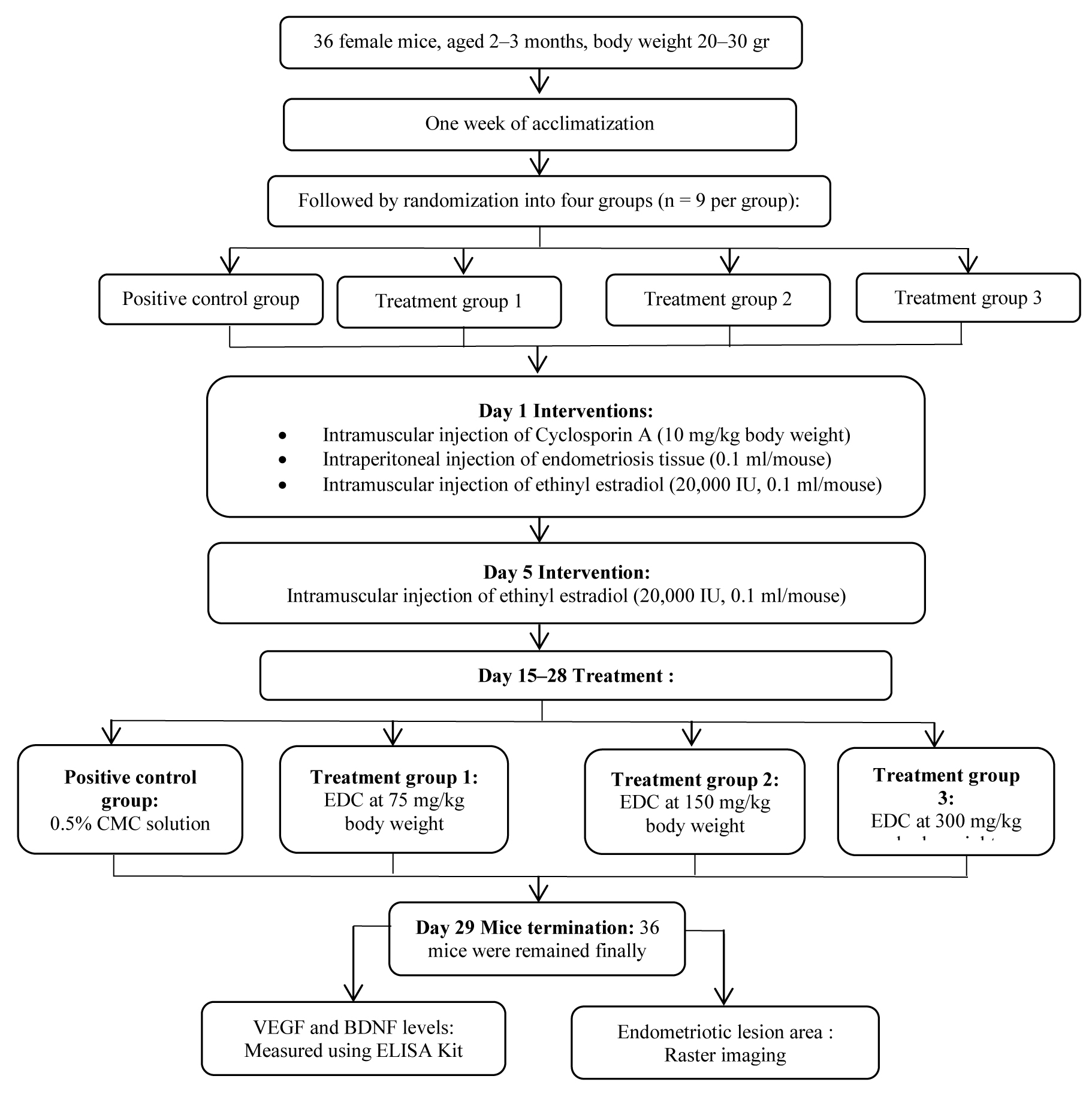
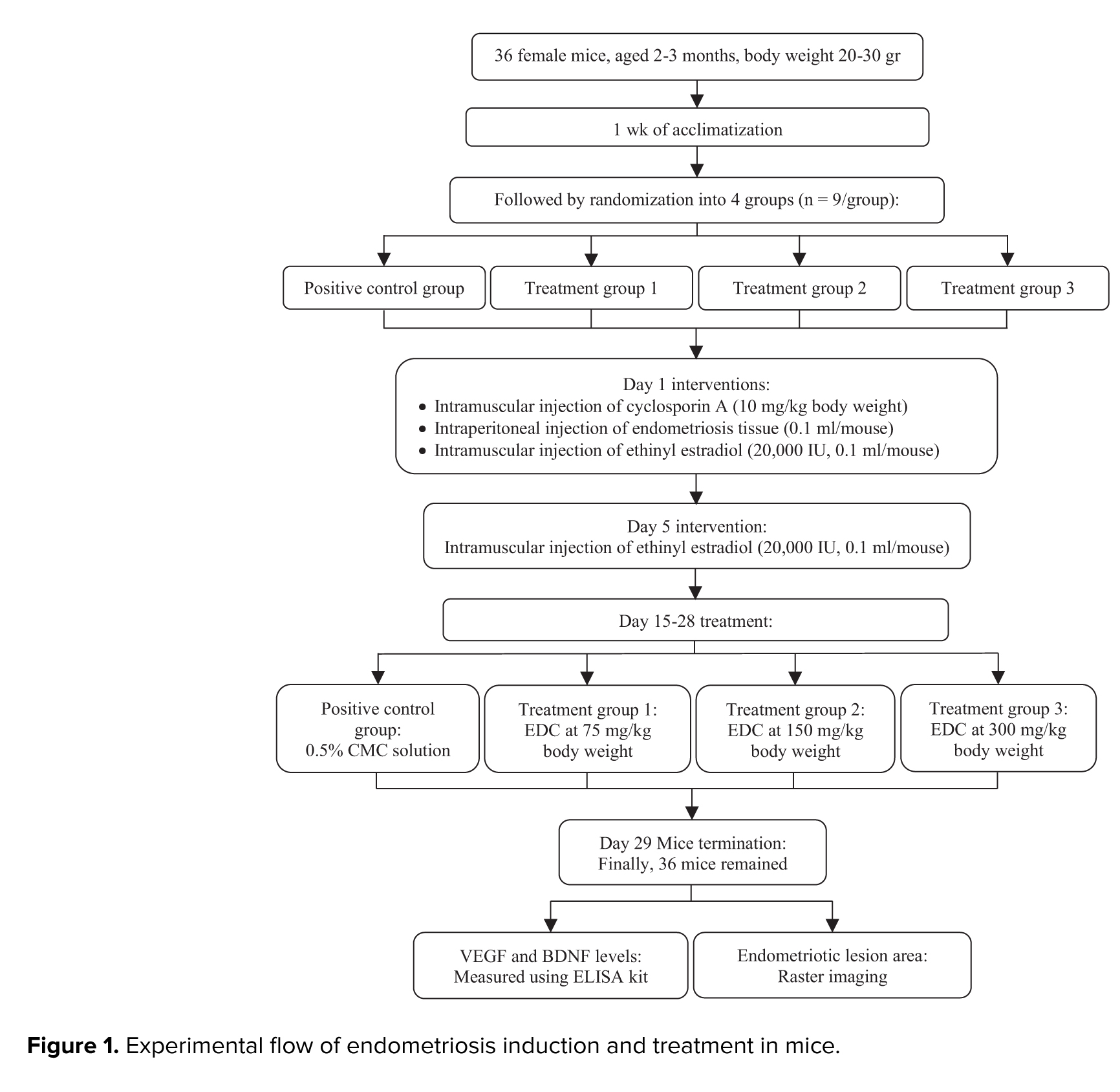
Estrogen in the form of 17α-ethinyl estradiolat dose of 20 μg manufactured by Sigma-Aldrich, Cat. No. E4876 was administered on day 5 to enhance ectopic tissue proliferation. Then, graded doses were administered to evaluate the effects of treatment on serum VEGF and BDNF levels, as well as the extent of endometriotic lesions. The treatment period lasted for 14 days. Clove leaf extract was administered orally using a feeding tube 3 times per day. Groups P1, P2, and P3 received clove leaf extract at doses of 75, 150, and 300 mg/kg body weight, respectively, while the untreated control group (K⁺) received 0.5% sodium carboxymethyl cellulose (Na CMC) as vehicle. The doses were selected based on previous toxicological and pharmacological studies of clove extract in animal models, including Indonesian studies that evaluated clove leaf extract toxicity and bioactivity (23). Meanwhile, in another study, physiological and immunological effects were found in mouse models (24). These studies support the relevance of the dose range we selected to evaluate the activity of clove leaf extract in vivo. Then, euthanasia was performed on the 29th day. Peritoneal tissue and blood were then analyzed. The experimental flow is illustrated in figure 1.
Inclusion criteria comprised healthy female mice aged 8-10 wk with body weight 20-25 gr; exclusion criteria were death during treatment or incomplete data collection. These criteria were established a priori, and no animals or data points were excluded. Final analysis included 9 mice per group (K⁺, P1, P2, P3). Blind administration of treatment was not possible due to staffing limitations, so the same staff members were responsible for daily management and dosing. However, outcome assessment was performed under blind conditions. Blood and endometriosis tissue sampling was performed by one operator, while VEGF and BDNF measurements were performed by separate technicians who were not involved in treatment allocation. Histological samples are coded by independent staff members and evaluated by blinded assessors. This separation of roles minimizes potential observer bias during outcome evaluation. Outcomes assessed were serum VEGF and BDNF concentrations, measured by ELISA, and peritoneal histopathology to evaluate lesion development. Serum VEGF and BDNF served as primary outcomes (mechanistic), while lesion size was defined as the primary (morphological) outcome. A study protocol, including the research question, design, and analysis plan, was prepared prior to experimentation but not registered in a public repository.
2.3. Endometriosis lesion assessment
Endometriotic lesions were excised via open abdominal approach, rinsed with phosphate-buffered saline, positioned on a calibrated grid for standardized imaging, and subjected to morphometric analysis using the Motic Images program.
2.4. Sampling of blood and serum hormone assays
Intracardial blood was collected and centrifuged at 3000 rpm for 10-15 min to obtain serum, which was transferred into sterile, pre-labeled Eppendorf tubes. VEGF and BDNF concentrations were quantitatively analyzed using an ELISA kit.
2.5. Ethical Considerations
All procedures were conducted at the Experimental Animal Cage and Veterinary Pathology Laboratory, Faculty of Veterinary Medicine, Airlangga University, Indonesia, with approval from the ethical review committee and animal care and use committee (Code: 2.KEH.131.09.2024). BALB/c mice were handled gently under light anesthesia and monitored daily for health, weight, and behavior. No adverse events occurred. Humane endpoints were predefined, none were met, and the study was completed as planned.
2.6. Statistical Analysis
IBM SPSS Statistics 25.0 was used for analysis. Results are presented as mean ± SD. Endometriotic lesion severity and serum levels of VEGF and BDNF were compared using one-way ANOVA and least significant difference (LSD) post hoc tests. Normality and homogeneity assumptions were assessed using the Shapiro-Wilk and Levene’s tests. If assumptions of normality or homogeneity were not met, non-parametric tests like Kruskal-Wallis followed by Dunn’s post hoc test were applied. A p < 0.05 was considered statistically significant.
3. Results
2.1. Clove leaf extraction and qualitative phytochemical screening
Clove leaves from Kediri City, East Java, Indonesia, were harvested for extraction using the maceration method with 96% ethanol at the UPT (Unit Pelaksana Teknis) Herbal Malang City, East Java, Indonesia. Clove leaf extract was obtained by following the established solvent extraction method as described by previous studies (19, 20), with modifications to suit our experimental design. Leaves were dried at 30-35°C for 2 days, ground, sieved, and soaked in ethanol thrice for optimal extraction. The filtrate was evaporated, yielding 183 gr of extract from 1000 gr of clove leaves. The extract was freeze-dried for preservation and kept at -20°C. Before being used in experiments, it was brought to the intended concentration through dilution with distilled water. The authenticity of the specimen was confirmed by a botanist at UPT Laboratorium Herbal Medica Malang, with voucher specimen number 400.7.21.4/2496/102.20/2024 and sample code 240723.P.G.P740 deposited at the laboratory herbarium. We also conducted qualitative phytochemical screening with number 400.7.21.4/2505/102.20/2024 to confirm the presence of flavonoids and other major compounds. The clove leaf extract we used was proven to contain flavonoids, alkaloids, tannins/phenols, saponins, and terpenoids.
2.2. Animal and study design
A total of 36 female BALB/c mouse endometriosis models (2-3 months old, weighing 20-30 gr) induced by uterine endometrial implantation with estrogen supplementation were obtained from the Animal House, Faculty of Medicine, University of Airlangga, Surabaya, Indonesia. Animals were pathogen-free, immunocompetent, and had not undergone any previous experimental procedures. Sample size was chosen based on prior literature using a similar endometriosis model with 36 mice, and was reported to have adequate statistical power (21). In this study, the experimental unit was defined as a single mouse. Each mouse was treated and analyzed independently, and statistical analyses were performed using individual animal data. All animals were provided with unlimited access to food and water in a controlled laboratory environment (temperature: 22-25°C; adequate ventilation). The clove leaf extract treatment sequence was randomized between groups to avoid time bias. Temperature was measured every morning and evening to ensure there were no potentially disruptive changes in the study. Cage locations were randomized weekly to minimize bias due to environmental effects. In the endometriosis mouse model, nociceptor neurons showed higher activation. This causes an increase in neurotrophic signals that contribute to pain in the endometriosis model. This condition widely causes an increase in VEGF in lesion tissue and peritoneal fluid (22).
After 1-wk of acclimatization period, 36 mice were randomly assigned to 4 groups: an untreated control group (K⁺) and 3 treatment groups (P1, P2, and P3), with 9 mice per group. Allocation to the control and treatment groups was performed by random draw using lottery cards. Randomization was conducted by laboratory personnel independent of treatment administration. Random sequences were generated using random draw cards, and allocation concealment was maintained by keeping the cards in a closed container until assignment. The personnel administering treatment were not aware of the allocation sequence. Endometriosis was induced with 0.2 ml intramuscular cyclosporine A (Sigma-Aldrich, Cat. No. C1832) and 0.1 ml intraperitoneal endometrial tissue on day 1. Endometrial tissue was obtained from the uterus of women who underwent hysterectomy at Dr. Soetomo General hospital, Surabaya, Indonesia. The procedure was conducted in accordance with hospital protocols, and hysterectomy was performed by a board‑certified obstetrician-gynecologist. Histological confirmation of endometriosis lesions for induction in mice was performed using hematoxylin-eosin (H&E) staining. Lesions were identified based on the presence of endometrial glands and stroma surrounded by hemosiderin-containing macrophages, in accordance with the criteria for endometriosis models in rodents. Our study used a mouse endometrial implantation model, in which donor endometrial tissue was induced on the peritoneal surface, representing ectopic endometrial implantation.
Estrogen in the form of 17α-ethinyl estradiolat dose of 20 μg manufactured by Sigma-Aldrich, Cat. No. E4876 was administered on day 5 to enhance ectopic tissue proliferation. Then, graded doses were administered to evaluate the effects of treatment on serum VEGF and BDNF levels, as well as the extent of endometriotic lesions. The treatment period lasted for 14 days. Clove leaf extract was administered orally using a feeding tube 3 times per day. Groups P1, P2, and P3 received clove leaf extract at doses of 75, 150, and 300 mg/kg body weight, respectively, while the untreated control group (K⁺) received 0.5% sodium carboxymethyl cellulose (Na CMC) as vehicle. The doses were selected based on previous toxicological and pharmacological studies of clove extract in animal models, including Indonesian studies that evaluated clove leaf extract toxicity and bioactivity (23). Meanwhile, in another study, physiological and immunological effects were found in mouse models (24). These studies support the relevance of the dose range we selected to evaluate the activity of clove leaf extract in vivo. Then, euthanasia was performed on the 29th day. Peritoneal tissue and blood were then analyzed. The experimental flow is illustrated in figure 1.
Inclusion criteria comprised healthy female mice aged 8-10 wk with body weight 20-25 gr; exclusion criteria were death during treatment or incomplete data collection. These criteria were established a priori, and no animals or data points were excluded. Final analysis included 9 mice per group (K⁺, P1, P2, P3). Blind administration of treatment was not possible due to staffing limitations, so the same staff members were responsible for daily management and dosing. However, outcome assessment was performed under blind conditions. Blood and endometriosis tissue sampling was performed by one operator, while VEGF and BDNF measurements were performed by separate technicians who were not involved in treatment allocation. Histological samples are coded by independent staff members and evaluated by blinded assessors. This separation of roles minimizes potential observer bias during outcome evaluation. Outcomes assessed were serum VEGF and BDNF concentrations, measured by ELISA, and peritoneal histopathology to evaluate lesion development. Serum VEGF and BDNF served as primary outcomes (mechanistic), while lesion size was defined as the primary (morphological) outcome. A study protocol, including the research question, design, and analysis plan, was prepared prior to experimentation but not registered in a public repository.
2.3. Endometriosis lesion assessment
Endometriotic lesions were excised via open abdominal approach, rinsed with phosphate-buffered saline, positioned on a calibrated grid for standardized imaging, and subjected to morphometric analysis using the Motic Images program.
2.4. Sampling of blood and serum hormone assays
Intracardial blood was collected and centrifuged at 3000 rpm for 10-15 min to obtain serum, which was transferred into sterile, pre-labeled Eppendorf tubes. VEGF and BDNF concentrations were quantitatively analyzed using an ELISA kit.
2.5. Ethical Considerations
All procedures were conducted at the Experimental Animal Cage and Veterinary Pathology Laboratory, Faculty of Veterinary Medicine, Airlangga University, Indonesia, with approval from the ethical review committee and animal care and use committee (Code: 2.KEH.131.09.2024). BALB/c mice were handled gently under light anesthesia and monitored daily for health, weight, and behavior. No adverse events occurred. Humane endpoints were predefined, none were met, and the study was completed as planned.
2.6. Statistical Analysis
IBM SPSS Statistics 25.0 was used for analysis. Results are presented as mean ± SD. Endometriotic lesion severity and serum levels of VEGF and BDNF were compared using one-way ANOVA and least significant difference (LSD) post hoc tests. Normality and homogeneity assumptions were assessed using the Shapiro-Wilk and Levene’s tests. If assumptions of normality or homogeneity were not met, non-parametric tests like Kruskal-Wallis followed by Dunn’s post hoc test were applied. A p < 0.05 was considered statistically significant.
3. Results
3.1. Effect of clove leaf extract on VEGF levels
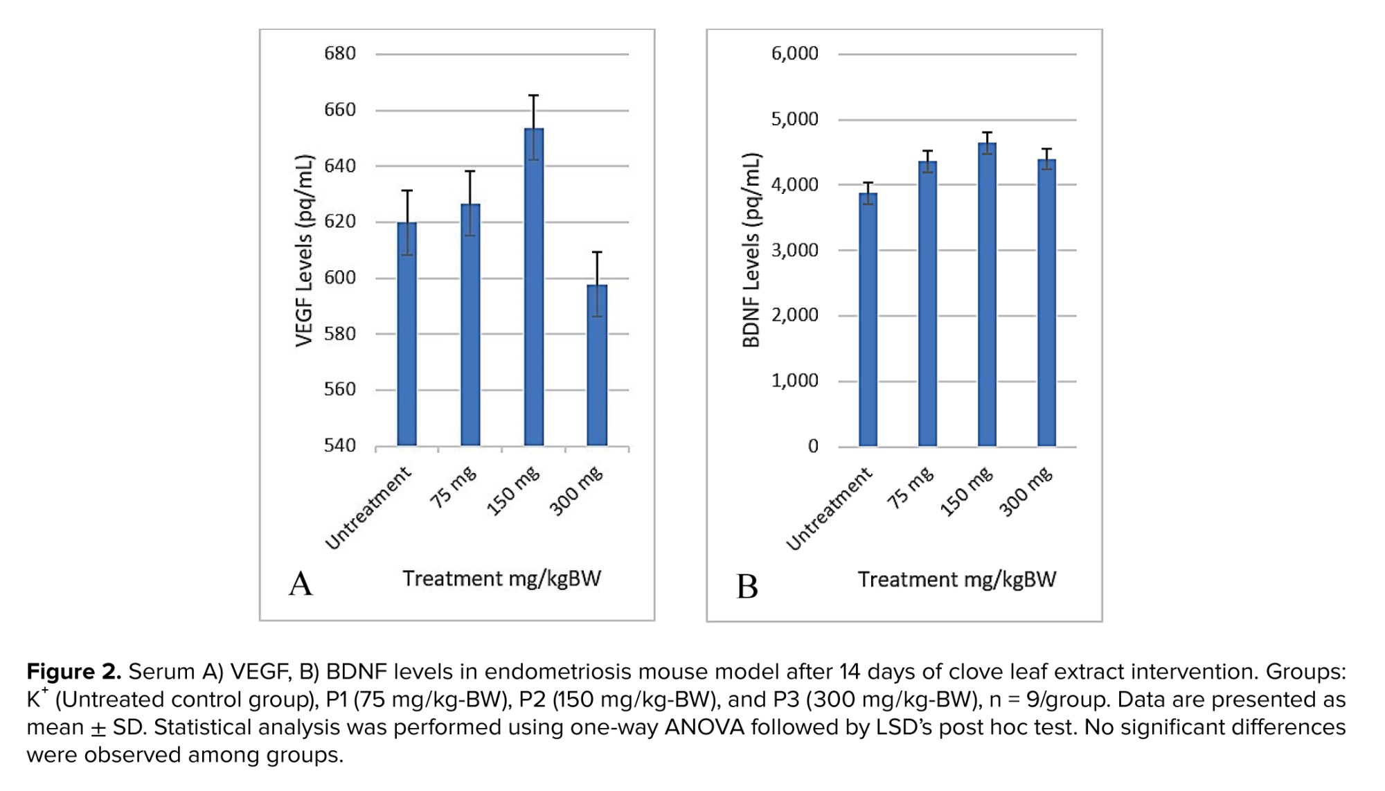
ELISA analysis showed that clove leaf extract did not significantly alter VEGF levels. As shown in figure 2A, administration of clove leaf extract at low dose (75 mg/kg-BW) and high dose (300 mg/kg-BW) to mouse for 14 days showed no statistically significant differences in VEGF levels compared to the control group (p > 0.05). Group P3 (300 mg/kg-BW) had the lowest average VEGF concentration (597.82 pg/mL), while group P2 (150 mg/kg-BW) had the highest average VEGF (653.78 pg/mL). Statistical analysis showed no significant differences between any group comparisons after 14 days of intervention (all p > 0.05). These findings indicate that clove leaf extract does not significantly affect VEGF expression under the conditions tested, although a slight decrease was observed at higher doses.
3.2. Effect of clove leaf extract on BDNF levels
ELISA analysis showed that clove leaf extract did not significantly reduce BDNF levels. As shown in figure 2B, administration of clove leaf extract at low dose (75 mg/kg-BW) and high dose (300 mg/kg-BW) to mouse for 14 days showed no statistically significant differences in BDNF levels compared to the control group (p > 0,05). The untreated control group (K⁺) had the lowest average BDNF concentration (3.876 pg/mL), while group P2 (150 mg/kg-BW) had the highest average BDNF (4.644 pg/mL). Statistical analysis showed no significant differences between any group comparisons after 14 days of intervention (all p > 0.05). These findings indicate that clove leaf extract did not significantly affect BDNF expression under the conditions tested.
3.3. Effect of clove leaf extract on endometriotic lesions
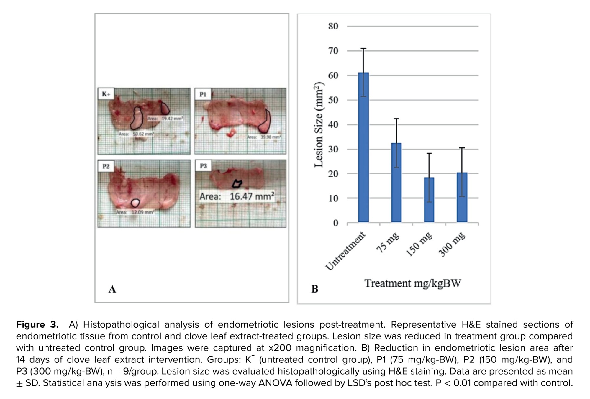
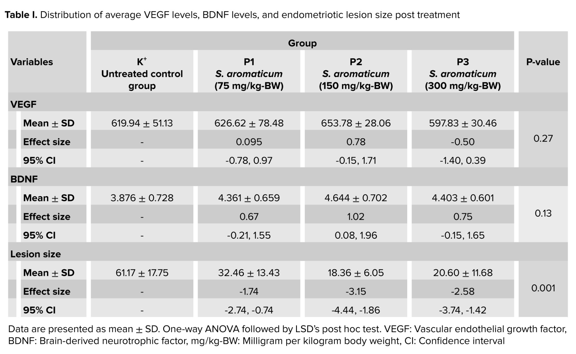
Microscopic analysis demonstrated persistent inflammation, characterized by dense microvasculature, neovascular development, and accelerated cell proliferation (Figure 3). The untreated control group (K⁺) exhibited the largest lesions with an average 61.17 mm2, while the P2 group (150 mg/kg-BW) showed the most significant reduction in lesion size with average 18.36 mm2. These results were significantly better than both clove leaf extract in low- (75 mg/kg-BW) and high-dose (300 mg/kg-BW) groups. The low- and high-dose groups also exhibited significant reduction relative to control. Statistical analysis confirmed significant differences in lesion size between groups (p < 0.001), with P2 (150 mg/kg-BW) having significantly smaller lesions compared to K⁺ (p < 0.001) and P1 (p < 0.05), but did not differ significantly from P3 (p > 0.05) (Table I). Table I summarizes these findings, highlighting that clove leaf extract at a dose of 150 mg/kg-BW produced the greatest reduction in lesion size under the tested conditions. Although serum VEGF and BDNF levels did not change significantly after 14 days, the reduction in lesion size remained evident. These results provide preliminary in vivo evidence that clove leaf extract may inhibit lesion development, particularly at moderate doses.
4. Discussion
ELISA analysis showed that clove leaf extract did not significantly alter VEGF levels. As shown in figure 2A, administration of clove leaf extract at low dose (75 mg/kg-BW) and high dose (300 mg/kg-BW) to mouse for 14 days showed no statistically significant differences in VEGF levels compared to the control group (p > 0.05). Group P3 (300 mg/kg-BW) had the lowest average VEGF concentration (597.82 pg/mL), while group P2 (150 mg/kg-BW) had the highest average VEGF (653.78 pg/mL). Statistical analysis showed no significant differences between any group comparisons after 14 days of intervention (all p > 0.05). These findings indicate that clove leaf extract does not significantly affect VEGF expression under the conditions tested, although a slight decrease was observed at higher doses.
3.2. Effect of clove leaf extract on BDNF levels
ELISA analysis showed that clove leaf extract did not significantly reduce BDNF levels. As shown in figure 2B, administration of clove leaf extract at low dose (75 mg/kg-BW) and high dose (300 mg/kg-BW) to mouse for 14 days showed no statistically significant differences in BDNF levels compared to the control group (p > 0,05). The untreated control group (K⁺) had the lowest average BDNF concentration (3.876 pg/mL), while group P2 (150 mg/kg-BW) had the highest average BDNF (4.644 pg/mL). Statistical analysis showed no significant differences between any group comparisons after 14 days of intervention (all p > 0.05). These findings indicate that clove leaf extract did not significantly affect BDNF expression under the conditions tested.
3.3. Effect of clove leaf extract on endometriotic lesions
Microscopic analysis demonstrated persistent inflammation, characterized by dense microvasculature, neovascular development, and accelerated cell proliferation (Figure 3). The untreated control group (K⁺) exhibited the largest lesions with an average 61.17 mm2, while the P2 group (150 mg/kg-BW) showed the most significant reduction in lesion size with average 18.36 mm2. These results were significantly better than both clove leaf extract in low- (75 mg/kg-BW) and high-dose (300 mg/kg-BW) groups. The low- and high-dose groups also exhibited significant reduction relative to control. Statistical analysis confirmed significant differences in lesion size between groups (p < 0.001), with P2 (150 mg/kg-BW) having significantly smaller lesions compared to K⁺ (p < 0.001) and P1 (p < 0.05), but did not differ significantly from P3 (p > 0.05) (Table I). Table I summarizes these findings, highlighting that clove leaf extract at a dose of 150 mg/kg-BW produced the greatest reduction in lesion size under the tested conditions. Although serum VEGF and BDNF levels did not change significantly after 14 days, the reduction in lesion size remained evident. These results provide preliminary in vivo evidence that clove leaf extract may inhibit lesion development, particularly at moderate doses.
4. Discussion
In this study, treatment with clove leaf extract significantly reduced endometriotic lesion size, particularly at 150 mg/kg body weight dose. The observed reduction suggests potential pro-apoptotic or anti-proliferative effects. Within 2 wk of intervention, VEGF and BDNF expression remained unchanged. Although clove extract did not significantly alter VEGF and BDNF, its anti-inflammatory properties via nuclear factor kappa-light-chain-enhancer of activated B cells (NFκB)-mediated apoptosis warrant further investigation, highlighting its potential role in modulating cell growth and survival. These findings suggest that clove extract exerts therapeutic effects through anti-inflammatory, pro-apoptotic, or anti-proliferative pathways rather than direct modulation of angiogenic or neurotrophic signaling.
Recent studies in experimental models and patient tissues show that VEGF as a central angiogenic mediator in endometriosis, activates VEGFR2 to drive endothelial proliferation, vascular permeability, and stromal expansion (22, 25-27). Neuroinflammatory factors, notably BDNF, influence female reproduction by activating tropomyosin receptor kinase B and NFκB signaling, thereby promoting ectopic endometrial proliferation and sustaining inflammation (12, 23).
Our histological analysis of endometriotic tissue revealed dense microvascular networks and enhanced stromal proliferation, consistent with angiogenic activity. VEGF and BDNF levels did not decrease, suggesting that clove leaf extract reduces lesion size through mechanisms other than VEGF/BDNF modulation. However, our findings align with previous studies, in which papaya leaf extract containing flavonoids modulated VEGF levels without statistical significance (28). Similar observations have also been reported (22), showing that neither VEGF neutralization nor vascular endothelial growth factor receptor 1 signal blockade effectively reduced lesion size or endometriosis-associated pain, suggesting that VEGF may not be the sole driver of disease progression (25).
Based on prior literature, several alternative pathways may hypothetically contribute to the observed reduction in lesion size. These included nerve growth factor (NGF)-mediated neurogenesis, inflammatory cytokines such as interleukin 6, tumor necrosis factor alpha, and oxidative stress (17, 29). Interleukin 6 has been implicated in regulating inflammation and immunity, while tumor necrosis factor alpha produced by activated macrophages, promotes inflammation and angiogenesis. Anti‑TNF therapy has been shown to reduce lesion development (30, 31). Elevated estrogen has also been reported to amplify BDNF expression in endometrial tissue, supporting its potential as a biomarker (13, 23). Although therapeutic targeting of BDNF has shown limited efficacy. In contrast, NGF-tropomyosin receptor kinase A (NGF-TrkA) signaling demonstrated a more pronounced role in endometriosis pathology, suggesting that BDNF may serve better as a biomarker than as a therapeutic target (22). In our study, clove leaf extract did not significantly alter VEGF or BDNF within 2 wk, yet markedly reduced lesion severity, most pronounced at 150 mg/kg-BW. Higher doses showed no added benefit. These findings may hypothetically reflect mechanisms involving inflammatory modulation, neuroplasticity, or growth factor regulation, rather than direct VEGF/BDNF suppression (32). Previous research has reported that clove extract can suppress inflammatory cytokines and inhibit fibroblast transformation (33), which may provide a reasonable explanation for our observations. However, this mechanistic interpretation remains speculative, as apoptosis, proliferation index, NFκB activity, cytokine profile, NGF, and infiltration of macrophages were not assessed directly in this research.
The activity clove extract is consistent with its bioactive profile, particularly eugenol, which disrupts oxidative phosphorylation, induces apoptosis, and limits abnormal proliferation (34). Other compounds such as flavonoids, alkaloids, tannins, saponins, and terpenoids (17, 35, 36), have anti-inflammatory and analgesic properties that have been tested in previous studies (19), contributing to a reduction in the extent of lesions in endometriosis.
Under current conditions, multiple signaling pathways may regulate VEGF, BDNF, and lesion progression, warranting further study. The limitations of our study include the short duration of the intervention (14 days) and the use of only one extract; no assessments of pain behavior, functional fertility, or pharmacokinetic and safety parameters were performed. In addition, VEGF and BDNF were measured only in serum, without corresponding peritoneal or tissue-level markers. In future studies, researchers are advised to use a combination of various extracts or pharmacological drugs so that their long-term effects can be compared and broader insights can be gained.
Recent studies indicate that clove extract significantly reduces the severity of peritoneal adhesion formation postsurgery. The observed effect correlates with reductions in adhesiometric indices, pro-inflammatory cytokine levels, oxidative stress markers, fibrotic tissue accumulation, and angiogenesis-associated biomolecules. These findings further support the therapeutic potential of clove extract in mitigating endometriotic lesion progression (17).
Although lesion size reduction occurred without detectable changes in systemic VEGF/BDNF levels over 14 days, our findings overall indicate a beneficial effect of clove leaf extract on lesion area reduction, particularly at a dose of 150 mg/kg body weight. These findings may differ for other strains, depending on immune response. Furthermore, the dose-dependent effect requires further validation in subsequent studies. Despite limitations in sample size and duration, this study provides a foundation for further research on clove leaf extract in endometriosis. The mechanism still needs to be confirmed, and further studies are needed before discussing clinical applications.
5. Conclusion
Recent studies in experimental models and patient tissues show that VEGF as a central angiogenic mediator in endometriosis, activates VEGFR2 to drive endothelial proliferation, vascular permeability, and stromal expansion (22, 25-27). Neuroinflammatory factors, notably BDNF, influence female reproduction by activating tropomyosin receptor kinase B and NFκB signaling, thereby promoting ectopic endometrial proliferation and sustaining inflammation (12, 23).
Our histological analysis of endometriotic tissue revealed dense microvascular networks and enhanced stromal proliferation, consistent with angiogenic activity. VEGF and BDNF levels did not decrease, suggesting that clove leaf extract reduces lesion size through mechanisms other than VEGF/BDNF modulation. However, our findings align with previous studies, in which papaya leaf extract containing flavonoids modulated VEGF levels without statistical significance (28). Similar observations have also been reported (22), showing that neither VEGF neutralization nor vascular endothelial growth factor receptor 1 signal blockade effectively reduced lesion size or endometriosis-associated pain, suggesting that VEGF may not be the sole driver of disease progression (25).
Based on prior literature, several alternative pathways may hypothetically contribute to the observed reduction in lesion size. These included nerve growth factor (NGF)-mediated neurogenesis, inflammatory cytokines such as interleukin 6, tumor necrosis factor alpha, and oxidative stress (17, 29). Interleukin 6 has been implicated in regulating inflammation and immunity, while tumor necrosis factor alpha produced by activated macrophages, promotes inflammation and angiogenesis. Anti‑TNF therapy has been shown to reduce lesion development (30, 31). Elevated estrogen has also been reported to amplify BDNF expression in endometrial tissue, supporting its potential as a biomarker (13, 23). Although therapeutic targeting of BDNF has shown limited efficacy. In contrast, NGF-tropomyosin receptor kinase A (NGF-TrkA) signaling demonstrated a more pronounced role in endometriosis pathology, suggesting that BDNF may serve better as a biomarker than as a therapeutic target (22). In our study, clove leaf extract did not significantly alter VEGF or BDNF within 2 wk, yet markedly reduced lesion severity, most pronounced at 150 mg/kg-BW. Higher doses showed no added benefit. These findings may hypothetically reflect mechanisms involving inflammatory modulation, neuroplasticity, or growth factor regulation, rather than direct VEGF/BDNF suppression (32). Previous research has reported that clove extract can suppress inflammatory cytokines and inhibit fibroblast transformation (33), which may provide a reasonable explanation for our observations. However, this mechanistic interpretation remains speculative, as apoptosis, proliferation index, NFκB activity, cytokine profile, NGF, and infiltration of macrophages were not assessed directly in this research.
The activity clove extract is consistent with its bioactive profile, particularly eugenol, which disrupts oxidative phosphorylation, induces apoptosis, and limits abnormal proliferation (34). Other compounds such as flavonoids, alkaloids, tannins, saponins, and terpenoids (17, 35, 36), have anti-inflammatory and analgesic properties that have been tested in previous studies (19), contributing to a reduction in the extent of lesions in endometriosis.
Under current conditions, multiple signaling pathways may regulate VEGF, BDNF, and lesion progression, warranting further study. The limitations of our study include the short duration of the intervention (14 days) and the use of only one extract; no assessments of pain behavior, functional fertility, or pharmacokinetic and safety parameters were performed. In addition, VEGF and BDNF were measured only in serum, without corresponding peritoneal or tissue-level markers. In future studies, researchers are advised to use a combination of various extracts or pharmacological drugs so that their long-term effects can be compared and broader insights can be gained.
Recent studies indicate that clove extract significantly reduces the severity of peritoneal adhesion formation postsurgery. The observed effect correlates with reductions in adhesiometric indices, pro-inflammatory cytokine levels, oxidative stress markers, fibrotic tissue accumulation, and angiogenesis-associated biomolecules. These findings further support the therapeutic potential of clove extract in mitigating endometriotic lesion progression (17).
Although lesion size reduction occurred without detectable changes in systemic VEGF/BDNF levels over 14 days, our findings overall indicate a beneficial effect of clove leaf extract on lesion area reduction, particularly at a dose of 150 mg/kg body weight. These findings may differ for other strains, depending on immune response. Furthermore, the dose-dependent effect requires further validation in subsequent studies. Despite limitations in sample size and duration, this study provides a foundation for further research on clove leaf extract in endometriosis. The mechanism still needs to be confirmed, and further studies are needed before discussing clinical applications.
5. Conclusion
Clove leaf ethanol extract, administered at 150 mg/kg-BW, significantly reduced lesion size. Further studies should incorporate additional biomarkers such as NFκB, NGF, and macrophage count providing a broader perspective on its role in preventing endometriosis.
Data Availability
Data supporting the findings of this study are available upon reasonable request from the corresponding author.
Author Contributions
FU. Mahardika and H. Hendarto designed the study and developed the research concept. FU. Mahardika conducted the research and collected the data under the supervision of Widjiati. Widjiati and H. Hendarto monitored, evaluated, and analyzed the results of the study. FU. Mahardika drafted the manuscript. H. Hendarto supervised overall research design and manuscript preparation. All authors critically revised the manuscript for important intellectual content, approved the final version, and take responsibility for the integrity of the data and the accuracy of the analysis.
Acknowledgements
We acknowledge UPT Herbal Materia Medica, Batu, Malang, for providing laboratory facilities. We would like to express our gratitude to the Faculty of Medicine, Universitas Airlangga, Surabaya; the Department of Obstetrics and Gynecology at Dr. Soetomo General hospital, Surabaya; and the laboratory team at the Faculty of Veterinary Medicine, Airlangga University, Surabaya, Indonesia. This study was supported by the Indonesian Ministry of Education, Culture, Research, and Technology (Grant number: 0459/E5/PG.02.00/2024). Artificial intelligence tools (Microsoft Copilot GPT-4) were used for language editing and grammar check.
Conflict of Interest
The authors declare that there is no conflict of interest.
Data Availability
Data supporting the findings of this study are available upon reasonable request from the corresponding author.
Author Contributions
FU. Mahardika and H. Hendarto designed the study and developed the research concept. FU. Mahardika conducted the research and collected the data under the supervision of Widjiati. Widjiati and H. Hendarto monitored, evaluated, and analyzed the results of the study. FU. Mahardika drafted the manuscript. H. Hendarto supervised overall research design and manuscript preparation. All authors critically revised the manuscript for important intellectual content, approved the final version, and take responsibility for the integrity of the data and the accuracy of the analysis.
Acknowledgements
We acknowledge UPT Herbal Materia Medica, Batu, Malang, for providing laboratory facilities. We would like to express our gratitude to the Faculty of Medicine, Universitas Airlangga, Surabaya; the Department of Obstetrics and Gynecology at Dr. Soetomo General hospital, Surabaya; and the laboratory team at the Faculty of Veterinary Medicine, Airlangga University, Surabaya, Indonesia. This study was supported by the Indonesian Ministry of Education, Culture, Research, and Technology (Grant number: 0459/E5/PG.02.00/2024). Artificial intelligence tools (Microsoft Copilot GPT-4) were used for language editing and grammar check.
Conflict of Interest
The authors declare that there is no conflict of interest.
Type of Study: Original Article |
Subject:
Cellular and Molecular Biology of Reproduction
Send email to the article author
| Rights and permissions | |
 |
This work is licensed under a Creative Commons Attribution-NonCommercial 4.0 International License. |