
International Journal of
Reproductive Biomedicine
Mon, Oct 12, 2026
[Archive]
Volume 24, Issue 6 (June 2026)
IJRM 2026, 24(6): 537-546 |
Back to browse issues page



Ethics code: IR.ACECR.AVICENNA.REC.1403.018


Download citation:
BibTeX | RIS | EndNote | Medlars | ProCite | Reference Manager | RefWorks
Send citation to:


BibTeX | RIS | EndNote | Medlars | ProCite | Reference Manager | RefWorks
Send citation to:
Ghadirkhomi E, Norouzi A. Accuracy of non-invasive prenatal screening compared with amniocentesis: A retrospective study. IJRM 2026; 24 (6) :537-546
URL: http://ijrm.ir/article-1-3793-en.html
URL: http://ijrm.ir/article-1-3793-en.html


1- Department of Genetics in Reproductive Disorders, Academic Center for Education, Culture, and Research (ACECR), Tabriz, Iran. , ghadirkhomi@acecr.ac.ir
2- Department of Genetics in Reproductive Disorders, Academic Center for Education, Culture, and Research (ACECR), Tabriz, Iran.
2- Department of Genetics in Reproductive Disorders, Academic Center for Education, Culture, and Research (ACECR), Tabriz, Iran.
Keywords: Prenatal screening, Amniocentesis, Chromosomal abnormalities, Prenatal diagnosis, Genetic counseling.
Full-Text [PDF 437 kb]
(316 Downloads)
| Abstract (HTML) (377 Views)
Full-Text: (51 Views)
1. Introduction
Chromosomal anomalies during pregnancy are a significant concern; the most prevalent conditions include Down syndrome (trisomy 21), Edwards syndrome (trisomy 18), and Patau syndrome (trisomy 13). These disorders can substantially affect fetal development and quality of life for affected children and their families. Early diagnosis enables informed decision-making and appropriate pregnancy management. Therefore, screening tests have become standard tools for assessing the risk of chromosomal abnormalities during the first and second trimesters of pregnancy (1-3).
First-trimester screening typically involves noninvasive blood tests and ultrasound measurements, including nuchal translucency (NT), free β-human chorionic gonadotropin, and pregnancy-associated plasma protein A. These markers, taken together with maternal age and gestational age, provide an estimate of the risk of trisomy 21, trisomy 18, and other genetic conditions (4, 5). First-trimester screening identifies approximately 90% of Down syndrome cases, although sensitivity can differ based on the combination of markers used (2, 6). Although cell-free DNA screening has demonstrated higher detection rates for common aneuploidies, its widespread use remains limited in some settings because of cost and accessibility constraints (5, 7-9). Second-trimester screening provides additional risk assessment through maternal serum markers and complements first-trimester findings (10). Invasive diagnostic testing, however, may be recommended where screening tests indicate a high risk of chromosomal abnormalities. Among invasive prenatal diagnostic methods, amniocentesis is one of the most frequently performed procedures involving the retrieval of amniotic fluid from the fetal environment. This fluid contains fetal cells that can be analyzed to confirm the presence of chromosomal abnormalities, such as trisomy 21, with a nearly 99% accuracy rate (11). Despite its high diagnostic accuracy, amniocentesis carries a small risk of complications, such as miscarriage, and is typically only performed after abnormal screening results.
Given the need for accurate prenatal risk assessment, this study compared conventional first- and second-trimester screening results with amniocentesis findings in an Iranian referral population to evaluate their clinical predictive value.
2. Materials and Methods
2.1. Study design and setting
This retrospective descriptive-analytical study was conducted using medical records of 490 pregnant women who underwent amniocentesis at the ACECR Genetic Center, Tabriz, Iran, between March 2018 and April 2023.
2.2. Sample size
The sample size was calculated using Cochran’s formula for estimating proportions:

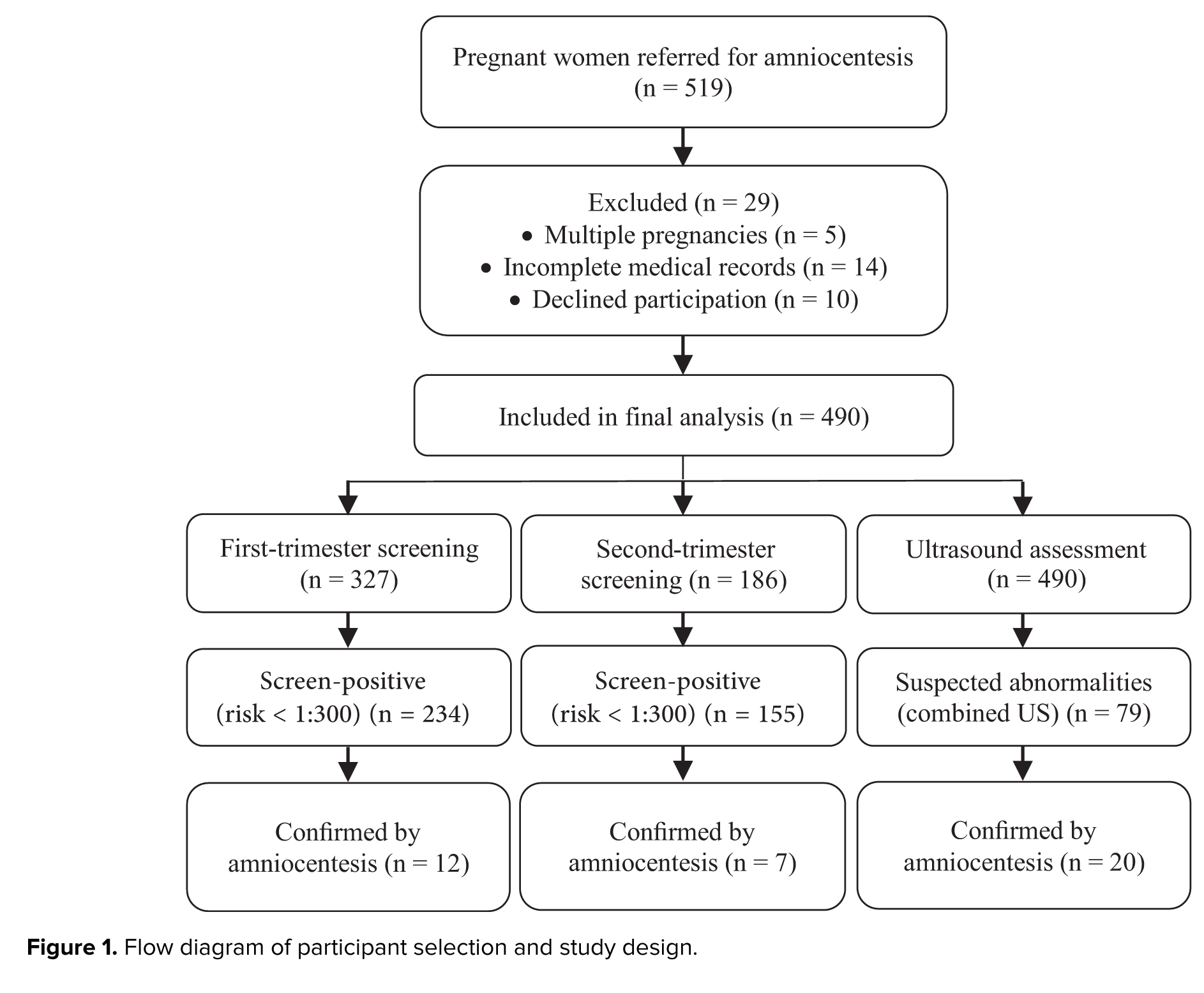
where Z represents the standard normal variate corresponding to a 95% confidence level (Z = 1.96), p is the estimated proportion based on previous similar studies, q = 1 - p, and d is the margin of error set at 0.05. Based on these assumptions, the minimum required sample size was calculated to be 214 participants. To improve the reliability of the findings and account for potential incomplete records, all eligible cases available during the study period were included, resulting in a final sample size of 490 participants. A flow diagram of participant selection is presented in figure 1.
2.3. Sample collection and criteria
Amniotic fluid samples were collected through standard amniocentesis procedures. Participants included pregnant women who met the inclusion criteria and provided informed consent. Inclusion criteria included pregnant women who were referred to the center by a gynecologist for amniocentesis due to various medical reasons (including the age of the mother, family history of genetic abnormalities, abnormal results of blood screenings, etc.). All referrals for amniocentesis were made in accordance with the Iranian Ministry of Health and Medical Education's national guidelines for prenatal diagnosis, according to the national prenatal diagnosis protocols and clinical recommendations in effect during the study period (2018-2023). The participants were between 19 and 43 yr old and were 15-20 wk pregnant. First- and second-trimester blood screening and ultrasound were performed for all participants. Individuals who did not wish to participate, as well as those with multiple pregnancies, were excluded from the study.
2.4. Ethical Considerations
This study was retrospective and based on previously collected medical records obtained during routine clinical care between 2018 and 2023. Ethical approval for retrospective data analysis and publication was obtained from the research ethics committee of Avicenna Research Institute, Tehran, Iran (Code: IR.ACECR.AVICENNA.REC.1403.018). All participants' data were anonymized, and no identifying information was used. The study protocol complied with the principles of the Declaration of Helsinki.
2.5. Statistical Analysis
For the data analysis, results from chromosomal tests, ultrasound parameters, blood screenings, and any relevant clinical history were gathered. SPSS software (version 27.0.1) was used to analyze the data. Continuous variables (e.g., maternal age, NT, crown-rump length [CRL], fetal heart rate [FHR]) were treated as numerical data, while categorical variables (e.g., risk categories, referral reasons, karyotype results) were expressed as frequencies and percentages. Descriptive statistics are presented as mean ± standard deviation or frequency (percentage). The independent samples t test was used to compare continuous variables between groups. A two-tailed p < 0.05 was considered statistically significant, and 95% confidence intervals (CIs) were calculated for all mean differences.
3. Results
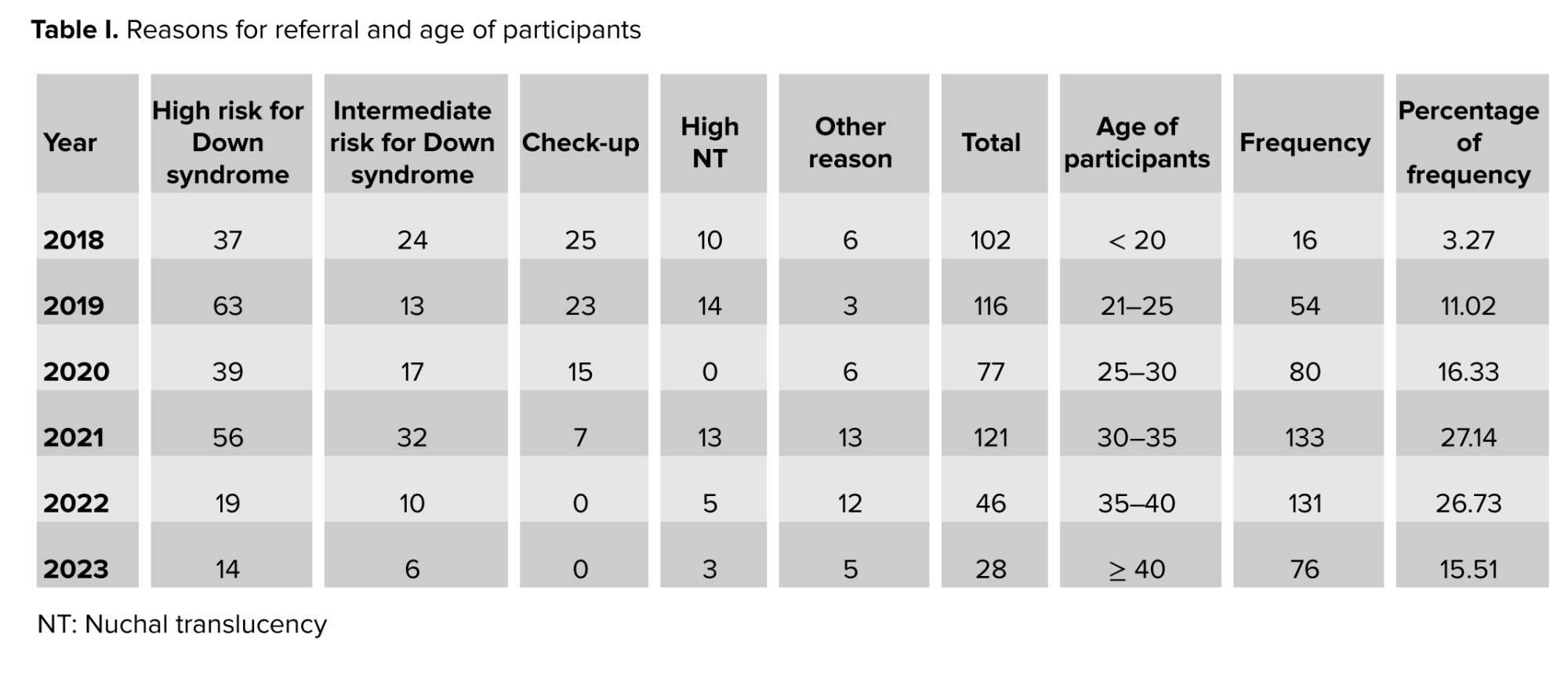
330 participants were referred to the laboratory due to high-risk findings in first- or second-trimester screening, 70 for check-ups (due to advanced maternal age or their request), 45 due to high NT levels, and 45 for various reasons, including genetic abnormalities. Table I presents the reasons for referral and the age of those referred to the laboratory.
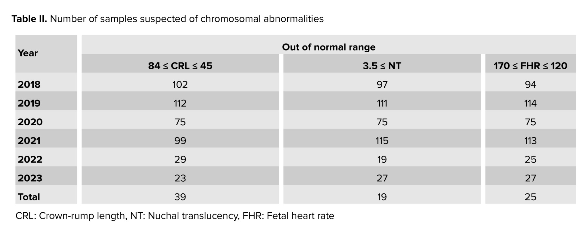
3.27% of the participants were < 20 yr old, 11.02% of the participants were between 20 and 25 yr old, 16.33% of the participants were between 25 and 30 yr old, 17.14% of the participants were between 30 and 35 yr old, 26.73% of the participants were between 35 and 40 yr old, and 15.51% of them were > 40 yr old. First, the ultrasound results from the first-trimester screening, including NT, CRL, and FHR, were examined. Table II presents the number of samples suspected of Down syndrome or other chromosomal abnormalities.
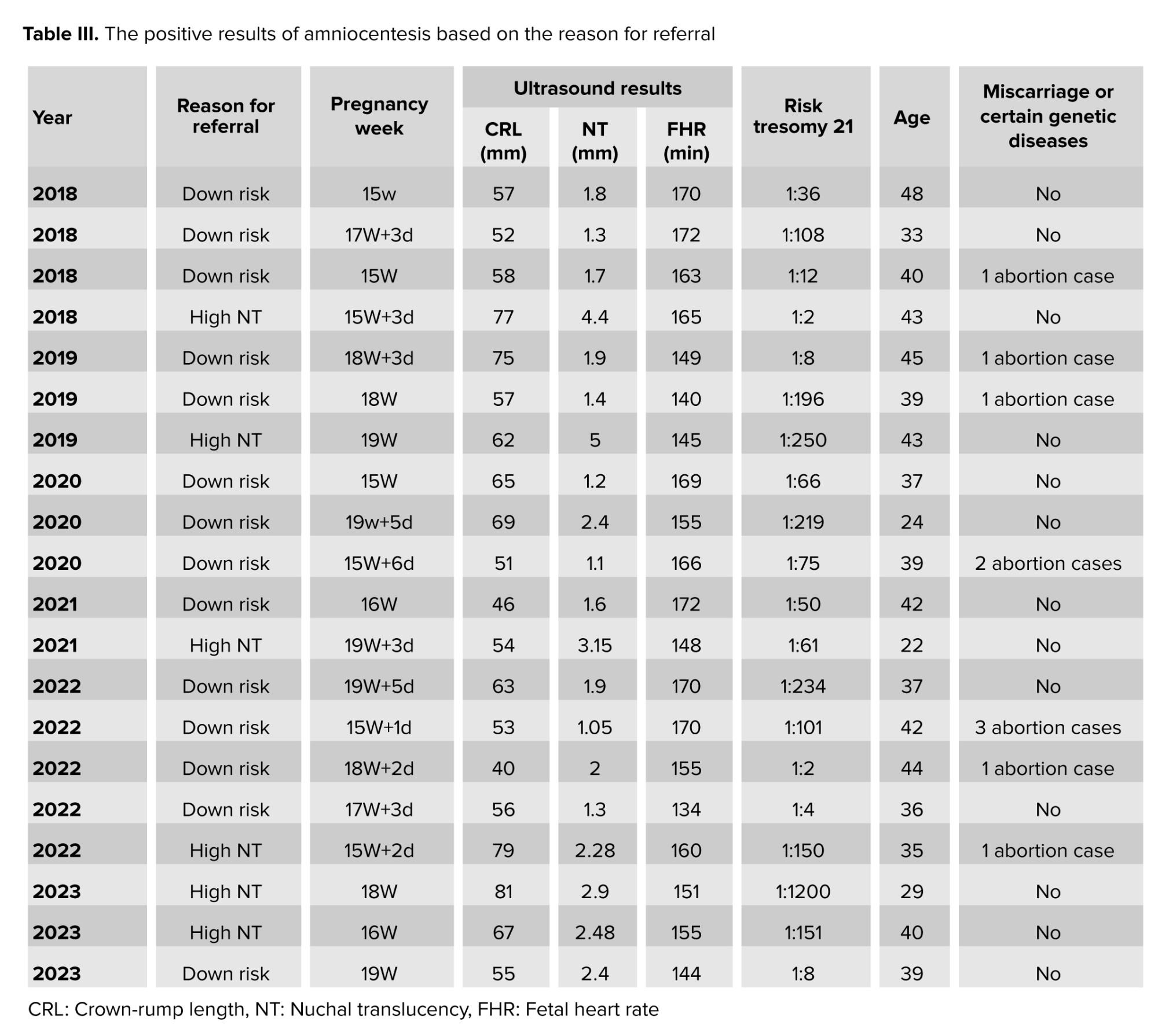
According to the results in table II, 39 samples based on CRL value, 19 samples based on NT value, and 25 samples based on FHR value can be suspected of chromosomal abnormalities. Based on the combined assessment of 3 ultrasound criteria, a total of 79 samples were suspected of chromosomal abnormalities. Cytogenetic analysis of amniotic fluid in these samples confirmed the presence of chromosomal abnormalities in only 20 samples (Table III).
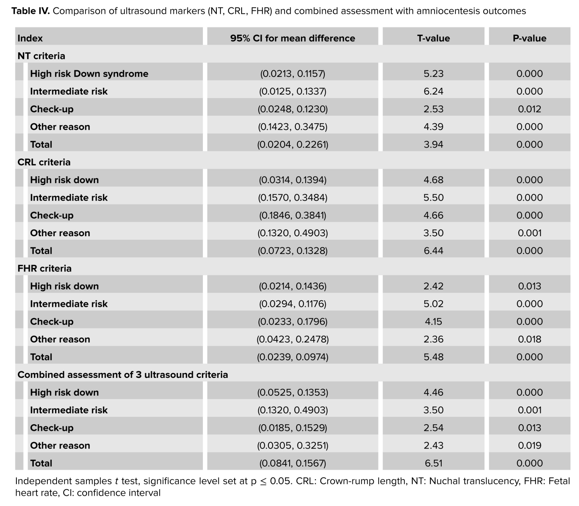
Table IV illustrates the correlation between ultrasound-based suspected chromosomal abnormality results and those obtained from amniocentesis.
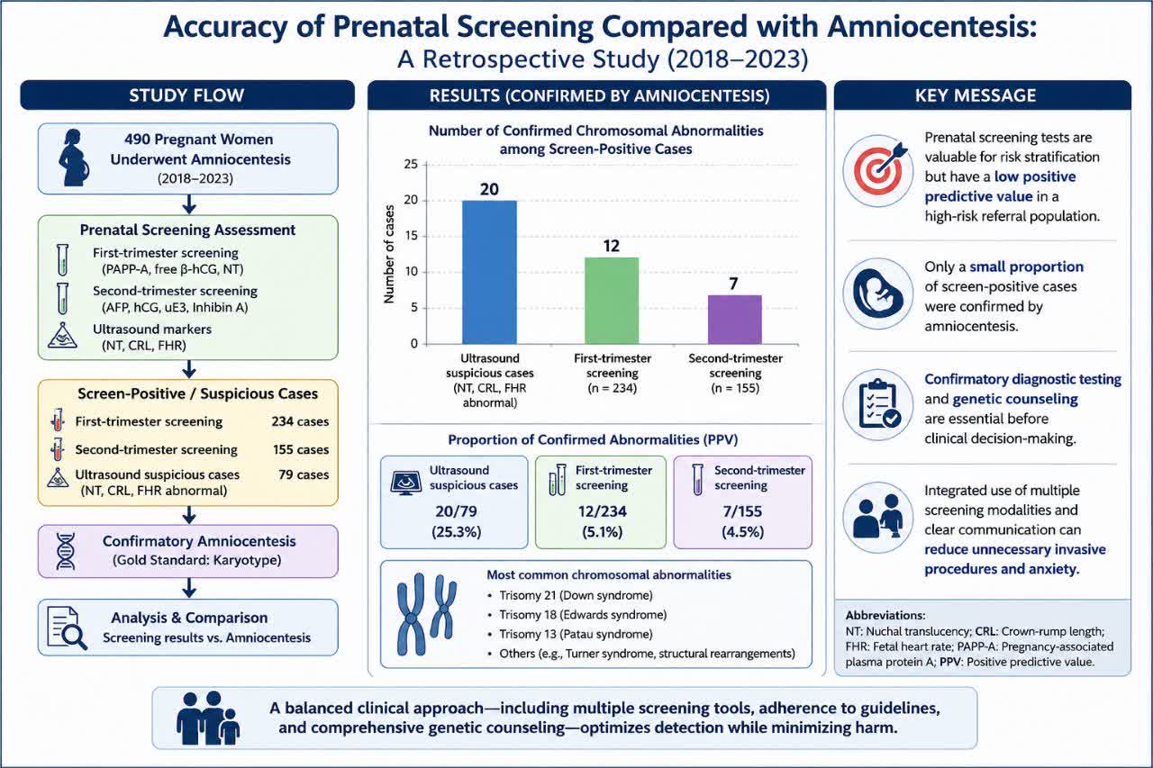
The results presented in table IV indicate that the p < 0.05 at the 95% CI, suggesting a statistically significant difference between the ultrasound criteria and the amniocentesis results. These findings imply that relying solely on ultrasound criteria may not be sufficient for accurate screening of chromosomal abnormalities. This discrepancy is particularly evident among participants who underwent amniocentesis due to elevated NT measurements, where only 5 out of 45 suspected cases were confirmed positive by amniocentesis. The inaccuracy and inability of ultrasound results to diagnose chromosomal abnormalities have led doctors to rely on the results of blood screening tests to make decisions. At this stage, based on the results of pregnancy-associated plasma protein A as well as other indicators, such as the mother's age, the risk of chromosomal abnormalities is determined. By reviewing the relevant data, 327 samples have undergone first-trimester screening tests, and 186 samples have undergone second-trimester screening tests. Among the 327 women who underwent first-trimester screening and subsequent amniocentesis, 93 women (28.4%) had a calculated risk in the low-risk range (≥ 1:300). The remaining 234 women (71.6%) had either an elevated risk (< 1:300) or an indeterminate result warranting further investigation. It is this latter group of 234 women that constituted the clinically relevant screen-positive population. Of these 234 women, only 12 (5.1%) had confirmed chromosomal abnormalities on karyotyping (positive predictive value [PPV] = 5.1%, 95% CI: 2.8-8.7).
For the 186 women who underwent second-trimester screening, 31 (16.7%) had a low-risk result (≥ 1:300). The remaining 155 women (83.3%) had elevated or indeterminate risk and underwent amniocentesis. In this group, 7 cases (4.5%) were confirmed positive (PPV = 4.5%, 95% CI: 2.0-9.0).
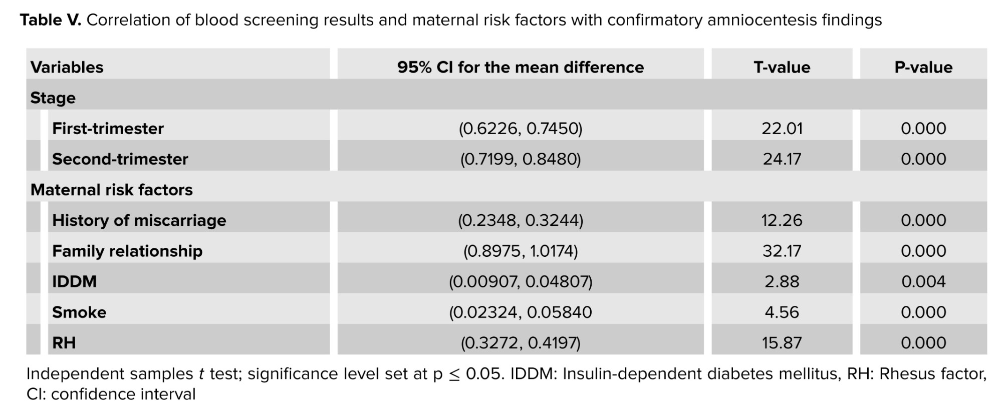
Table V shows that a significant difference was observed between the risk of the first and second-trimester screening tests and the results of the amniocentesis test. This means that the presence of chromosomal abnormalities cannot be confirmed with certainty based on the high risk of blood screening. In general, a significant difference in variables were observed in the results of the first and second-trimester screening and amniocentesis test. Additionally, by considering other variables that influence chromosomal disorders (Table V), it was found that there was a significant difference between the results, indicating that the presence of chromosomal abnormalities cannot be determined solely from an individual's history.
4. Discussion
This study examined the effectiveness of noninvasive screening methods, such as blood tests and ultrasounds performed during the first and second trimesters, in predicting chromosomal abnormalities compared to the more invasive procedure of amniocentesis. This issue has generated significant conversation in prenatal care regarding the dependability of these screening techniques in relation to conventional diagnostic tests. Primarily, the research emphasizes the restricted diagnostic precision of ultrasound indicators, especially NT, CRL, and FHR. Despite being widely used in the first trimester, ultrasound-based assessments yielded a significant number of false positives. Only 20 out of 79 cases flagged as suspicious by ultrasound were confirmed to have chromosomal abnormalities through amniocentesis, underscoring its low PPV. This aligns with prior research that cautions against over-reliance on ultrasound findings alone without confirmatory testing (2, 11). The blood tests from the first and second trimesters seem more specific than ultrasound, but they still have their downsides. For those identified as high-risk based on serum markers, only a few confirmed chromosomal issues through amniocentesis (12 in the first trimester and 7 in the second). This finding is consistent with previous large-scale real-world studies, which indicate that although first- and second-trimester serum screening tests can detect a substantial proportion of trisomy 21 cases, they are insufficient for a definitive diagnosis (5, 6).
The psychological toll of false-positive screening results is substantial and well-documented. Maternal anxiety following a high-risk screening result can persist throughout pregnancy and may adversely affect prenatal attachment and postpartum mental health (12-14). In the present cohort, over 75% of invasive procedures performed for abnormal ultrasound findings, and approximately 87% of those performed for high-risk serum screening, yielded normal karyotypes. This high false-positive rate underscores the critical need for robust pre-test counseling and shared decision-making to mitigate iatrogenic psychological harm.
In fact, the majority of high-risk cases identified in this study were not confirmed by amniocentesis, illustrating the emotional and ethical damage caused by premature or poorly communicated screening results. While ultrasound and laboratory tests have some connection to amniocentesis results, they don’t solely guarantee accurate diagnoses. This discrepancy highlights the need for clear and compassionate communication when discussing screening results with individuals. The use of terms like "high risk" must be carefully contextualized, with clinicians offering absolute risk estimates and explaining the probabilistic nature of screening, not deterministic outcomes. Without this clarity, individuals may misinterpret the results as a definitive diagnosis, leading to undue stress or premature decisions (14, 15). That’s why it’s important to look at a mix of clinical info, lab results, and imaging together. This method lines up with the best practices and the latest prenatal screening guidelines (5).
What is really clear is that no single factor, like a woman's age or past miscarriages, can reliably predict chromosomal issues on its own. Even though non-invasive methods have their limits, amniocentesis is still the go-to for prenatal testing, with almost 99% accuracy in finding chromosomal problems (2). Since it is invasive and carries a small risk of miscarriage, it’s usually recommended only when tests point to a high chance of chromosomal issues. This way, we get the best certainty in diagnosis while keeping risks low. It is noteworthy that, as demonstrated in table III, all confirmed cases of chromosomal abnormalities in this cohort were identified among women with either a high-risk screening result (risk < 1:300) or an elevated NT measurement. This observation underscores the fundamental value of prenatal screening programs in effectively triaging patients for invasive diagnostic testing. Conversely, the performance of amniocentesis in women with low-risk screening results (risk ≥ 1:300) or those referred solely for maternal age without additional risk factors highlights a potential deviation from evidence-based practice. Such practices expose women to the procedure-related risk of miscarriage, estimated at approximately 1 in 500 to 1 in 1000 in contemporary practice (16) without a proportionate increase in diagnostic yield. Strict adherence to national guideline thresholds for offering invasive testing is therefore essential to minimize iatrogenic harm and optimize the risk-benefit ratio of prenatal care.
Based on the study findings, several key recommendations can enhance the accuracy, effectiveness, and emotional safety of prenatal screening practices.
First, clinicians should adopt a combined risk assessment strategy, integrating maternal age, medical history, ultrasound findings, and blood screening results to better estimate the likelihood of chromosomal abnormalities before recommending invasive procedures like amniocentesis. Second, genetic counseling must be embedded in prenatal care, especially following abnormal screening outcomes, to ensure individuals fully understand the distinction between screening and diagnostic tests and can make informed decisions. To further aid understanding, healthcare providers should implement decision-support tools, such as visual aids or interactive risk calculators, which help individuals contextualize risk in relation to population norms. Additionally, where resources allow, non-invasive prenatal testing should be considered as a second-tier screening method, beneficial for reducing unnecessary amniocentesis in low-risk pregnancies (5, 17). Finally, mental health support and psychological reassurance should be included in prenatal consultations to help manage the anxiety and stress often triggered by uncertain or high-risk screening results.
This study has several important limitations. First, its retrospective design relies on the accuracy and completeness of medical records. Second, and most critically, the study cohort consists exclusively of women referred for invasive diagnostic testing, introducing significant selection (verification) bias. Women with screen-negative results who did not undergo amniocentesis and subsequently delivered healthy infants are not represented in this analysis. Consequently, the true negative predictive value of screening cannot be assessed, and the findings are not generalizable to the general low-risk obstetric population.
5. Conclusion
While first- and second-trimester screening are essential components of prenatal care for early risk stratification, their limited specificity can lead to significant parental anxiety and unnecessary invasive procedures. A balanced clinical approach (integrating multiple screening modalities, adhering to evidence-based guidelines, and providing comprehensive genetic counseling) is essential to optimize detection of chromosomal abnormalities while minimizing psychological and procedural harms.
Data Availability
Data supporting the findings of this study are available upon reasonable request from the corresponding author.
Author Contributions
E. Ghadirkhomi and A. Norouzi designed the study and conducted the research. They also collected and evaluated data from medical records and analyzed the study data. A. Norouzi drafted the manuscript, and E. Ghadirkhomi critically reviewed and revised the manuscript for important intellectual content. Both authors approved the final version of the manuscript and take responsibility for the integrity and accuracy of the work.
Acknowledgments
The authors would like to thank the staff of the ACECR Genetic Center, Tabriz, Iran, for their assistance in data collection and technical support. This study received no specific grant from any funding agency in the public, commercial, or not-for-profit sectors. ChatGPT (OpenAI) was used solely for language editing, grammar correction, and improvement of manuscript readability. All scientific content, data analysis, interpretation of results, and final manuscript approval were performed by the authors. The authors also thank all individuals who contributed to this study but did not meet the criteria for authorship.
Conflict of Interest
The authors declare that there is no conflict of interest.
Chromosomal anomalies during pregnancy are a significant concern; the most prevalent conditions include Down syndrome (trisomy 21), Edwards syndrome (trisomy 18), and Patau syndrome (trisomy 13). These disorders can substantially affect fetal development and quality of life for affected children and their families. Early diagnosis enables informed decision-making and appropriate pregnancy management. Therefore, screening tests have become standard tools for assessing the risk of chromosomal abnormalities during the first and second trimesters of pregnancy (1-3).
First-trimester screening typically involves noninvasive blood tests and ultrasound measurements, including nuchal translucency (NT), free β-human chorionic gonadotropin, and pregnancy-associated plasma protein A. These markers, taken together with maternal age and gestational age, provide an estimate of the risk of trisomy 21, trisomy 18, and other genetic conditions (4, 5). First-trimester screening identifies approximately 90% of Down syndrome cases, although sensitivity can differ based on the combination of markers used (2, 6). Although cell-free DNA screening has demonstrated higher detection rates for common aneuploidies, its widespread use remains limited in some settings because of cost and accessibility constraints (5, 7-9). Second-trimester screening provides additional risk assessment through maternal serum markers and complements first-trimester findings (10). Invasive diagnostic testing, however, may be recommended where screening tests indicate a high risk of chromosomal abnormalities. Among invasive prenatal diagnostic methods, amniocentesis is one of the most frequently performed procedures involving the retrieval of amniotic fluid from the fetal environment. This fluid contains fetal cells that can be analyzed to confirm the presence of chromosomal abnormalities, such as trisomy 21, with a nearly 99% accuracy rate (11). Despite its high diagnostic accuracy, amniocentesis carries a small risk of complications, such as miscarriage, and is typically only performed after abnormal screening results.
Given the need for accurate prenatal risk assessment, this study compared conventional first- and second-trimester screening results with amniocentesis findings in an Iranian referral population to evaluate their clinical predictive value.
2. Materials and Methods
2.1. Study design and setting
This retrospective descriptive-analytical study was conducted using medical records of 490 pregnant women who underwent amniocentesis at the ACECR Genetic Center, Tabriz, Iran, between March 2018 and April 2023.
2.2. Sample size
The sample size was calculated using Cochran’s formula for estimating proportions:
where Z represents the standard normal variate corresponding to a 95% confidence level (Z = 1.96), p is the estimated proportion based on previous similar studies, q = 1 - p, and d is the margin of error set at 0.05. Based on these assumptions, the minimum required sample size was calculated to be 214 participants. To improve the reliability of the findings and account for potential incomplete records, all eligible cases available during the study period were included, resulting in a final sample size of 490 participants. A flow diagram of participant selection is presented in figure 1.
2.3. Sample collection and criteria
Amniotic fluid samples were collected through standard amniocentesis procedures. Participants included pregnant women who met the inclusion criteria and provided informed consent. Inclusion criteria included pregnant women who were referred to the center by a gynecologist for amniocentesis due to various medical reasons (including the age of the mother, family history of genetic abnormalities, abnormal results of blood screenings, etc.). All referrals for amniocentesis were made in accordance with the Iranian Ministry of Health and Medical Education's national guidelines for prenatal diagnosis, according to the national prenatal diagnosis protocols and clinical recommendations in effect during the study period (2018-2023). The participants were between 19 and 43 yr old and were 15-20 wk pregnant. First- and second-trimester blood screening and ultrasound were performed for all participants. Individuals who did not wish to participate, as well as those with multiple pregnancies, were excluded from the study.
2.4. Ethical Considerations
This study was retrospective and based on previously collected medical records obtained during routine clinical care between 2018 and 2023. Ethical approval for retrospective data analysis and publication was obtained from the research ethics committee of Avicenna Research Institute, Tehran, Iran (Code: IR.ACECR.AVICENNA.REC.1403.018). All participants' data were anonymized, and no identifying information was used. The study protocol complied with the principles of the Declaration of Helsinki.
2.5. Statistical Analysis
For the data analysis, results from chromosomal tests, ultrasound parameters, blood screenings, and any relevant clinical history were gathered. SPSS software (version 27.0.1) was used to analyze the data. Continuous variables (e.g., maternal age, NT, crown-rump length [CRL], fetal heart rate [FHR]) were treated as numerical data, while categorical variables (e.g., risk categories, referral reasons, karyotype results) were expressed as frequencies and percentages. Descriptive statistics are presented as mean ± standard deviation or frequency (percentage). The independent samples t test was used to compare continuous variables between groups. A two-tailed p < 0.05 was considered statistically significant, and 95% confidence intervals (CIs) were calculated for all mean differences.
3. Results
330 participants were referred to the laboratory due to high-risk findings in first- or second-trimester screening, 70 for check-ups (due to advanced maternal age or their request), 45 due to high NT levels, and 45 for various reasons, including genetic abnormalities. Table I presents the reasons for referral and the age of those referred to the laboratory.
3.27% of the participants were < 20 yr old, 11.02% of the participants were between 20 and 25 yr old, 16.33% of the participants were between 25 and 30 yr old, 17.14% of the participants were between 30 and 35 yr old, 26.73% of the participants were between 35 and 40 yr old, and 15.51% of them were > 40 yr old. First, the ultrasound results from the first-trimester screening, including NT, CRL, and FHR, were examined. Table II presents the number of samples suspected of Down syndrome or other chromosomal abnormalities.
According to the results in table II, 39 samples based on CRL value, 19 samples based on NT value, and 25 samples based on FHR value can be suspected of chromosomal abnormalities. Based on the combined assessment of 3 ultrasound criteria, a total of 79 samples were suspected of chromosomal abnormalities. Cytogenetic analysis of amniotic fluid in these samples confirmed the presence of chromosomal abnormalities in only 20 samples (Table III).
Table IV illustrates the correlation between ultrasound-based suspected chromosomal abnormality results and those obtained from amniocentesis.
The results presented in table IV indicate that the p < 0.05 at the 95% CI, suggesting a statistically significant difference between the ultrasound criteria and the amniocentesis results. These findings imply that relying solely on ultrasound criteria may not be sufficient for accurate screening of chromosomal abnormalities. This discrepancy is particularly evident among participants who underwent amniocentesis due to elevated NT measurements, where only 5 out of 45 suspected cases were confirmed positive by amniocentesis. The inaccuracy and inability of ultrasound results to diagnose chromosomal abnormalities have led doctors to rely on the results of blood screening tests to make decisions. At this stage, based on the results of pregnancy-associated plasma protein A as well as other indicators, such as the mother's age, the risk of chromosomal abnormalities is determined. By reviewing the relevant data, 327 samples have undergone first-trimester screening tests, and 186 samples have undergone second-trimester screening tests. Among the 327 women who underwent first-trimester screening and subsequent amniocentesis, 93 women (28.4%) had a calculated risk in the low-risk range (≥ 1:300). The remaining 234 women (71.6%) had either an elevated risk (< 1:300) or an indeterminate result warranting further investigation. It is this latter group of 234 women that constituted the clinically relevant screen-positive population. Of these 234 women, only 12 (5.1%) had confirmed chromosomal abnormalities on karyotyping (positive predictive value [PPV] = 5.1%, 95% CI: 2.8-8.7).
For the 186 women who underwent second-trimester screening, 31 (16.7%) had a low-risk result (≥ 1:300). The remaining 155 women (83.3%) had elevated or indeterminate risk and underwent amniocentesis. In this group, 7 cases (4.5%) were confirmed positive (PPV = 4.5%, 95% CI: 2.0-9.0).
Table V shows that a significant difference was observed between the risk of the first and second-trimester screening tests and the results of the amniocentesis test. This means that the presence of chromosomal abnormalities cannot be confirmed with certainty based on the high risk of blood screening. In general, a significant difference in variables were observed in the results of the first and second-trimester screening and amniocentesis test. Additionally, by considering other variables that influence chromosomal disorders (Table V), it was found that there was a significant difference between the results, indicating that the presence of chromosomal abnormalities cannot be determined solely from an individual's history.
4. Discussion
This study examined the effectiveness of noninvasive screening methods, such as blood tests and ultrasounds performed during the first and second trimesters, in predicting chromosomal abnormalities compared to the more invasive procedure of amniocentesis. This issue has generated significant conversation in prenatal care regarding the dependability of these screening techniques in relation to conventional diagnostic tests. Primarily, the research emphasizes the restricted diagnostic precision of ultrasound indicators, especially NT, CRL, and FHR. Despite being widely used in the first trimester, ultrasound-based assessments yielded a significant number of false positives. Only 20 out of 79 cases flagged as suspicious by ultrasound were confirmed to have chromosomal abnormalities through amniocentesis, underscoring its low PPV. This aligns with prior research that cautions against over-reliance on ultrasound findings alone without confirmatory testing (2, 11). The blood tests from the first and second trimesters seem more specific than ultrasound, but they still have their downsides. For those identified as high-risk based on serum markers, only a few confirmed chromosomal issues through amniocentesis (12 in the first trimester and 7 in the second). This finding is consistent with previous large-scale real-world studies, which indicate that although first- and second-trimester serum screening tests can detect a substantial proportion of trisomy 21 cases, they are insufficient for a definitive diagnosis (5, 6).
The psychological toll of false-positive screening results is substantial and well-documented. Maternal anxiety following a high-risk screening result can persist throughout pregnancy and may adversely affect prenatal attachment and postpartum mental health (12-14). In the present cohort, over 75% of invasive procedures performed for abnormal ultrasound findings, and approximately 87% of those performed for high-risk serum screening, yielded normal karyotypes. This high false-positive rate underscores the critical need for robust pre-test counseling and shared decision-making to mitigate iatrogenic psychological harm.
In fact, the majority of high-risk cases identified in this study were not confirmed by amniocentesis, illustrating the emotional and ethical damage caused by premature or poorly communicated screening results. While ultrasound and laboratory tests have some connection to amniocentesis results, they don’t solely guarantee accurate diagnoses. This discrepancy highlights the need for clear and compassionate communication when discussing screening results with individuals. The use of terms like "high risk" must be carefully contextualized, with clinicians offering absolute risk estimates and explaining the probabilistic nature of screening, not deterministic outcomes. Without this clarity, individuals may misinterpret the results as a definitive diagnosis, leading to undue stress or premature decisions (14, 15). That’s why it’s important to look at a mix of clinical info, lab results, and imaging together. This method lines up with the best practices and the latest prenatal screening guidelines (5).
What is really clear is that no single factor, like a woman's age or past miscarriages, can reliably predict chromosomal issues on its own. Even though non-invasive methods have their limits, amniocentesis is still the go-to for prenatal testing, with almost 99% accuracy in finding chromosomal problems (2). Since it is invasive and carries a small risk of miscarriage, it’s usually recommended only when tests point to a high chance of chromosomal issues. This way, we get the best certainty in diagnosis while keeping risks low. It is noteworthy that, as demonstrated in table III, all confirmed cases of chromosomal abnormalities in this cohort were identified among women with either a high-risk screening result (risk < 1:300) or an elevated NT measurement. This observation underscores the fundamental value of prenatal screening programs in effectively triaging patients for invasive diagnostic testing. Conversely, the performance of amniocentesis in women with low-risk screening results (risk ≥ 1:300) or those referred solely for maternal age without additional risk factors highlights a potential deviation from evidence-based practice. Such practices expose women to the procedure-related risk of miscarriage, estimated at approximately 1 in 500 to 1 in 1000 in contemporary practice (16) without a proportionate increase in diagnostic yield. Strict adherence to national guideline thresholds for offering invasive testing is therefore essential to minimize iatrogenic harm and optimize the risk-benefit ratio of prenatal care.
Based on the study findings, several key recommendations can enhance the accuracy, effectiveness, and emotional safety of prenatal screening practices.
First, clinicians should adopt a combined risk assessment strategy, integrating maternal age, medical history, ultrasound findings, and blood screening results to better estimate the likelihood of chromosomal abnormalities before recommending invasive procedures like amniocentesis. Second, genetic counseling must be embedded in prenatal care, especially following abnormal screening outcomes, to ensure individuals fully understand the distinction between screening and diagnostic tests and can make informed decisions. To further aid understanding, healthcare providers should implement decision-support tools, such as visual aids or interactive risk calculators, which help individuals contextualize risk in relation to population norms. Additionally, where resources allow, non-invasive prenatal testing should be considered as a second-tier screening method, beneficial for reducing unnecessary amniocentesis in low-risk pregnancies (5, 17). Finally, mental health support and psychological reassurance should be included in prenatal consultations to help manage the anxiety and stress often triggered by uncertain or high-risk screening results.
This study has several important limitations. First, its retrospective design relies on the accuracy and completeness of medical records. Second, and most critically, the study cohort consists exclusively of women referred for invasive diagnostic testing, introducing significant selection (verification) bias. Women with screen-negative results who did not undergo amniocentesis and subsequently delivered healthy infants are not represented in this analysis. Consequently, the true negative predictive value of screening cannot be assessed, and the findings are not generalizable to the general low-risk obstetric population.
5. Conclusion
While first- and second-trimester screening are essential components of prenatal care for early risk stratification, their limited specificity can lead to significant parental anxiety and unnecessary invasive procedures. A balanced clinical approach (integrating multiple screening modalities, adhering to evidence-based guidelines, and providing comprehensive genetic counseling) is essential to optimize detection of chromosomal abnormalities while minimizing psychological and procedural harms.
Data Availability
Data supporting the findings of this study are available upon reasonable request from the corresponding author.
Author Contributions
E. Ghadirkhomi and A. Norouzi designed the study and conducted the research. They also collected and evaluated data from medical records and analyzed the study data. A. Norouzi drafted the manuscript, and E. Ghadirkhomi critically reviewed and revised the manuscript for important intellectual content. Both authors approved the final version of the manuscript and take responsibility for the integrity and accuracy of the work.
Acknowledgments
The authors would like to thank the staff of the ACECR Genetic Center, Tabriz, Iran, for their assistance in data collection and technical support. This study received no specific grant from any funding agency in the public, commercial, or not-for-profit sectors. ChatGPT (OpenAI) was used solely for language editing, grammar correction, and improvement of manuscript readability. All scientific content, data analysis, interpretation of results, and final manuscript approval were performed by the authors. The authors also thank all individuals who contributed to this study but did not meet the criteria for authorship.
Conflict of Interest
The authors declare that there is no conflict of interest.
Type of Study: Original Article |
Subject:
Reproductive Biology
Send email to the article author
| Rights and permissions | |
 |
This work is licensed under a Creative Commons Attribution-NonCommercial 4.0 International License. |