
International Journal of
Reproductive Biomedicine
Sat, Jun 13, 2026
[Archive]
Volume 24, Issue 3 (March 2026)
IJRM 2026, 24(3): 257-264 |
Back to browse issues page


Ethics code: IR.SSU.MEDICINE.REC.1402.097
![]()
![]()
![]()
Download citation:
BibTeX | RIS | EndNote | Medlars | ProCite | Reference Manager | RefWorks
Send citation to:


BibTeX | RIS | EndNote | Medlars | ProCite | Reference Manager | RefWorks
Send citation to:
Nematollahi R, Samadi M, Montazeri F, Kalantar S M, Shams A. Endometrial expression of T-cell immunoreceptor with Ig and ITIM domains and cluster of differentiation 155: A case-control study of a novel immunomodulatory axis in endometriosis. IJRM 2026; 24 (3) :257-264
URL: http://ijrm.ir/article-1-3598-en.html
URL: http://ijrm.ir/article-1-3598-en.html


1- Department of Immunology, Faculty of Medicine, Shahid Sadoughi University of Medical Sciences, Yazd, Iran. & Abortion Research Center, Yazd Reproductive Sciences Institute, Shahid Sadoughi University of Medical Sciences, Yazd, Iran.
2- Department of Immunology, School of Medicine, Isfahan University of Medical Sciences, Isfahan, Iran.
3- Abortion Research Center, Yazd Reproductive Sciences Institute, Shahid Sadoughi University of Medical Sciences, Yazd, Iran.
4- Department of Immunology, Faculty of Medicine, Shahid Sadoughi University of Medical Sciences, Yazd, Iran. ,alis743@yahoo.com; Alishams@ssu.ac.ir
2- Department of Immunology, School of Medicine, Isfahan University of Medical Sciences, Isfahan, Iran.
3- Abortion Research Center, Yazd Reproductive Sciences Institute, Shahid Sadoughi University of Medical Sciences, Yazd, Iran.
4- Department of Immunology, Faculty of Medicine, Shahid Sadoughi University of Medical Sciences, Yazd, Iran. ,
Full-Text [PDF 421 kb]
(152 Downloads)
| Abstract (HTML) (135 Views)
Full-Text: (18 Views)
1. Introduction
Research has indicated the vital involvement of endocrine and immune system dysregulations in highlighting the complexity of this gynecologic disease (1). Macrophages and natural killer cells in endometriosis cases exhibit reduced capacity to clear endometrial cells in the peritoneal cavity. The dysregulation of T-cell subsets contributes to inflammation and abnormal cytokine secretion, ultimately facilitating the expansion of endometriotic lesions (2). Endometriosis disease has been considered a specific immune response because of high levels of cytokines associated with T helper2 (Th2) lymphocytes found in the plasma and peritoneal fluid of women suffering from endometriosis. Still, as elevated levels of Th1 are seen in the peripheral blood of cases, the explanation of studies is complicated (3-5). Understanding the functions of co-inhibitory receptors in regulatory T-cells and effector T-cells is essential because current therapeutic strategies for inflammatory diseases increasingly focus on targeting these receptors (6).
Cluster of differentiation 155 (CD155), also known as the poliovirus receptor, is a type of adhesion molecule naturally present at low levels in various tissues, including vascular endothelial cells, dendritic cells, and macrophages (7). The CD155 gene is frequently overexpressed in various types of tumors. This overexpression facilitates cell migration and proliferation and has been linked to a poor prognosis in human cancers of different types (8).
T-cell immunoreceptor with immunoglobulin and immunoreceptor tyrosine-based inhibitory motif domains (TIGIT), an inhibitory receptor of the immunoglobulin superfamily, is detected in several lymphoid cells, including activated T cells, natural killer cells, and regulatory T cells. It plays a crucial role in controlling the immune responses (9, 10). Moreover, TIGIT is a surface receptor that has an inhibitory function by interacting with CD155, CD122, and CD113 (7, 11). TIGIT affects immune responses both directly and indirectly by inhibiting T cell proliferation and activation, as well as stimulating the secretion of interleukin-10 (IL-10), consequently affecting Th17 and Th1 cell responses (12, 13) and enhancing Th2-type immunity (14). The TIGIT gene has been upregulated in several types of cancers, highlighting its significant role in cancer immunology. It can be considered a valuable immunotherapy target (15, 16).
This study aims to investigate the levels of TIGIT and CD155 genes in endometriosis cases, with the ultimate goal of elucidating the probable role of this pathway in the onset and progression of endometriosis.
2. Materials and Methods
2.1. Study design
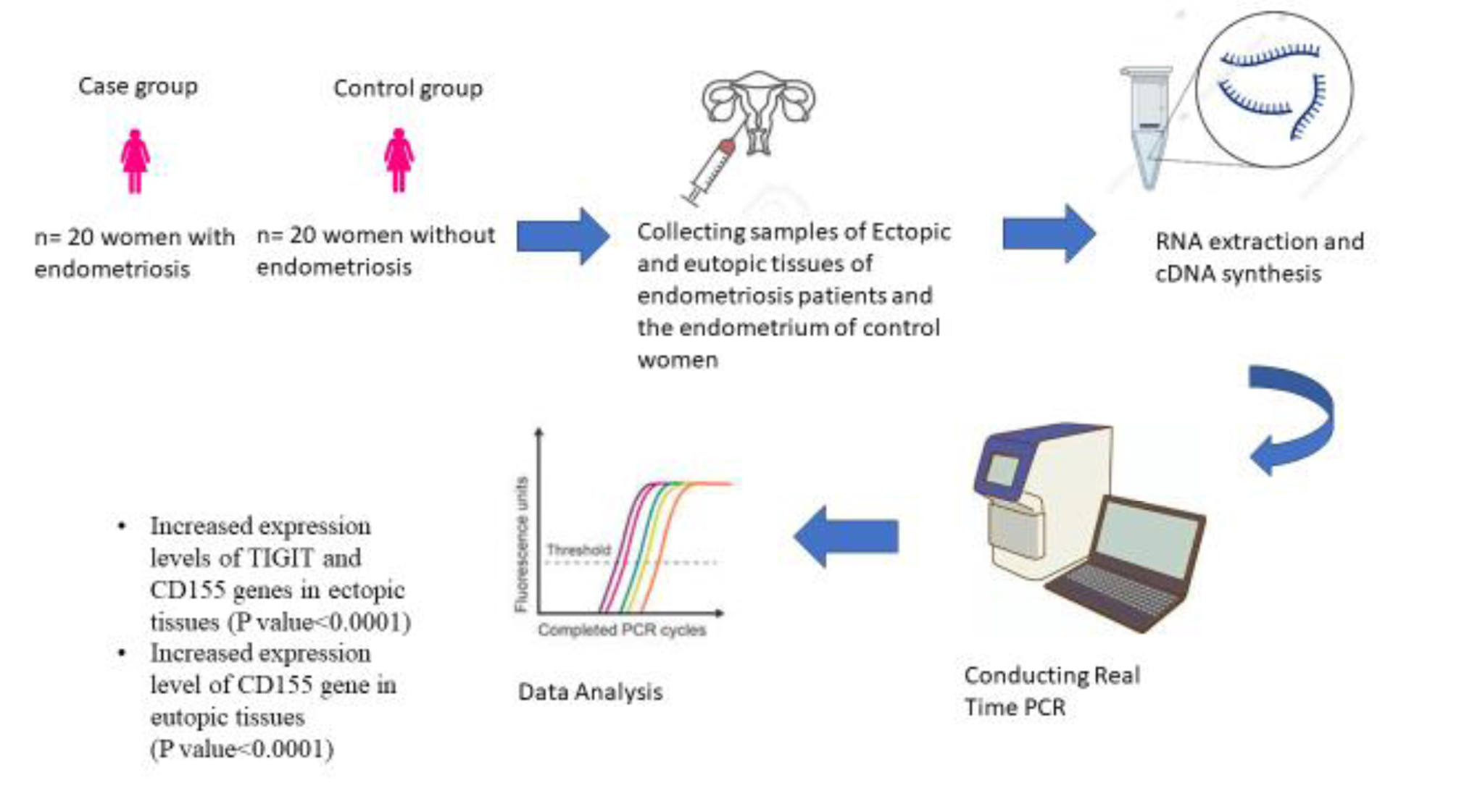
This case-control study was conducted at the Abortion Research Center, Yazd Reproductive Sciences Institute, Shahid Sadoughi University of Medical Sciences, Yazd, Iran. The study aimed to compare TIGIT and CD155 in 20 women with endometriosis to 20 women without this condition. Participant recruitment, tissue sampling, and data collection were performed over a 6-month period, from August 2023 to January 2024.
2.2. Participants
Eligible participants were women aged between 25 and 40 who had not used any hormonal therapy in the past 3 months. Women with endometrial carcinoma, hyperplasia, human papillomavirus infection, or benign endometrial tumors were excluded (17). The case group consisted of 20 women with a definitive diagnosis of endometriosis. Diagnosis was established by laparoscopy and subsequently confirmed by histopathological examination. The control group consisted of 20 women with benign gynecological conditions confirmed to have no endometriotic lesions. Clinical symptoms, participants' history, ultrasound mapping of pelvic sites, ureteral and rectal involvement, surgical indications, and magnetic resonance imaging findings were assessed.
2.3. Variables
Clinical and demographic variables, such as age, miscarriage history, and menstrual cycle regularity, were collected through standardized patient interviews and a review of medical records. The primary outcome variables were the relative gene expression levels of TIGIT and CD155 in tissue samples.
2.4. Sample size
The sample size was calculated using a standard formula for comparing 2 means. In addition, relevant published articles were reviewed to ensure the appropriate sample size for this study.

The calculation incorporated the desired significance level (α), statistical power (1-β), estimated standard deviation (S), and the anticipated mean difference between groups. There were no interim analyses or stopping guidelines applied in this study.
2.5. Tissue sample collection
Tissue sampling was conducted from August 2023 to January 2024. Ectopic and eutopic lesions were collected via laparoscopy from cases, while endometrial tissue from controls was obtained via curettage for non-endometriosis indications. Samples were gathered in RNA Later at -80°C until the time for RNA extraction.
2.6. RNA extraction and real-time polymerase chain reaction
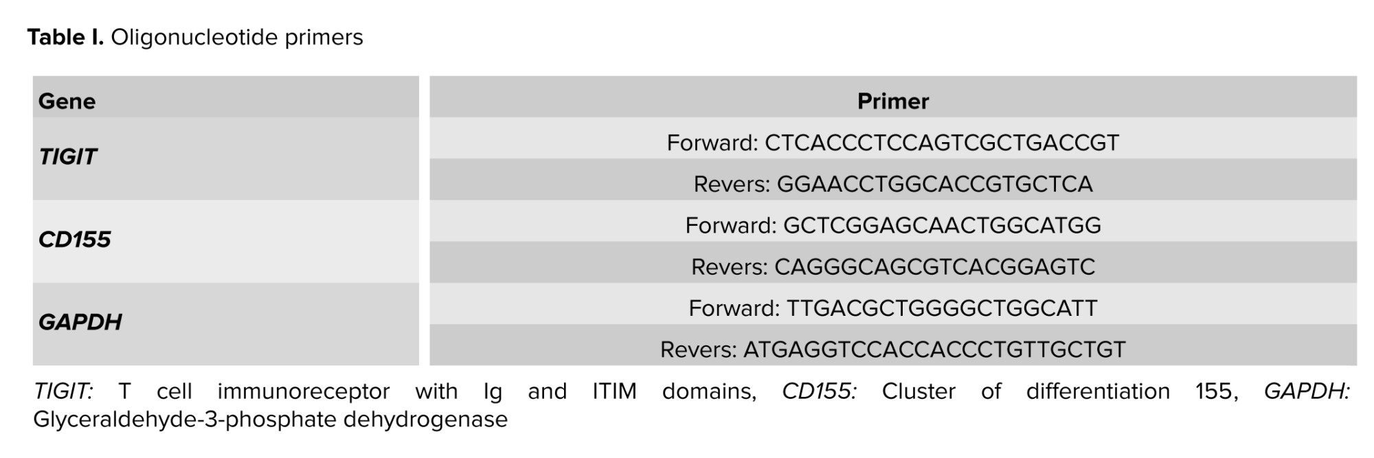
The TRIPURE isolation reagent was used to extract total RNA. All samples were analyzed for RNA purity and concentration using the Thermo Fisher NanoDrop. cDNA was synthesized immediately after RNA extraction. To assess the TIGIT and CD155 gene expression levels, a Fermentase cDNA synthesis kit was used with a thermocycler system. The real-time polymerase chain reaction was conducted using the synthesized cDNA in earlier steps as a template, along with the matched primers, with the help of a thermal cycler (Applied Biosystem, ABI, Step One Plus, USA). Glyceraldehyde‐3‐phosphate dehydrogenase was used as a reference gene to check the TIGIT and CD155 gene expression (Table Ⅰ).
2.7. Ethical Considerations
The ethics committee of Shahid Sadoughi University of Medical Sciences, Yazd, Iran, granted ethical approval for this research study (Code: IR.SSU.MEDICINE.REC.1402.097). Written informed consent was obtained from all participants for using their tissue samples in the research project and publishing their clinical data.
2.8. Statistical Analysis
The study included 2 quantitative variables: CD155 and TIGIT gene expression levels. The relative quantification ΔCt method (2−ΔCt) was used to assess changes in gene expression. The expression levels of TIGIT and CD155 genes were compared between ectopic and eutopic tissue of case and control groups.
The results were analyzed using GraphPad Prism software. At first, the normality of data distribution was assessed utilizing the one-sample Kolmogorov-Smirnov test. The findings indicated that the data distribution for TIGIT and CD155 gene expression did not follow a normal distribution. Because of the non-normal distribution of samples, to compare 3 groups, the Kruskal-Wallis test was used to compare 3 groups and Mann-Whitney U test was used to compare 2 groups. All findings were reported with statistically significant levels at a p < 0.05.
3. Results
A total of 70 women were initially enrolled, with 30 assigned to the case group and 40 to the control group. After excluding participants due to malignancy, infection, age, or other medical conditions, data from 20 women in each group were included in the final analysis.
The demographic data from both groups show comparable age ranges, between 25 and 40 yr. In the case group, several participants experienced recurrence of endometriosis, which is absent in the control group by definition. Infertility and irregular menstrual cycles were observed in some participants from both groups.
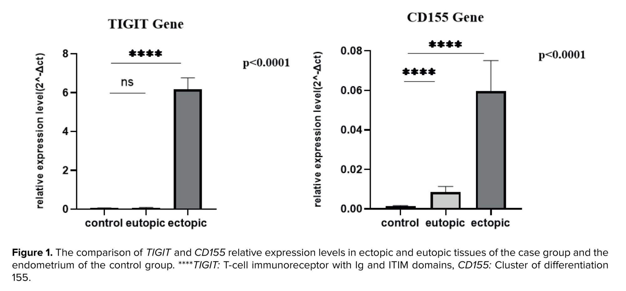
The research results indicated a notable upregulation of TIGIT gene expression in the ectopic tissue of cases compared to controls' endometrial tissue (p < 0001). In contrast, no substantial difference was observed in TIGIT gene expression between the eutopic endometrial tissue of cases and that of the control group (p = 0.49).
Data analysis revealed that CD155 gene expression was significantly elevated in the ectopic tissue of cases compared to their eutopic tissue and the endometrial tissue of controls (p < 0.0001). Moreover, the eutopic tissue of individuals in the case group demonstrated higher CD155 gene expression than the control group (p < 0.0001). The study's findings highlighted substantial differences in CD155 gene expression among the ectopic and eutopic tissues of cases, as well as the endometrial tissue of the control group (Figure 1).
4. Discussion
This study demonstrates increased expression of TIGIT and CD155 in the abnormal tissues of cases with endometriosis as well as upregulation of CD155 in the eutopic endometrium. Newly discovered evidence has shown the potential connection between dysregulated expression of immune checkpoints and the underlying causes of endometriosis (18). For example, the expression of programmed cell death protein 1 and programmed cell death ligand 1 has illustrated a significant increase in both ectopic and eutopic endometrium (19). Moreover, the amount of T-cell immunoglobulin and mucin-domain containing-3 expression in eutopic endometrium and also ectopic tissues was upregulated compared to the endometrium of controls (20).
TIGIT, a newly discovered inhibitory immune checkpoint, and its ligand CD155 make a pathway that has essential effects on immune regulation and many disease pathogenesis. For example, in colorectal cancer cases, the expression levels of CD155 and TIGIT genes were significantly elevated in tumor tissues compared to other normal tissues. Furthermore, individuals with higher CD155 gene expression experienced markedly worse clinical outcomes compared to those with low CD155 gene expression (21).
In cases with cervical cancer, TIGIT and CD155 genes are significantly overexpressed. Moreover, CD155 gene overexpression negatively correlates with the infiltration levels of CD8+ T cells (22).
In endometriosis, a shift toward M2 macrophage polarization and Th2 immune responses has been documented. Individuals' peritoneal fluid exhibits increased IL-10 and decreased Interferon-γ concentrations (2). The TIGIT-CD155 interaction facilitates the generation of tolerogenic dendritic cells, enhancing IL-10 production while suppressing IL-12. TIGIT binding to its ligand triggers regulatory T-cells to secrete IL-10 and transforming growth factor-beta, promoting T cell polarization towards Th2. Our findings demonstrate a significant upregulation of TIGIT-CD155 gene expression in endometriosis lesions (p < 0.0001). This heightened TIGIT-CD155 presence could potentially account for the elevated levels of M2 macrophages, Th2-associated cytokines, and IL-10 observed in endometriosis (23). The expression level of the CD155 gene was also elevated in the eutopic endometrium of the case group compared to the controls. The expression level of the TIGIT gene in the eutopic endometrium was not remarkably different. TIGIT is an immunological marker expressed on immune cells, and the inflammatory microenvironment and immunological conditions influence its expression. Also, the eutopic endometrium typically maintains a more homeostatic immune profile, which may not result in increased TIGIT expression to the same extent. Notably, a study examining TIGIT expression on CD4+ T cells in peripheral blood reported no significant difference between endometriosis cases and the control group (24).
5. Conclusion
In conclusion, the TIGIT/CD155 signaling pathway displays elevated activity in individuals with endometriosis. Additional research could provide deeper insights into the relations between the expression of TIGIT and CD155 genes and endometriosis, potentially offering them for disease diagnosis.
Data Availability
Data supporting the findings of this study are available upon reasonable request from the corresponding author.
Author Contributions
R. Nematollahi and A. Shams designed the study and conducted the research. M. Samadi and F. Montazeri and SM. Kalantar monitored, evaluated and analyzed the result of the study. All authors have reviewed and approved the final manuscript and assume full responsibility for the accuracy and integrity of the data presented.
Acknowledgments
This research received financial support from Shahid Sadoughi University of Medical Sciences, Yazd, Iran, and the Abortion Research Center, Yazd Reproductive Sciences Institute, Shahid Sadoughi University of Medical Sciences, Yazd, Iran. The artificial intelligence tool Paperpal was used only for grammar checking and language revision in this research.
Conflict of Interest
The authors declares that there is no conflict of interest.
Research has indicated the vital involvement of endocrine and immune system dysregulations in highlighting the complexity of this gynecologic disease (1). Macrophages and natural killer cells in endometriosis cases exhibit reduced capacity to clear endometrial cells in the peritoneal cavity. The dysregulation of T-cell subsets contributes to inflammation and abnormal cytokine secretion, ultimately facilitating the expansion of endometriotic lesions (2). Endometriosis disease has been considered a specific immune response because of high levels of cytokines associated with T helper2 (Th2) lymphocytes found in the plasma and peritoneal fluid of women suffering from endometriosis. Still, as elevated levels of Th1 are seen in the peripheral blood of cases, the explanation of studies is complicated (3-5). Understanding the functions of co-inhibitory receptors in regulatory T-cells and effector T-cells is essential because current therapeutic strategies for inflammatory diseases increasingly focus on targeting these receptors (6).
Cluster of differentiation 155 (CD155), also known as the poliovirus receptor, is a type of adhesion molecule naturally present at low levels in various tissues, including vascular endothelial cells, dendritic cells, and macrophages (7). The CD155 gene is frequently overexpressed in various types of tumors. This overexpression facilitates cell migration and proliferation and has been linked to a poor prognosis in human cancers of different types (8).
T-cell immunoreceptor with immunoglobulin and immunoreceptor tyrosine-based inhibitory motif domains (TIGIT), an inhibitory receptor of the immunoglobulin superfamily, is detected in several lymphoid cells, including activated T cells, natural killer cells, and regulatory T cells. It plays a crucial role in controlling the immune responses (9, 10). Moreover, TIGIT is a surface receptor that has an inhibitory function by interacting with CD155, CD122, and CD113 (7, 11). TIGIT affects immune responses both directly and indirectly by inhibiting T cell proliferation and activation, as well as stimulating the secretion of interleukin-10 (IL-10), consequently affecting Th17 and Th1 cell responses (12, 13) and enhancing Th2-type immunity (14). The TIGIT gene has been upregulated in several types of cancers, highlighting its significant role in cancer immunology. It can be considered a valuable immunotherapy target (15, 16).
This study aims to investigate the levels of TIGIT and CD155 genes in endometriosis cases, with the ultimate goal of elucidating the probable role of this pathway in the onset and progression of endometriosis.
2. Materials and Methods
2.1. Study design
This case-control study was conducted at the Abortion Research Center, Yazd Reproductive Sciences Institute, Shahid Sadoughi University of Medical Sciences, Yazd, Iran. The study aimed to compare TIGIT and CD155 in 20 women with endometriosis to 20 women without this condition. Participant recruitment, tissue sampling, and data collection were performed over a 6-month period, from August 2023 to January 2024.
2.2. Participants
Eligible participants were women aged between 25 and 40 who had not used any hormonal therapy in the past 3 months. Women with endometrial carcinoma, hyperplasia, human papillomavirus infection, or benign endometrial tumors were excluded (17). The case group consisted of 20 women with a definitive diagnosis of endometriosis. Diagnosis was established by laparoscopy and subsequently confirmed by histopathological examination. The control group consisted of 20 women with benign gynecological conditions confirmed to have no endometriotic lesions. Clinical symptoms, participants' history, ultrasound mapping of pelvic sites, ureteral and rectal involvement, surgical indications, and magnetic resonance imaging findings were assessed.
2.3. Variables
Clinical and demographic variables, such as age, miscarriage history, and menstrual cycle regularity, were collected through standardized patient interviews and a review of medical records. The primary outcome variables were the relative gene expression levels of TIGIT and CD155 in tissue samples.
2.4. Sample size
The sample size was calculated using a standard formula for comparing 2 means. In addition, relevant published articles were reviewed to ensure the appropriate sample size for this study.
The calculation incorporated the desired significance level (α), statistical power (1-β), estimated standard deviation (S), and the anticipated mean difference between groups. There were no interim analyses or stopping guidelines applied in this study.
2.5. Tissue sample collection
Tissue sampling was conducted from August 2023 to January 2024. Ectopic and eutopic lesions were collected via laparoscopy from cases, while endometrial tissue from controls was obtained via curettage for non-endometriosis indications. Samples were gathered in RNA Later at -80°C until the time for RNA extraction.
2.6. RNA extraction and real-time polymerase chain reaction
The TRIPURE isolation reagent was used to extract total RNA. All samples were analyzed for RNA purity and concentration using the Thermo Fisher NanoDrop. cDNA was synthesized immediately after RNA extraction. To assess the TIGIT and CD155 gene expression levels, a Fermentase cDNA synthesis kit was used with a thermocycler system. The real-time polymerase chain reaction was conducted using the synthesized cDNA in earlier steps as a template, along with the matched primers, with the help of a thermal cycler (Applied Biosystem, ABI, Step One Plus, USA). Glyceraldehyde‐3‐phosphate dehydrogenase was used as a reference gene to check the TIGIT and CD155 gene expression (Table Ⅰ).
2.7. Ethical Considerations
The ethics committee of Shahid Sadoughi University of Medical Sciences, Yazd, Iran, granted ethical approval for this research study (Code: IR.SSU.MEDICINE.REC.1402.097). Written informed consent was obtained from all participants for using their tissue samples in the research project and publishing their clinical data.
2.8. Statistical Analysis
The study included 2 quantitative variables: CD155 and TIGIT gene expression levels. The relative quantification ΔCt method (2−ΔCt) was used to assess changes in gene expression. The expression levels of TIGIT and CD155 genes were compared between ectopic and eutopic tissue of case and control groups.
The results were analyzed using GraphPad Prism software. At first, the normality of data distribution was assessed utilizing the one-sample Kolmogorov-Smirnov test. The findings indicated that the data distribution for TIGIT and CD155 gene expression did not follow a normal distribution. Because of the non-normal distribution of samples, to compare 3 groups, the Kruskal-Wallis test was used to compare 3 groups and Mann-Whitney U test was used to compare 2 groups. All findings were reported with statistically significant levels at a p < 0.05.
3. Results
A total of 70 women were initially enrolled, with 30 assigned to the case group and 40 to the control group. After excluding participants due to malignancy, infection, age, or other medical conditions, data from 20 women in each group were included in the final analysis.
The demographic data from both groups show comparable age ranges, between 25 and 40 yr. In the case group, several participants experienced recurrence of endometriosis, which is absent in the control group by definition. Infertility and irregular menstrual cycles were observed in some participants from both groups.
The research results indicated a notable upregulation of TIGIT gene expression in the ectopic tissue of cases compared to controls' endometrial tissue (p < 0001). In contrast, no substantial difference was observed in TIGIT gene expression between the eutopic endometrial tissue of cases and that of the control group (p = 0.49).
Data analysis revealed that CD155 gene expression was significantly elevated in the ectopic tissue of cases compared to their eutopic tissue and the endometrial tissue of controls (p < 0.0001). Moreover, the eutopic tissue of individuals in the case group demonstrated higher CD155 gene expression than the control group (p < 0.0001). The study's findings highlighted substantial differences in CD155 gene expression among the ectopic and eutopic tissues of cases, as well as the endometrial tissue of the control group (Figure 1).
4. Discussion
This study demonstrates increased expression of TIGIT and CD155 in the abnormal tissues of cases with endometriosis as well as upregulation of CD155 in the eutopic endometrium. Newly discovered evidence has shown the potential connection between dysregulated expression of immune checkpoints and the underlying causes of endometriosis (18). For example, the expression of programmed cell death protein 1 and programmed cell death ligand 1 has illustrated a significant increase in both ectopic and eutopic endometrium (19). Moreover, the amount of T-cell immunoglobulin and mucin-domain containing-3 expression in eutopic endometrium and also ectopic tissues was upregulated compared to the endometrium of controls (20).
TIGIT, a newly discovered inhibitory immune checkpoint, and its ligand CD155 make a pathway that has essential effects on immune regulation and many disease pathogenesis. For example, in colorectal cancer cases, the expression levels of CD155 and TIGIT genes were significantly elevated in tumor tissues compared to other normal tissues. Furthermore, individuals with higher CD155 gene expression experienced markedly worse clinical outcomes compared to those with low CD155 gene expression (21).
In cases with cervical cancer, TIGIT and CD155 genes are significantly overexpressed. Moreover, CD155 gene overexpression negatively correlates with the infiltration levels of CD8+ T cells (22).
In endometriosis, a shift toward M2 macrophage polarization and Th2 immune responses has been documented. Individuals' peritoneal fluid exhibits increased IL-10 and decreased Interferon-γ concentrations (2). The TIGIT-CD155 interaction facilitates the generation of tolerogenic dendritic cells, enhancing IL-10 production while suppressing IL-12. TIGIT binding to its ligand triggers regulatory T-cells to secrete IL-10 and transforming growth factor-beta, promoting T cell polarization towards Th2. Our findings demonstrate a significant upregulation of TIGIT-CD155 gene expression in endometriosis lesions (p < 0.0001). This heightened TIGIT-CD155 presence could potentially account for the elevated levels of M2 macrophages, Th2-associated cytokines, and IL-10 observed in endometriosis (23). The expression level of the CD155 gene was also elevated in the eutopic endometrium of the case group compared to the controls. The expression level of the TIGIT gene in the eutopic endometrium was not remarkably different. TIGIT is an immunological marker expressed on immune cells, and the inflammatory microenvironment and immunological conditions influence its expression. Also, the eutopic endometrium typically maintains a more homeostatic immune profile, which may not result in increased TIGIT expression to the same extent. Notably, a study examining TIGIT expression on CD4+ T cells in peripheral blood reported no significant difference between endometriosis cases and the control group (24).
5. Conclusion
In conclusion, the TIGIT/CD155 signaling pathway displays elevated activity in individuals with endometriosis. Additional research could provide deeper insights into the relations between the expression of TIGIT and CD155 genes and endometriosis, potentially offering them for disease diagnosis.
Data Availability
Data supporting the findings of this study are available upon reasonable request from the corresponding author.
Author Contributions
R. Nematollahi and A. Shams designed the study and conducted the research. M. Samadi and F. Montazeri and SM. Kalantar monitored, evaluated and analyzed the result of the study. All authors have reviewed and approved the final manuscript and assume full responsibility for the accuracy and integrity of the data presented.
Acknowledgments
This research received financial support from Shahid Sadoughi University of Medical Sciences, Yazd, Iran, and the Abortion Research Center, Yazd Reproductive Sciences Institute, Shahid Sadoughi University of Medical Sciences, Yazd, Iran. The artificial intelligence tool Paperpal was used only for grammar checking and language revision in this research.
Conflict of Interest
The authors declares that there is no conflict of interest.
Type of Study: Original Article |
Subject:
Reproductive Genetics
Send email to the article author
| Rights and permissions | |
 |
This work is licensed under a Creative Commons Attribution-NonCommercial 4.0 International License. |
