International Journal of
Reproductive Biomedicine
Tue, Sep 29, 2026
[Archive]
Volume 24, Issue 5 (May 2026)
IJRM 2026, 24(5): 433-444 |
Back to browse issues page



Ethics code: IR.ARAKU.REC.1402.094


Download citation:
BibTeX | RIS | EndNote | Medlars | ProCite | Reference Manager | RefWorks
Send citation to:


BibTeX | RIS | EndNote | Medlars | ProCite | Reference Manager | RefWorks
Send citation to:
Soleimani Mehranjani M, Garavand S, Ahmadi S. Selenium mitigates cyclophosphamide-induced toxicity on sperm quality in mice: An experimental study. IJRM 2026; 24 (5) :433-444
URL: http://ijrm.ir/article-1-3735-en.html
URL: http://ijrm.ir/article-1-3735-en.html
Selenium mitigates cyclophosphamide-induced toxicity on sperm quality in mice: An experimental study


1- Department of Biology, Faculty of Science, Arak University, Arak, Iran. , m-soleimani@araku.ac.ir
2- Department of Biology, Faculty of Science, Arak University, Arak, Iran.
2- Department of Biology, Faculty of Science, Arak University, Arak, Iran.
Full-Text [PDF 1955 kb]
(419 Downloads)
| Abstract (HTML) (451 Views)
Full-Text: (54 Views)
1. Introduction
Cyclophosphamide (CP) is a widely used chemotherapy drug for treating various cancers and some autoimmune diseases (1). In the body, it is metabolized by cytochrome P450 enzymes into active metabolites, including 4-hydroxycyclophosphamide. This unstable metabolite is further converted to aldophosphamide and then to phosphoramide mustard and acrolein. Phosphoramide mustard is primarily responsible for DNA damage, whereas acrolein is a toxic metabolite that induces inflammation and tissue injury, especially in the reproductive tract (2). Acrolein promotes oxidative stress by depleting glutathione and increasing lipid peroxidation (3). Elevated oxidative stress has been shown to reduce intracellular antioxidants in Leydig cells, impair testosterone synthesis and secretion, decrease reproductive organ weight, disrupt spermatogenesis, and ultimately lower sperm count (1). Selenium, an essential micronutrient and potent antioxidant, plays a key role in improving sperm parameters and supporting normal spermatogenesis (4). It is a structural component of glutathione peroxidase, which, together with glutathione, neutralizes reactive oxygen species (ROS) such as hydrogen peroxide and lipid peroxides, protecting DNA, cell membranes, and proteins in testicular tissue, and supporting overall testicular health and function (5). Experimental studies have confirmed selenium’s protective effects. For instance, co-administration of selenium with doxorubicin reduced oxidative stress and apoptosis, improved sperm quality, and preserved normal spermatogenesis in mice (6). Another study showed that selenium mitigated acrylamide-induced testicular damage by reducing oxidative stress, regulating steroidogenesis-related gene expression, and enhancing tissue quality (7). Additionally, selenium protected testicular structure against the adverse effects of venlafaxine hydrochloride in male mice by strengthening antioxidant defenses and lowering apoptotic protein expression (8). This study aimed to evaluate the effects of selenium on sperm quality and related parameters in mice following treatment with CP.
2. Materials and Methods
2.1. Animals and treatment
In this experimental study, each mouse was considered an experimental unit. Mice were chosen for their well-established reproductive physiology and widespread use as a model organism in biomedical research, allowing for extensive comparative data. The NMRI strain is a commonly used outbred stock in toxicology and reproductive studies, known for its robustness and predictable response to various chemical agents.
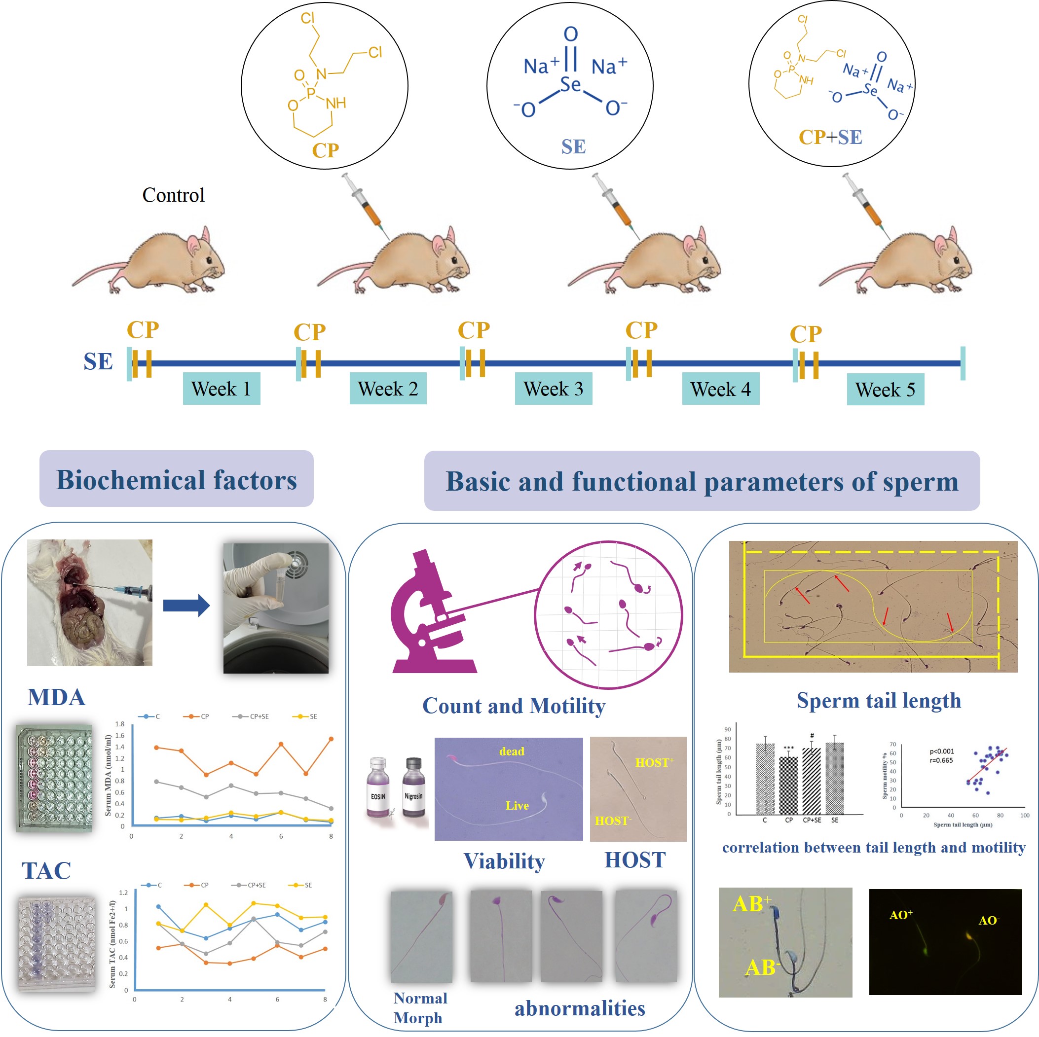
32 adult male NMRI mice (6 wk, 36 ± 2 gr) were obtained from the animal house of Arak University, Arak, Iran and kept under favorable conditions (21°C, 12-hr dark/light cycle). This study did not establish specific humane endpoints for experimental animals. The health and well-being of the mice were monitored throughout the study. The animals were randomly divided into 4 groups (n = 8/each): control (mice in this group received no treatment. They were maintained under identical conditions to the experimental groups), CP (100 mg/kg/wk, Sigma, Cat. No: C0768) (9), selenium (1 mg/kg/day, sodium selenite; Sigma, Cat. No: 6214485) (10) and CP + selenium. The treatments were administered intraperitoneally for 35 days.
All experiments of this study were conducted from April to December 2024 in the research laboratory of the Department of Biology, Faculty of Basic Sciences, Arak University, Arak, Iran.
2.1.1. Sample size
A priori power analysis was conducted to estimate the minimum sample size required to detect a statistically significant effect. Assuming an effect size, α = 0.05, and power (1-β) = 0.8, the analysis indicated that n = 8/group would be sufficient to achieve adequate statistical power. These calculations were performed using the Cochran table.
Randomization was used to allocate experimental units to control and treatment groups. After determining the required sample size (n = 8/group), the 32 purchased mice were randomly assigned to 4 groups of 8, ensuring no significant differences in initial average body weight among the groups.
To minimize potential confounders related to the order of treatments and measurements or animal location and handling, the following strategies were employed: 1) animals were weighed once weekly. The timing of weight measurement was consistent across all groups and study days. Doses for drugs were calculated and adjusted based on individual animal’s body weight recorded during the weekly measurement. 2) cages were randomly assigned to racks within the animal facility, and their positions were rotated weekly to account for any potential micro-environmental variations. 3) all procedures, including weighing and treatment administration, were performed by the trained researcher to ensure consistency and minimize handling-related variability.
Furthermore, to minimize potential bias, a double-blinding strategy was implemented throughout the experiment.
2.2. Animal dissection
At the end of the treatment period, the mice were anesthetized with ketamine and xylazine, and blood was collected from the heart through cardiac puncture after opening the chest cavity. Blood samples were centrifuged in 2 consecutive steps at 13,000 rpm for 10 and 5 min, respectively. The resulting serum was collected and stored at -80°C for the evaluation of malondialdehyde (MDA) and total antioxidant capacity (TAC) levels. Then, by opening the sclerotome, the left testicle was separated from the body and stored in a nitrogen tank (liquid nitrogen) to calculate daily sperm production. The caudal epididymis was cut in Hams’ F10 medium to release spermatozoa for sperm parameter analysis.
2.3. Basic sperm parameters
Following sperm release into the culture medium, 10 μL of the sperm-containing suspension was transferred to a Neubauer slide. The percentages of progressive motile sperms, non-progressive motile sperms, and immotile sperms were then determined by analyzing a minimum of 200 sperm across at least 5 microscopic fields, using a light microscope (Olympus BH- 2, Japan) at 400× magnification (11). For sperm counting, 100 μL of the suspension was mixed with 900 μL of a fixative solution containing 5% sodium bicarbonate and 2% formaldehyde. Subsequently, 10 μL of this mixture was applied to a Neubauer slide. Spermatozoa within the 4 large corner squares and the central square of the hemocytometer were counted using a light microscope at 400× magnification (11). To calculate the sperm count at 1 ml, the following formula was used:
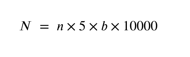
Where: n is the number of sperm in 5 squares of the Neubauer slide, b is the dilution factor, and 10,000 is the result of dividing 1 ml (1000 µl) by 0.1 µl, the volume of one of the squares.
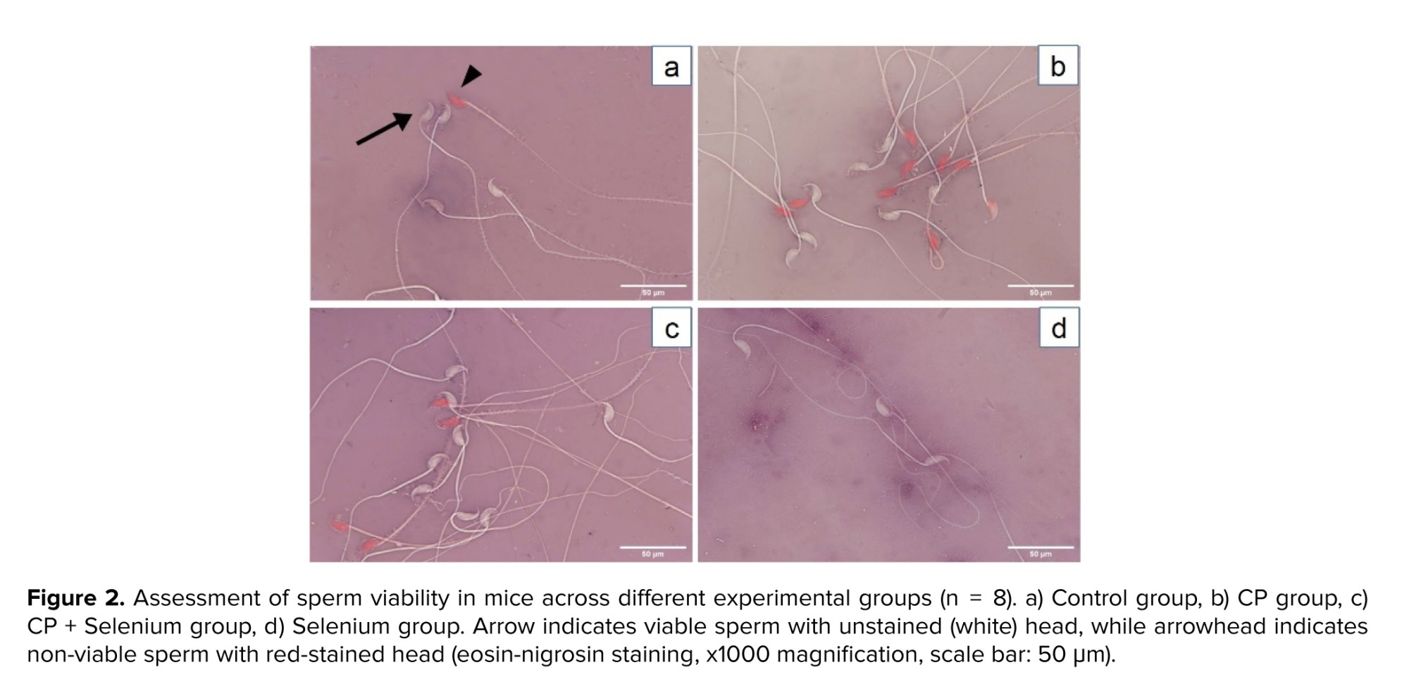
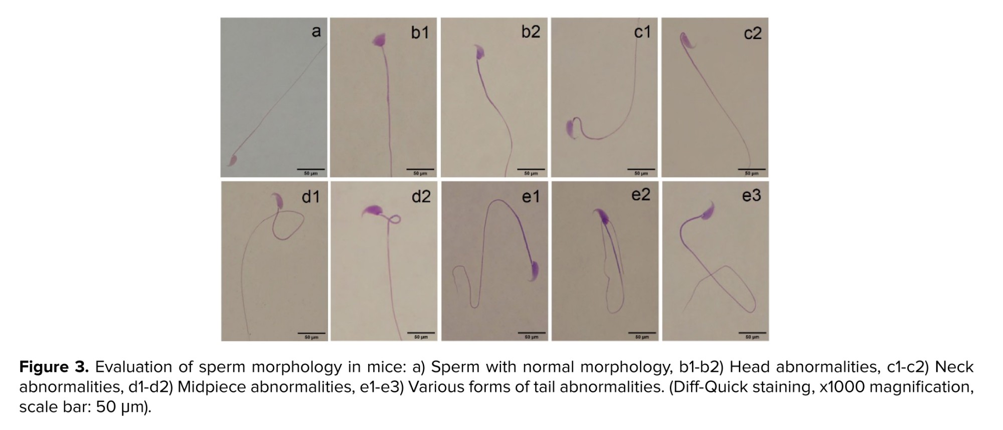
Sperm viability was assessed using 1% eosin and 10% nigrosin staining. One volume of sperm suspension was mixed with 2 volumes of eosin solution and 3 volumes of nigrosin solution, and a thin smear was prepared. After the smear dried, 200 sperm cells were examined using a light microscope at 1000× magnification. Live sperm heads appeared white to pale pink, while dead sperm heads appeared pink to red. To assess sperm morphology, the Diff-Quik staining kit (Ideh Varzan Farda Co., Iran) was used. Finally, the morphology of 200 sperm cells for each sample was examined by light microscopy at 1000× magnification, and the percentage of abnormalities was calculated and recorded (12).
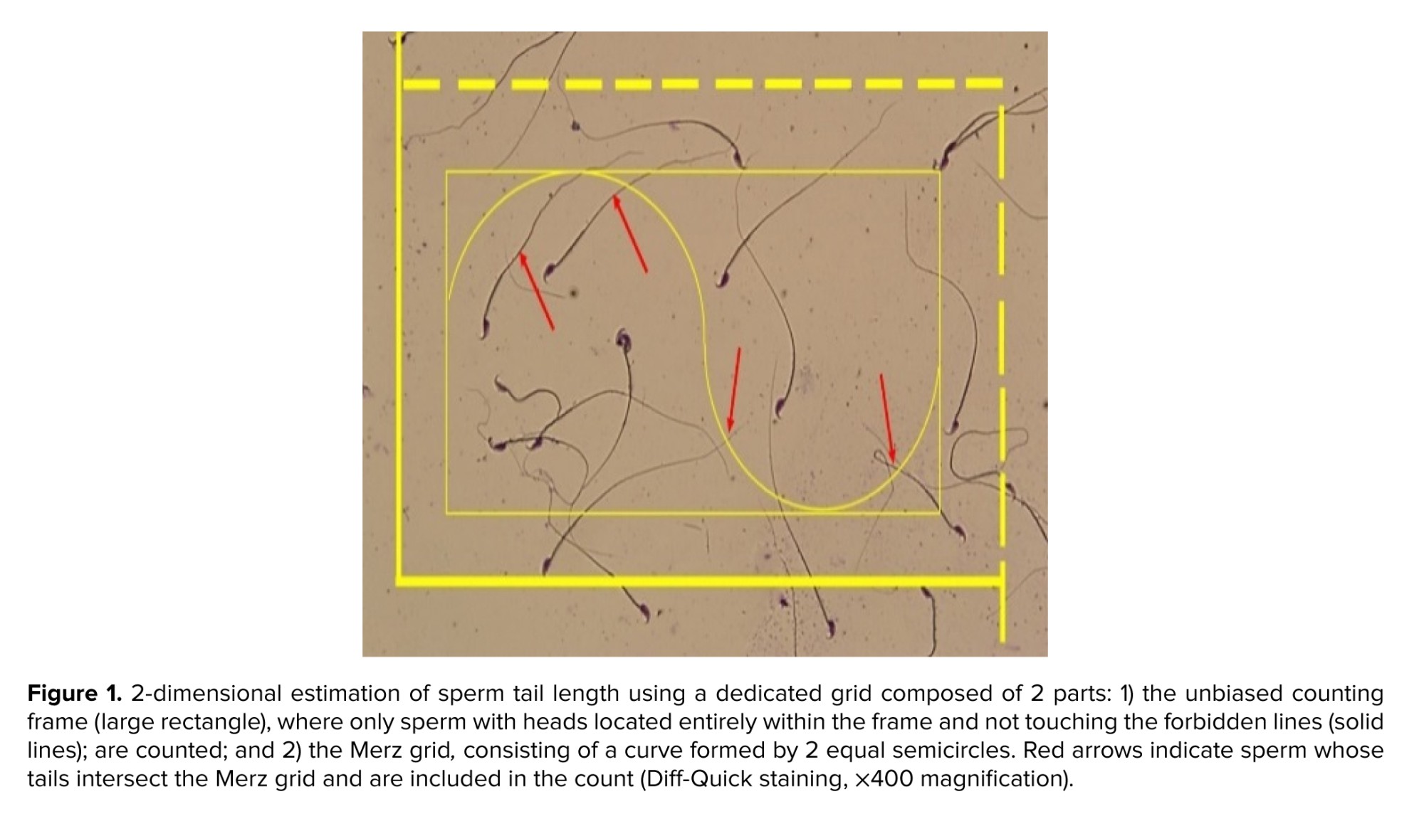
To estimate sperm tail length, Diff-Quick-stained slides were prepared, and 150-200 sperm were evaluated per slide. The measurement was performed in 2 dimensions using a specialized grid composed of 2 components. The first component was the unbiased counting frame, where only sperms with heads located entirely within the frame not touching the forbidden lines were included. The second component was the Merz grid, consisting of a curved structure with 2 equal semicircles. In this section, sperms were counted if their tails intersected the curve (Figure 1). Sperm tail length was then calculated in micrometers using the following formula:
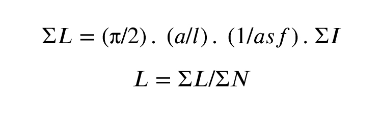
Here, a/l represents the stability coefficient of the Merz grid, asf indicates the ratio of the small frame area to the total area of the counting frame, ΣI is the total number of sperm tail contacts with the Merz grid, and ΣN is the total number of sperm counted within the counting frame (13, 14).
2.4. Sperm functional parameters
Sperm plasma membrane integrity was assessed using the Hypo-Osmotic Swelling test. Briefly, 50 μL of the sperm suspension was mixed with 500 μL of a pre-warmed hypo-osmotic solution (containing 0.735 gr sodium citrate and 1.351 gr fructose in 100 mL distilled water). The mixture was incubated at 37°C for 30 min. Subsequently, 10 μL of the incubated solution was placed on a slide, covered with a cover glass, and 200 sperm cells were counted using a light microscope at 400× magnification. The number of sperm with coiled or swollen tails, representing sperm with intact membranes, was expressed as the percentage response to osmotic environment (15).
To assess sperm nuclear maturity, the aniline blue staining method was used. In this staining procedure, prepared smears were fixed with 4% formalin solution for 10 min. After washing with water, staining was performed with a 5% aniline blue and 4% acetic acid solution. Subsequently, 200 sperm cells were examined using a light microscope at 1000× magnification. The percentage of sperm with pale blue heads was reported as the percentage of mature sperm (9).
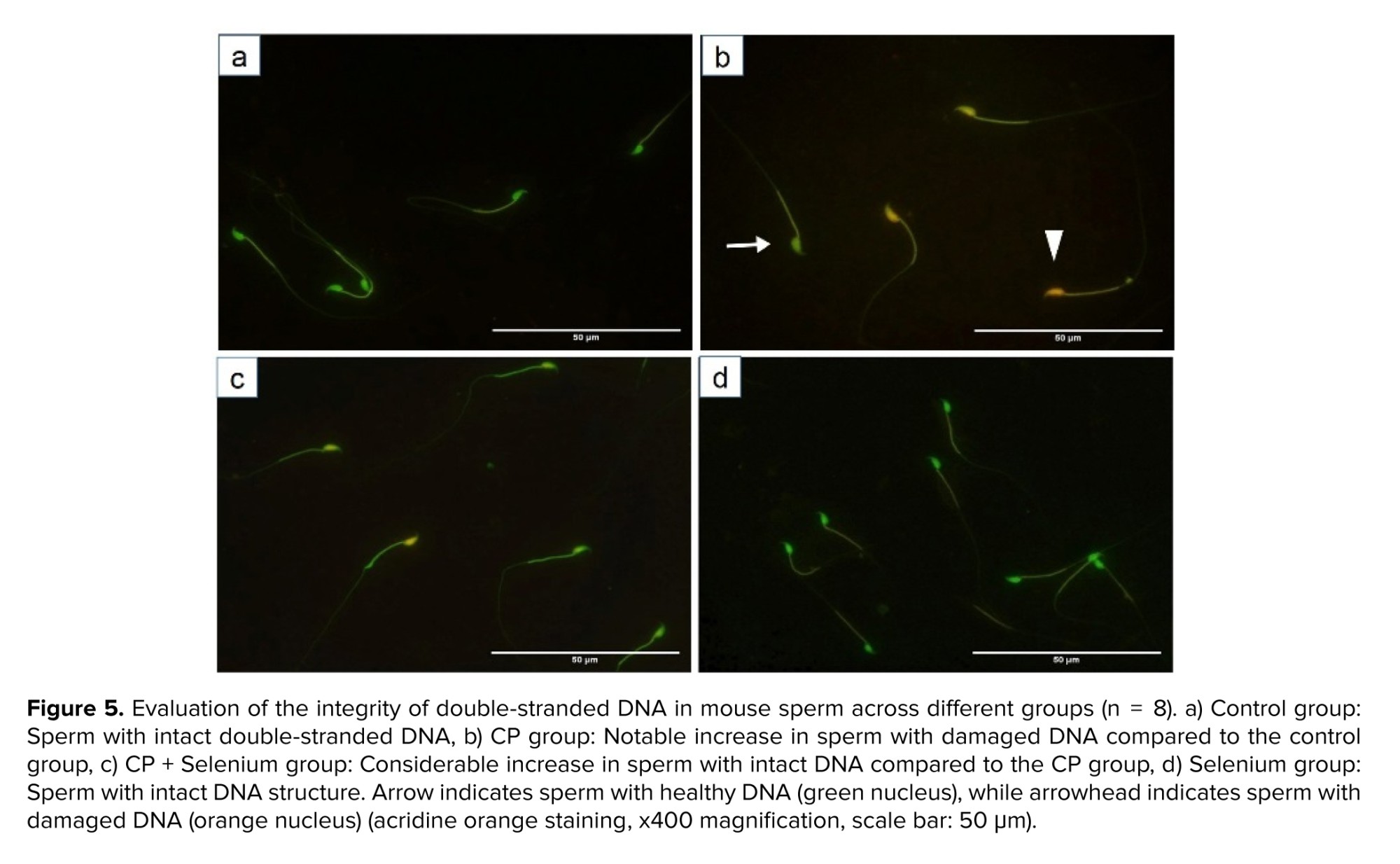
To assess the extent of sperm DNA damage, acridine orange staining was used. Smears were incubated in a methanol: glacial acetic acid solution (3:1 v/v) for 14 hr at 4°C. Subsequently, staining was performed with acridine orange solution. The percentages of sperm exhibiting orange and red fluorescence, indicative of damaged DNA, were recorded and reported using fluorescence microscopy (9).
MDA and TAC levels in serum were measured using the NalondiTM Lipid Peroxidation (MDA) Assay kit and NaxiferTM TAC Assay kit, respectively, according to the kits’ protocols. Based on the standard curve formula, MDA and TAC levels in serum were calculated. Finally, MDA concentrations were reported in nmol/ml, and TAC concentrations were reported in mmol Fe2+/L.
2.5. Daily sperm production
To assess daily sperm production, the testicular parenchyma was homogenized in normal saline containing 0.05% Triton X-100 after decapsulation. A few drops of 1% eosin were added to the solution for staining spermatids. Subsequently, 10 μL of the resulting suspension was placed on a Neubauer chamber, and the number of spermatids was counted using a light microscope at 400x magnification. Daily sperm production was then calculated by dividing the number of spermatids per gram of testicular tissue by 4.84 (the duration for which spermatids are in stages 14-16 of the seminiferous epithelium cycle before transforming into sperm) (9, 13).
2.6. Ethical Considerations
Ethical approval for the animal experiments was obtained from the ethics committee of Arak University, Arak, Iran (Code: IR.ARAKU.REC.1402.094). All ethical guidelines for the care and use of laboratory animals were followed.
2.7. Statistical Analysis
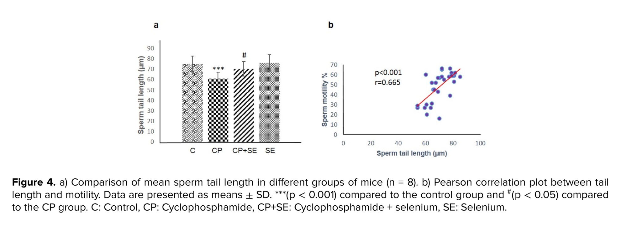
Data were statistically analyzed using SPSS software (version 22), one-way ANOVA, and Tukey’s post-hoc test. The Kolmogorov-Smirnov and Shapiro-Wilk tests were used to assess the normality of the data. The correlation between values of motility and sperm tail length was assessed by bivariate Pearson’s correlation. Differences between means were considered significant at p < 0.05.
3. Results
3.1. Evaluation of basic sperm parameters
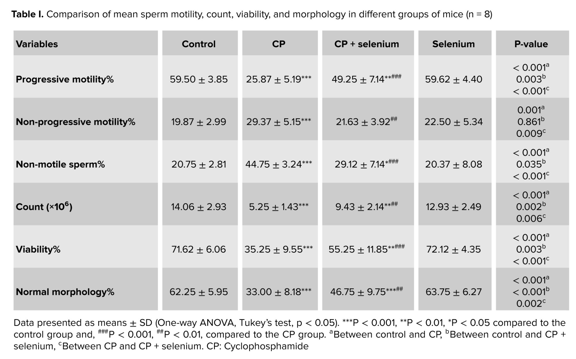
The CP group showed significantly reduced progressive motility and increased non-progressive and immotile sperm compared with controls (p < 0.001). Conversely, the CP + selenium group exhibited a higher percentage of progressively motile sperm (p < 0.001) and lower non-progressive (p < 0.01) and immotile sperm (p < 0.001) than the CP group. Sperm count decreased significantly in the CP group vs. control (p < 0.001) and increased significantly with selenium treatment (p < 0.01) (Table I).
The CP group showed significantly lower sperm viability, normal morphology, and tail length compared with the control group (p < 0.001). Selenium co‑administration significantly increased sperm viability in the CP + selenium group compared with the CP group (p < 0.001). Normal morphology also improved in the CP + selenium group relative to CP (p < 0.01). In addition, sperm tail length was significantly greater in the CP + selenium group (p < 0.05). Pearson correlation analysis revealed a significant positive correlation between progressive motility and tail length (r = 0.665, p < 0.001) (Figures 2-4 and Table I).
3.2. Evaluation of sperm functional parameters
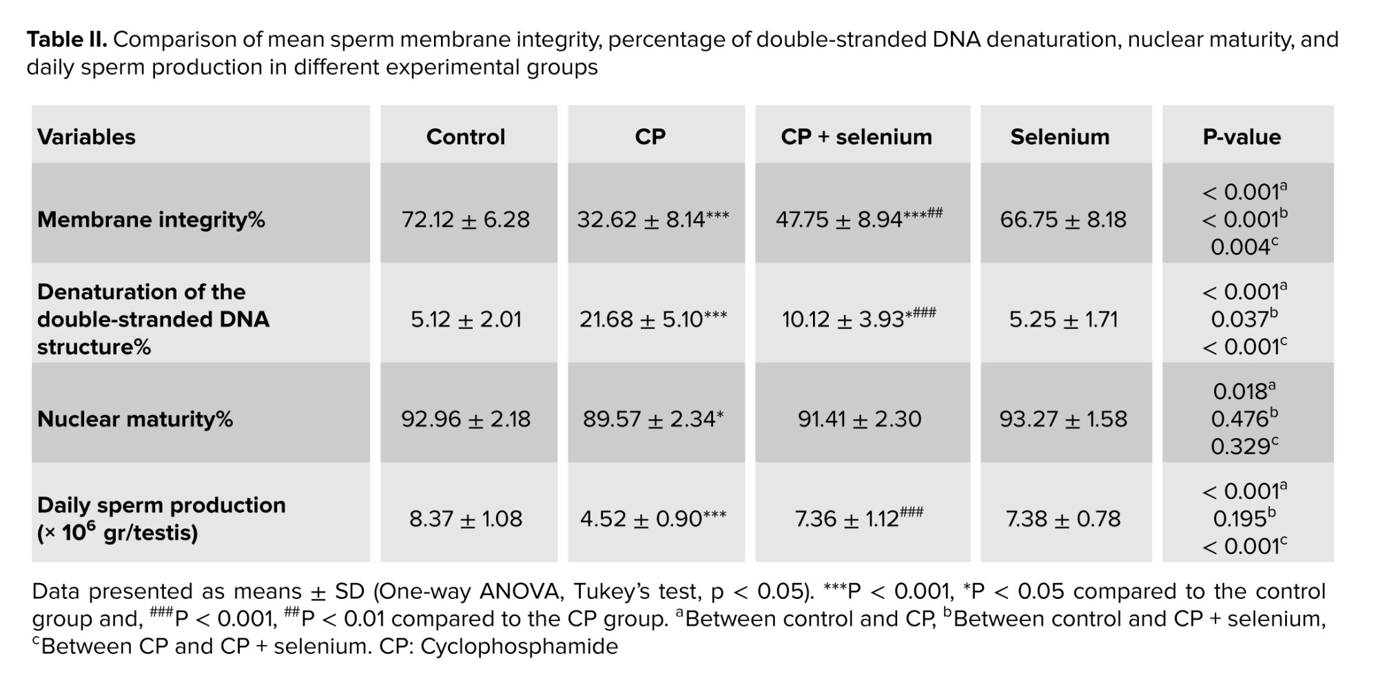
The CP group showed a significant decrease in sperm with intact membranes (p < 0.001) and nuclear maturity (p < 0.05) compared with control. The CP + selenium group exhibited a significant increase in intact membranes relative to the CP group (p < 0.01). The percentage of sperm with double-stranded DNA damage was significantly higher in the CP group than in control (p < 0.001), while this damage was significantly reduced in the CP + selenium group compared with the CP group (p < 0.001; Figure 5 and Table II).
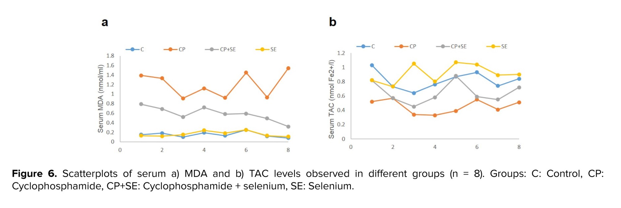
The mean serum MDA level was significantly increased in the CP group than in the control group (p < 0.001). The CP + selenium group significantly reduced serum MDA levels in the CP + selenium group compared with the CP group (p < 0.001). Serum TAC levels were significantly decreased in the CP group compared with the control group (p < 0.001). In contrast, TAC levels were significantly increased in the CP + selenium group relative to the CP group (p < 0.05), (Figure 6a and b).
3.3. Daily sperm production
The average daily sperm production was significantly reduced in the CP group compared with the control group (p < 0.001). Mean daily sperm production was significantly increased in the CP + selenium group relative to the CP group (p < 0.001) and reached control group level (Table II).
4. Discussion
In the present study, CP administration significantly reduced sperm count, motility, viability, normal morphology, membrane integrity, and daily sperm production compared with controls. These findings agree with previous studies showing that CP markedly decreases sperm count, motility, and viability in adult male mice (9, 16). CP disrupts meiosis by reducing expression of meiosis-related proteins such as synaptonemal complex protein 3, leading to abnormal sperm formation (17). Additionally, CP induces oxidative stress that damages sperm mitochondria, essential for ATP production, motility, and membrane integrity, thereby reducing sperm viability (18). Active CP metabolites, particularly acrolein, increase ROS, causing lipid peroxidation, membrane damage, and decreased motility (9). Conversely, selenium co-administration alleviated CP-induced reproductive toxicity. Mice treated with CP and selenium showed improvements in sperm count, motility, viability, morphology, membrane integrity, and daily sperm production compared with the CP group. These results align with previous studies reporting selenium’s protective effects. Selenium supplementation improved sperm parameters in a rat model of varicocele (19) and enhanced sperm count, motility, and viability in male mice exposed to bisphenol A (20). As a component of antioxidant enzymes like glutathione peroxidase, selenium reduces oxidative stress, protects sperm membranes and DNA, enhances mitochondrial function, and improves motility and fertilization potential (19). Selenium has been shown to promote spermatogenesis by increasing testosterone levels and stimulating sperm production (20). It is also essential for antioxidant enzymes such as catalase, which eliminate hydrogen peroxide, protect sperm from structural damage, and support improvements in sperm morphology, quality, and function (21). In the our study, the mean tail length of sperm in the CP-treated group was significantly reduced compared with controls, and correlation analysis revealed a direct relationship between tail length and sperm motility. CP induces free radical production, causing lipid peroxidation of sperm plasma membranes, damaging mitochondrial structures, and reducing ATP levels, which leads to impaired motility and viability (18, 22). The mid-piece, containing the mitochondrial sheath, is critical for energy generation required for motility (23). Therefore, oxidative stress and mitochondrial damage in the CP group are likely major contributors to reduced motility. In contrast, selenium co-treatment significantly increased sperm tail length and improved motility. Selenium enhances the antioxidant defense system, reducing oxidative damage to membranes, preventing structural abnormalities, and activating enzymes that maintain membrane integrity (24, 25). Selenium deficiency is associated with mid-piece fragility and reduced motility (26). By supporting these protective mechanisms, selenium preserves mid-piece structure and sustains sperm motility under oxidative stress (24, 25).
In the this study, the CP-treated group showed a significant increase in DNA double-strand denaturation and a marked decrease in sperm nuclear maturation compared with controls. These findings align with previous reports showing that CP reduces the expression of DNA double-strand break repair genes in male mice (16) and significantly decreases sperm nuclear maturation (27). CP, an alkylating agent, induces DNA damage by attaching alkyl groups to DNA bases and generating ROS (28). If unrepaired, this damage can lead to double-strand breaks. Normally, sperm nuclear maturation involves replacing primary nuclear proteins with protamines and other sperm-specific proteins. CP disrupts this process, resulting in retention of abnormal nuclear proteins and increased aniline blue staining, indicative of chromatin immaturity (9). Conversely, co-treatment with CP and selenium significantly reduced DNA double-strand denaturation compared with CP group. Selenium, a key component of antioxidant enzymes such as glutathione peroxidase, reduces oxidative stress by neutralizing free radicals and peroxides, thereby protecting DNA. Selenium also reduces nitric oxide levels, another DNA-damaging factor, and may enhance DNA repair mechanisms through its antioxidant properties (4). These protective effects preserve sperm nuclear integrity and support proper chromatin maturation under CP-induced oxidative stress. We also found that, CP treatment significantly increased serum MDA levels and decreased TAC compared with controls. These findings are consistent with previous studies (16, 29). The active CP metabolite acrolein generates ROS that attack membrane lipids, causing lipid peroxidation and elevated MDA levels (18). CP also reduces the activity of key antioxidant enzymes, including superoxide dismutase, catalase, and glutathione peroxidase, leading to ROS accumulation and further lipid peroxidation (16, 29). In mice co-treated with CP and selenium, MDA levels significantly decreased, while TAC increased compared with the CP group. Similar studies have shown that selenium reduces the toxic effects of acrylamide in male rats by lowering MDA levels and enhancing antioxidant enzyme activity in testicular tissue (7). Selenium, as an essential trace element, is a structural component of antioxidant enzymes such as glutathione peroxidase, which neutralize lipid peroxides and prevent MDA formation (19). Additionally, selenium strengthens the body’s antioxidant defense by activating enzymes, including glutathione peroxidase and superoxide dismutase, increasing antioxidant vitamins such as vitamin E, and protecting against oxidative damage and lipid peroxidation (4). These mechanisms underlie selenium’s protective and antioxidant effects.
5. Conclusion
CP harms male reproductive health by increasing oxidative stress, inflammation, and DNA damage, reducing sperm quality. The results demonstrated that selenium co-treatment mitigates these effects by protecting DNA, reducing oxidative damage, and enhancing antioxidant capacity.
Data Availability
Data will be made available on request.
Author Contributions
M. Soleimani Mehranjani: Conceptualization, project administration, supervision, formal analysis, writing-review, and editing. S. Garavand: Methodology, data curation. S. Ahmadi: Methodology, data curation, formal analysis, and writing-original draft. All authors read and approved the final manuscript.
Acknowledgments
The authors would like to acknowledge financial support provided by the Deputy of Research and Technology and express their appreciation to the Research Laboratory of the Department of Biology, Faculty of Basic Sciences, Arak University, Arak, Iran. Artificial intelligence was not used in this study.
Conflict of Interest
The authors declare that there is no conflict of interest.
Cyclophosphamide (CP) is a widely used chemotherapy drug for treating various cancers and some autoimmune diseases (1). In the body, it is metabolized by cytochrome P450 enzymes into active metabolites, including 4-hydroxycyclophosphamide. This unstable metabolite is further converted to aldophosphamide and then to phosphoramide mustard and acrolein. Phosphoramide mustard is primarily responsible for DNA damage, whereas acrolein is a toxic metabolite that induces inflammation and tissue injury, especially in the reproductive tract (2). Acrolein promotes oxidative stress by depleting glutathione and increasing lipid peroxidation (3). Elevated oxidative stress has been shown to reduce intracellular antioxidants in Leydig cells, impair testosterone synthesis and secretion, decrease reproductive organ weight, disrupt spermatogenesis, and ultimately lower sperm count (1). Selenium, an essential micronutrient and potent antioxidant, plays a key role in improving sperm parameters and supporting normal spermatogenesis (4). It is a structural component of glutathione peroxidase, which, together with glutathione, neutralizes reactive oxygen species (ROS) such as hydrogen peroxide and lipid peroxides, protecting DNA, cell membranes, and proteins in testicular tissue, and supporting overall testicular health and function (5). Experimental studies have confirmed selenium’s protective effects. For instance, co-administration of selenium with doxorubicin reduced oxidative stress and apoptosis, improved sperm quality, and preserved normal spermatogenesis in mice (6). Another study showed that selenium mitigated acrylamide-induced testicular damage by reducing oxidative stress, regulating steroidogenesis-related gene expression, and enhancing tissue quality (7). Additionally, selenium protected testicular structure against the adverse effects of venlafaxine hydrochloride in male mice by strengthening antioxidant defenses and lowering apoptotic protein expression (8). This study aimed to evaluate the effects of selenium on sperm quality and related parameters in mice following treatment with CP.
2. Materials and Methods
2.1. Animals and treatment
In this experimental study, each mouse was considered an experimental unit. Mice were chosen for their well-established reproductive physiology and widespread use as a model organism in biomedical research, allowing for extensive comparative data. The NMRI strain is a commonly used outbred stock in toxicology and reproductive studies, known for its robustness and predictable response to various chemical agents.
32 adult male NMRI mice (6 wk, 36 ± 2 gr) were obtained from the animal house of Arak University, Arak, Iran and kept under favorable conditions (21°C, 12-hr dark/light cycle). This study did not establish specific humane endpoints for experimental animals. The health and well-being of the mice were monitored throughout the study. The animals were randomly divided into 4 groups (n = 8/each): control (mice in this group received no treatment. They were maintained under identical conditions to the experimental groups), CP (100 mg/kg/wk, Sigma, Cat. No: C0768) (9), selenium (1 mg/kg/day, sodium selenite; Sigma, Cat. No: 6214485) (10) and CP + selenium. The treatments were administered intraperitoneally for 35 days.
All experiments of this study were conducted from April to December 2024 in the research laboratory of the Department of Biology, Faculty of Basic Sciences, Arak University, Arak, Iran.
2.1.1. Sample size
A priori power analysis was conducted to estimate the minimum sample size required to detect a statistically significant effect. Assuming an effect size, α = 0.05, and power (1-β) = 0.8, the analysis indicated that n = 8/group would be sufficient to achieve adequate statistical power. These calculations were performed using the Cochran table.
Randomization was used to allocate experimental units to control and treatment groups. After determining the required sample size (n = 8/group), the 32 purchased mice were randomly assigned to 4 groups of 8, ensuring no significant differences in initial average body weight among the groups.
To minimize potential confounders related to the order of treatments and measurements or animal location and handling, the following strategies were employed: 1) animals were weighed once weekly. The timing of weight measurement was consistent across all groups and study days. Doses for drugs were calculated and adjusted based on individual animal’s body weight recorded during the weekly measurement. 2) cages were randomly assigned to racks within the animal facility, and their positions were rotated weekly to account for any potential micro-environmental variations. 3) all procedures, including weighing and treatment administration, were performed by the trained researcher to ensure consistency and minimize handling-related variability.
Furthermore, to minimize potential bias, a double-blinding strategy was implemented throughout the experiment.
2.2. Animal dissection
At the end of the treatment period, the mice were anesthetized with ketamine and xylazine, and blood was collected from the heart through cardiac puncture after opening the chest cavity. Blood samples were centrifuged in 2 consecutive steps at 13,000 rpm for 10 and 5 min, respectively. The resulting serum was collected and stored at -80°C for the evaluation of malondialdehyde (MDA) and total antioxidant capacity (TAC) levels. Then, by opening the sclerotome, the left testicle was separated from the body and stored in a nitrogen tank (liquid nitrogen) to calculate daily sperm production. The caudal epididymis was cut in Hams’ F10 medium to release spermatozoa for sperm parameter analysis.
2.3. Basic sperm parameters
Following sperm release into the culture medium, 10 μL of the sperm-containing suspension was transferred to a Neubauer slide. The percentages of progressive motile sperms, non-progressive motile sperms, and immotile sperms were then determined by analyzing a minimum of 200 sperm across at least 5 microscopic fields, using a light microscope (Olympus BH- 2, Japan) at 400× magnification (11). For sperm counting, 100 μL of the suspension was mixed with 900 μL of a fixative solution containing 5% sodium bicarbonate and 2% formaldehyde. Subsequently, 10 μL of this mixture was applied to a Neubauer slide. Spermatozoa within the 4 large corner squares and the central square of the hemocytometer were counted using a light microscope at 400× magnification (11). To calculate the sperm count at 1 ml, the following formula was used:
Where: n is the number of sperm in 5 squares of the Neubauer slide, b is the dilution factor, and 10,000 is the result of dividing 1 ml (1000 µl) by 0.1 µl, the volume of one of the squares.
Sperm viability was assessed using 1% eosin and 10% nigrosin staining. One volume of sperm suspension was mixed with 2 volumes of eosin solution and 3 volumes of nigrosin solution, and a thin smear was prepared. After the smear dried, 200 sperm cells were examined using a light microscope at 1000× magnification. Live sperm heads appeared white to pale pink, while dead sperm heads appeared pink to red. To assess sperm morphology, the Diff-Quik staining kit (Ideh Varzan Farda Co., Iran) was used. Finally, the morphology of 200 sperm cells for each sample was examined by light microscopy at 1000× magnification, and the percentage of abnormalities was calculated and recorded (12).
To estimate sperm tail length, Diff-Quick-stained slides were prepared, and 150-200 sperm were evaluated per slide. The measurement was performed in 2 dimensions using a specialized grid composed of 2 components. The first component was the unbiased counting frame, where only sperms with heads located entirely within the frame not touching the forbidden lines were included. The second component was the Merz grid, consisting of a curved structure with 2 equal semicircles. In this section, sperms were counted if their tails intersected the curve (Figure 1). Sperm tail length was then calculated in micrometers using the following formula:
Here, a/l represents the stability coefficient of the Merz grid, asf indicates the ratio of the small frame area to the total area of the counting frame, ΣI is the total number of sperm tail contacts with the Merz grid, and ΣN is the total number of sperm counted within the counting frame (13, 14).
2.4. Sperm functional parameters
Sperm plasma membrane integrity was assessed using the Hypo-Osmotic Swelling test. Briefly, 50 μL of the sperm suspension was mixed with 500 μL of a pre-warmed hypo-osmotic solution (containing 0.735 gr sodium citrate and 1.351 gr fructose in 100 mL distilled water). The mixture was incubated at 37°C for 30 min. Subsequently, 10 μL of the incubated solution was placed on a slide, covered with a cover glass, and 200 sperm cells were counted using a light microscope at 400× magnification. The number of sperm with coiled or swollen tails, representing sperm with intact membranes, was expressed as the percentage response to osmotic environment (15).
To assess sperm nuclear maturity, the aniline blue staining method was used. In this staining procedure, prepared smears were fixed with 4% formalin solution for 10 min. After washing with water, staining was performed with a 5% aniline blue and 4% acetic acid solution. Subsequently, 200 sperm cells were examined using a light microscope at 1000× magnification. The percentage of sperm with pale blue heads was reported as the percentage of mature sperm (9).
To assess the extent of sperm DNA damage, acridine orange staining was used. Smears were incubated in a methanol: glacial acetic acid solution (3:1 v/v) for 14 hr at 4°C. Subsequently, staining was performed with acridine orange solution. The percentages of sperm exhibiting orange and red fluorescence, indicative of damaged DNA, were recorded and reported using fluorescence microscopy (9).
MDA and TAC levels in serum were measured using the NalondiTM Lipid Peroxidation (MDA) Assay kit and NaxiferTM TAC Assay kit, respectively, according to the kits’ protocols. Based on the standard curve formula, MDA and TAC levels in serum were calculated. Finally, MDA concentrations were reported in nmol/ml, and TAC concentrations were reported in mmol Fe2+/L.
2.5. Daily sperm production
To assess daily sperm production, the testicular parenchyma was homogenized in normal saline containing 0.05% Triton X-100 after decapsulation. A few drops of 1% eosin were added to the solution for staining spermatids. Subsequently, 10 μL of the resulting suspension was placed on a Neubauer chamber, and the number of spermatids was counted using a light microscope at 400x magnification. Daily sperm production was then calculated by dividing the number of spermatids per gram of testicular tissue by 4.84 (the duration for which spermatids are in stages 14-16 of the seminiferous epithelium cycle before transforming into sperm) (9, 13).
2.6. Ethical Considerations
Ethical approval for the animal experiments was obtained from the ethics committee of Arak University, Arak, Iran (Code: IR.ARAKU.REC.1402.094). All ethical guidelines for the care and use of laboratory animals were followed.
2.7. Statistical Analysis
Data were statistically analyzed using SPSS software (version 22), one-way ANOVA, and Tukey’s post-hoc test. The Kolmogorov-Smirnov and Shapiro-Wilk tests were used to assess the normality of the data. The correlation between values of motility and sperm tail length was assessed by bivariate Pearson’s correlation. Differences between means were considered significant at p < 0.05.
3. Results
3.1. Evaluation of basic sperm parameters
The CP group showed significantly reduced progressive motility and increased non-progressive and immotile sperm compared with controls (p < 0.001). Conversely, the CP + selenium group exhibited a higher percentage of progressively motile sperm (p < 0.001) and lower non-progressive (p < 0.01) and immotile sperm (p < 0.001) than the CP group. Sperm count decreased significantly in the CP group vs. control (p < 0.001) and increased significantly with selenium treatment (p < 0.01) (Table I).
The CP group showed significantly lower sperm viability, normal morphology, and tail length compared with the control group (p < 0.001). Selenium co‑administration significantly increased sperm viability in the CP + selenium group compared with the CP group (p < 0.001). Normal morphology also improved in the CP + selenium group relative to CP (p < 0.01). In addition, sperm tail length was significantly greater in the CP + selenium group (p < 0.05). Pearson correlation analysis revealed a significant positive correlation between progressive motility and tail length (r = 0.665, p < 0.001) (Figures 2-4 and Table I).
3.2. Evaluation of sperm functional parameters
The CP group showed a significant decrease in sperm with intact membranes (p < 0.001) and nuclear maturity (p < 0.05) compared with control. The CP + selenium group exhibited a significant increase in intact membranes relative to the CP group (p < 0.01). The percentage of sperm with double-stranded DNA damage was significantly higher in the CP group than in control (p < 0.001), while this damage was significantly reduced in the CP + selenium group compared with the CP group (p < 0.001; Figure 5 and Table II).
The mean serum MDA level was significantly increased in the CP group than in the control group (p < 0.001). The CP + selenium group significantly reduced serum MDA levels in the CP + selenium group compared with the CP group (p < 0.001). Serum TAC levels were significantly decreased in the CP group compared with the control group (p < 0.001). In contrast, TAC levels were significantly increased in the CP + selenium group relative to the CP group (p < 0.05), (Figure 6a and b).
3.3. Daily sperm production
The average daily sperm production was significantly reduced in the CP group compared with the control group (p < 0.001). Mean daily sperm production was significantly increased in the CP + selenium group relative to the CP group (p < 0.001) and reached control group level (Table II).
4. Discussion
In the present study, CP administration significantly reduced sperm count, motility, viability, normal morphology, membrane integrity, and daily sperm production compared with controls. These findings agree with previous studies showing that CP markedly decreases sperm count, motility, and viability in adult male mice (9, 16). CP disrupts meiosis by reducing expression of meiosis-related proteins such as synaptonemal complex protein 3, leading to abnormal sperm formation (17). Additionally, CP induces oxidative stress that damages sperm mitochondria, essential for ATP production, motility, and membrane integrity, thereby reducing sperm viability (18). Active CP metabolites, particularly acrolein, increase ROS, causing lipid peroxidation, membrane damage, and decreased motility (9). Conversely, selenium co-administration alleviated CP-induced reproductive toxicity. Mice treated with CP and selenium showed improvements in sperm count, motility, viability, morphology, membrane integrity, and daily sperm production compared with the CP group. These results align with previous studies reporting selenium’s protective effects. Selenium supplementation improved sperm parameters in a rat model of varicocele (19) and enhanced sperm count, motility, and viability in male mice exposed to bisphenol A (20). As a component of antioxidant enzymes like glutathione peroxidase, selenium reduces oxidative stress, protects sperm membranes and DNA, enhances mitochondrial function, and improves motility and fertilization potential (19). Selenium has been shown to promote spermatogenesis by increasing testosterone levels and stimulating sperm production (20). It is also essential for antioxidant enzymes such as catalase, which eliminate hydrogen peroxide, protect sperm from structural damage, and support improvements in sperm morphology, quality, and function (21). In the our study, the mean tail length of sperm in the CP-treated group was significantly reduced compared with controls, and correlation analysis revealed a direct relationship between tail length and sperm motility. CP induces free radical production, causing lipid peroxidation of sperm plasma membranes, damaging mitochondrial structures, and reducing ATP levels, which leads to impaired motility and viability (18, 22). The mid-piece, containing the mitochondrial sheath, is critical for energy generation required for motility (23). Therefore, oxidative stress and mitochondrial damage in the CP group are likely major contributors to reduced motility. In contrast, selenium co-treatment significantly increased sperm tail length and improved motility. Selenium enhances the antioxidant defense system, reducing oxidative damage to membranes, preventing structural abnormalities, and activating enzymes that maintain membrane integrity (24, 25). Selenium deficiency is associated with mid-piece fragility and reduced motility (26). By supporting these protective mechanisms, selenium preserves mid-piece structure and sustains sperm motility under oxidative stress (24, 25).
In the this study, the CP-treated group showed a significant increase in DNA double-strand denaturation and a marked decrease in sperm nuclear maturation compared with controls. These findings align with previous reports showing that CP reduces the expression of DNA double-strand break repair genes in male mice (16) and significantly decreases sperm nuclear maturation (27). CP, an alkylating agent, induces DNA damage by attaching alkyl groups to DNA bases and generating ROS (28). If unrepaired, this damage can lead to double-strand breaks. Normally, sperm nuclear maturation involves replacing primary nuclear proteins with protamines and other sperm-specific proteins. CP disrupts this process, resulting in retention of abnormal nuclear proteins and increased aniline blue staining, indicative of chromatin immaturity (9). Conversely, co-treatment with CP and selenium significantly reduced DNA double-strand denaturation compared with CP group. Selenium, a key component of antioxidant enzymes such as glutathione peroxidase, reduces oxidative stress by neutralizing free radicals and peroxides, thereby protecting DNA. Selenium also reduces nitric oxide levels, another DNA-damaging factor, and may enhance DNA repair mechanisms through its antioxidant properties (4). These protective effects preserve sperm nuclear integrity and support proper chromatin maturation under CP-induced oxidative stress. We also found that, CP treatment significantly increased serum MDA levels and decreased TAC compared with controls. These findings are consistent with previous studies (16, 29). The active CP metabolite acrolein generates ROS that attack membrane lipids, causing lipid peroxidation and elevated MDA levels (18). CP also reduces the activity of key antioxidant enzymes, including superoxide dismutase, catalase, and glutathione peroxidase, leading to ROS accumulation and further lipid peroxidation (16, 29). In mice co-treated with CP and selenium, MDA levels significantly decreased, while TAC increased compared with the CP group. Similar studies have shown that selenium reduces the toxic effects of acrylamide in male rats by lowering MDA levels and enhancing antioxidant enzyme activity in testicular tissue (7). Selenium, as an essential trace element, is a structural component of antioxidant enzymes such as glutathione peroxidase, which neutralize lipid peroxides and prevent MDA formation (19). Additionally, selenium strengthens the body’s antioxidant defense by activating enzymes, including glutathione peroxidase and superoxide dismutase, increasing antioxidant vitamins such as vitamin E, and protecting against oxidative damage and lipid peroxidation (4). These mechanisms underlie selenium’s protective and antioxidant effects.
5. Conclusion
CP harms male reproductive health by increasing oxidative stress, inflammation, and DNA damage, reducing sperm quality. The results demonstrated that selenium co-treatment mitigates these effects by protecting DNA, reducing oxidative damage, and enhancing antioxidant capacity.
Data Availability
Data will be made available on request.
Author Contributions
M. Soleimani Mehranjani: Conceptualization, project administration, supervision, formal analysis, writing-review, and editing. S. Garavand: Methodology, data curation. S. Ahmadi: Methodology, data curation, formal analysis, and writing-original draft. All authors read and approved the final manuscript.
Acknowledgments
The authors would like to acknowledge financial support provided by the Deputy of Research and Technology and express their appreciation to the Research Laboratory of the Department of Biology, Faculty of Basic Sciences, Arak University, Arak, Iran. Artificial intelligence was not used in this study.
Conflict of Interest
The authors declare that there is no conflict of interest.
Type of Study: Original Article |
Subject:
Reproductive Biology
Send email to the article author
| Rights and permissions | |
 |
This work is licensed under a Creative Commons Attribution-NonCommercial 4.0 International License. |