
International Journal of
Reproductive Biomedicine
Fri, Jun 12, 2026
[Archive]
Volume 24, Issue 3 (March 2026)
IJRM 2026, 24(3): 181-192 |
Back to browse issues page


Ethics code: 2.KEH.158.11.2024
![]()
![]()
![]()
Download citation:
BibTeX | RIS | EndNote | Medlars | ProCite | Reference Manager | RefWorks
Send citation to:


BibTeX | RIS | EndNote | Medlars | ProCite | Reference Manager | RefWorks
Send citation to:
Herawati U N, Annas J Y, Widjiati W. Citrus limon peel extract versus metformin in testosterone-induced polycystic ovary syndrome: An experimental study. IJRM 2026; 24 (3) :181-192
URL: http://ijrm.ir/article-1-3751-en.html
URL: http://ijrm.ir/article-1-3751-en.html


1- Master Program of Reproductive Health Science, Faculty of Medicine, Universitas Airlangga, Surabaya, Indonesia. , ulfanadiya27@gmail.com; ulfa.nadiya.herawati-2022@fk.unair.ac.id
2- Master Program of Reproductive Health Science, Faculty of Medicine, Universitas Airlangga, Surabaya, Indonesia. & Department of Obstetrics and Gynecology, Faculty of Medicine, Universitas Airlangga, Surabaya, Indonesia.
3- Department of Veterinary Science, Faculty of Veterinary Medicine, Universitas Airlangga, Surabaya, Indonesia.
Full-Text [PDF 1060 kb]
(201 Downloads)
| Abstract (HTML) (165 Views)
Full-Text: (14 Views)
1. Introduction
Polycystic ovary syndrome (PCOS) is an endocrine disorder commonly found in women of reproductive age (15-49 yr) and accounts for 80% of female infertility factors (1, 2). Globally, PCOS cases have increased by 58% from 1990-2021 and are predicted to continue rising by 10.87% by 2036 (3). PCOS is not limited to endocrine disorders but also carries the risk of cardiometabolic disorders, especially in PCOS with a hyperandrogen phenotype (4). The development of comorbidities begins with insulin resistance, which occurs in up to 95% of women with PCOS and has been associated with low-grade inflammation (5).
Appropriate and sustained management is needed to prevent the long-term effects of PCOS. Metformin has long been used in the management of PCOS because it has been proven to increase insulin sensitivity, lower androgen levels, and reduce inflammation by decreasing pro-inflammatory cytokines, such as tumor necrosis factor-α (TNF-α) (6, 7). Although metformin has been proven effective, its use often causes gastrointestinal side effects such as nausea, bloating, vomiting, and loss of appetite, with approximately 75% of patients reporting these complaints, which can ultimately reduce treatment adherence (8, 9). Therefore, a safe alternative with minimal side effects but equivalent therapeutic efficacy is needed.
Since the 1960s, various PCOS animal models have been developed, with Wistar rats most widely used due to their genetic stability and reproductive characteristics. Induction with testosterone propionate (TP) is considered valid, as it elevates Cyp17a1 expression, promotes androgen overproduction, and leads to ovarian changes resembling human PCOS, including follicular cysts, reduced granulosa cells, absence of corpus luteum, and ovulatory disorders (10, 11).
One promising natural candidate is lemon peel (Citrus limon) because it contains various bioactive compounds that may reduce inflammation, increase glycogen synthesis, and improve symptoms of hyperandrogenism in PCOS (12, 13). Previous studies demonstrated that lemon peel extract (LPE) reduces nuclear factor kappa-β (NF-κB) expression and TNF-α levels in rats with premature ovarian failure and rheumatoid arthritis (14, 15). These studies explained that LPE has similar anti-inflammatory activity to metformin (16). It is plausible to hypothesize that LPE may have comparable potential to metformin, and their combination warrants further exploration for possible synergistic effects. Therefore, this study aims to investigate the effects of LPE, metformin, and their combination on inflammatory and glucose indicators in a rat model of PCOS, by measuring TNF-α levels, homeostatic model assessment of insulin resistance (HOMA-IR) scores, and fasting blood glucose (FBG).
2. Materials and Methods
2.1. Study design
This is an experimental study with a randomized post-test-only control group design, which aims to evaluate the effects of LPE, metformin extended release (XR), and their combination on TNF-α levels, HOMA-IR scores, and FBG in PCOS model rats (PCOS rats).
2.2. Sample size and randomization
2.2. Sample size and randomization
The sample size was determined using a power analysis approach. The standard deviation (σ) and the expected mean difference (Δ) were derived from a previously published study (17). Based on standard deviation of 1.41, a mean difference of 2.4, 2-sided significance level (α) of 0.05, and statistical power of 80%, the minimum required sample size was 8 subjects per group.
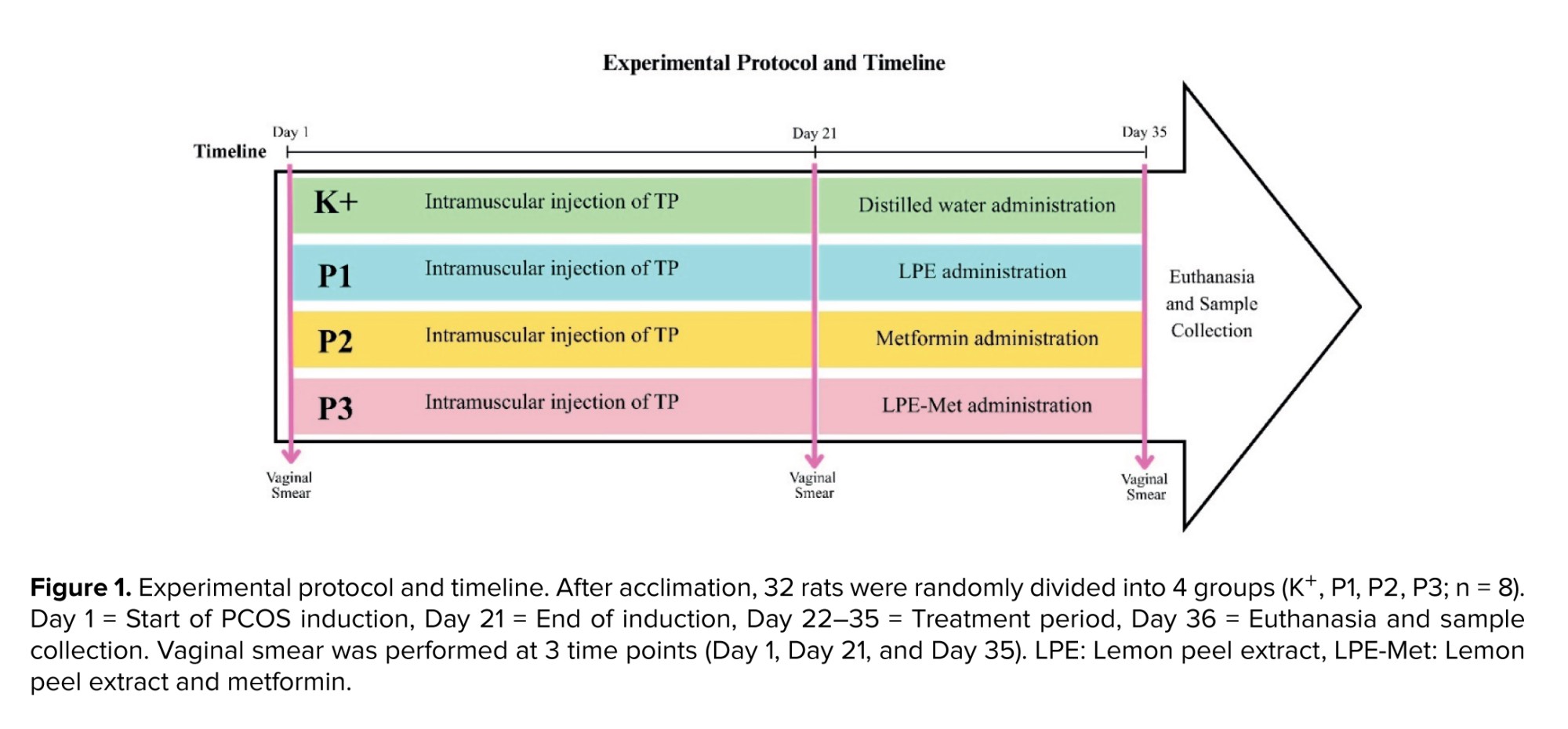
Therefore, 32 rats were used in this study. The technique used was simple randomization method by marking the rats with numbers 1-32, then randomly selecting them and dividing them into 4 groups (n = 8): 1) PCOS rats without treatment (K+), 2) PCOS rats with LPE therapy (P1), 3) PCOS rats with metformin XR therapy (P2), 4) PCOS rats with combination of LPE and metformin (LPE-Met) therapy (P3).
2.3. Experimental animals
The research subjects were Rattus norvegicus Wistar strain rats obtained from the Laboratory of the Faculty of Veterinary Medicine, Universitas Airlangga, Surabaya, Indonesia. The inclusion criteria were determined before the study to ensure appropriate subject selection. Therefore, no exclusion criteria were applied. The rats were selected based on the following criteria: 1) female, 2) age 12 wk, 3) body weight 130-170 gr, 4) normal activity and behavior. Rats that died or refused to eat during the study were excluded from the experiment, along with any previously recorded data.
2.4. Experimental procedures
24.1. Preparation of LPE
2.4. Experimental procedures
24.1. Preparation of LPE
Lemon fruits (Citrus limon) aged 4 months were obtained from the Mulyant farm, Kediri, Indonesia. The authenticity of those herbal materials was validated by the UPT Materia Medica Herbal Laboratory, Batu, Indonesia (Sample code: 241008.P.G.1015). The LPE process was subsequently carried out in the same laboratory. The extraction method was adjusted to the procedures applicable in the laboratory and based on BPOM Regulation No. 32 of 2019.
Fresh lemon was peeled, weighed, washed, and cut into small pieces, then dried in an oven at 50°C for 12 hr. After drying, the material was ground and filtered through a 60-mesh sieve to obtain powdered simplisia. The simplisia was extracted using the maceration method with 96% ethanol (1:10) for 3 days at room temperature, then evaporated using a rotary evaporator at 50°C for 15 hr to produce a thick extract.
2.4.2. Rats acclimation
Rats were acclimatized for 7 days in the same laboratory where the study was conducted and where the rats were obtained. They were housed in a comfortable, quiet laboratory with good ventilation, at room temperatures between 25 and 27°C and were placed in plastic cages lined with wood shavings. The rats were maintained on a 12-hr light/dark cycle and received ad libitum feed and water.
2.4.3. Induction of PCOS
2.4.3. Induction of PCOS
The PCOS rats were created by administering intramuscular injections of TP at a dose of 10 mg/kg for 21 days to all groups, including the control group. This method refers to previous research reporting that TP administration in female rats not only successfully induced the PCOS phenotype but also caused inflammation and metabolic disorders (17, 18). Vaginal swabs were taken from all groups before and after PCOS induction (on day 1 and day 21).
2.4.4. Treatment administration
2.4.4. Treatment administration
This study consisted of 4 therapy administrations, which began simultaneously after the 21 day PCOS induction was completed (Figure 1). Therapy administration started on day 22 and was given for 14 consecutive days with the following details: 1) the control group (K+) received 1 mL of distilled water, 2) group P1 received LPE at a dose of 500 mg/kg, 3) group P2 received metformin at a dose of 200 mg/kg, and 4) group P3 received a combination of 500 mg/kg LPE + 200 mg/kg metformin.
All treatments were administered orally using a feeding tube. The dosage and duration of LPE administration were based on a previous study, which recommended a safe dosage of 500 mg/kg for 14 days (19). Meanwhile, the metformin dosage was based on a previous study and on the safe limit for rats, 200 mg/kg (17). On day 35, vaginal swabs were performed on all groups to assess the estrous cycle status following the completion of treatment.
2.5. Outcome measures
On day 36, a fasting period of 12 hr was observed for rats with free access to water. After the fasting period, euthanasia procedures were carried out in accordance with ethical guidelines. Blood samples were then collected directly from the heart using a sterile syringe for biochemical analysis.
2.5.1. Estrous cycle
2.5.1. Estrous cycle
The estrous cycle in rats was assessed at 3 distinct time points: 1) day 1: before the induction of PCOS, 2) day 21: after PCOS induction with TP, and 3) day 35: after 14 days of treatment with LPE, metformin, and LPE-Met. This assessment was conducted using the vaginal swab method, which involves a microscopic examination of vaginal epithelial cells to identify the different phases of the estrous cycle.
2.5.2. TNF-α level
2.5.2. TNF-α level
The collected blood samples were placed in EDTA tubes. Centrifugation was then performed at 1500 rpm for 15 min to obtain a yellowish liquid layer at the top of the tube, known as plasma. The collected blood plasma samples were tested using the Elabscience Rat TNF-α ELISA kit produced by Elabscience Biotechnology Inc. (Code: E-EL-R2856), which employs a Sandwich ELISA principle.
2.5.3. HOMA-IR
2.5.3. HOMA-IR
Insulin resistance can be assessed using several methods; however, based on validity testing, HOMA-IR is a valid method for determining insulin resistance in Wistar rats (19). HOMA-IR is calculated from fasting insulin and fasting glucose levels using the following formula:
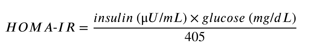
In a previous study, the average HOMA-IR score in healthy 3-4-month-old R. norvegicus rats was 1.23. Therefore, it can be used as a reference that insulin sensitivity is optimal in R. norvegicus rats if they have a HOMA-IR score of ≤ 1.2 (20).
Insulin levels were obtained from blood serum sample tests using the Elabscience Rat INS ELISA kit produced by Elabscience Biotechnology Inc. (Code: E-EL-R3034).
2.5.4. FBG
FBG measurement was performed using a glucometer by applying a blood sample to a glucose strip, with the result displayed on the glucometer screen.
2.6. Ethical Considerations
2.6. Ethical Considerations
All protocols in this study followed the ethical principles of the Faculty of Veterinary Medicine, Universitas Airlangga, Surabaya, Indonesia, namely 5F (free from hunger and thirst; free from discomfort; free from pain, injury, or illness; free to express normal behavior; free from fear and distress) in accordance with Indonesian national law and the "Guide to Care and Use of Laboratory Animals, 8th edition". This research permit was issued by the Animal Care and Use Committee, Faculty of Veterinary Medicine, Airlangga University, Indonesia (Code: 2.KEH.158.11.2024).
2.7. Statistical Analysis
2.7. Statistical Analysis
All data obtained were statistically analyzed using SPSS software (IBM SPSS Statistics 29.0). The initial stage involved performing a normality test using the Shapiro-Wilk test and a homogeneity test using Levene's test to evaluate TNF-α levels, HOMA-IR scores, and FBG. Data that satisfied the assumptions of normality and homogeneity were analyzed using one-way ANOVA test, followed by the Tukey’s post-hoc test.
3. Results
3. Results
3.1. Phytochemical analysis of LPE
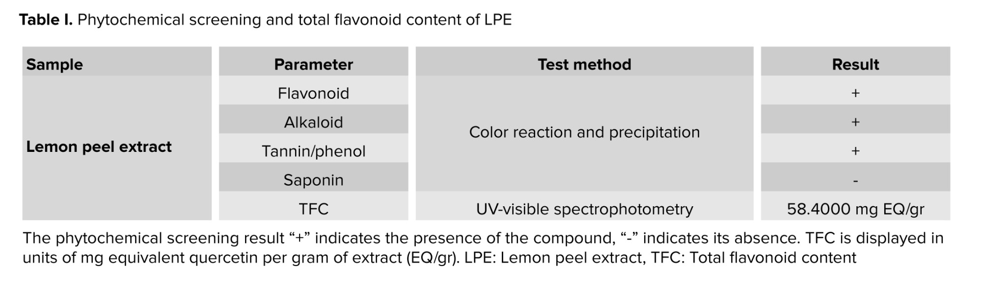
A total of 15 kg of fresh lemons were peeled, resulting in 6.15 kg of fresh lemon peel. The lemon peel was dried and had a dry weight of 1.57 kg. The extraction was performed using the maceration method, producing 340.5 gr of thick extract.
Phytochemical screening shows that LPE contains active compounds, including flavonoid, alkaloid, tannin/phenol (Table I). Further analysis shows that the total flavonoid content in LPE is 58.4 mg equivalent quercetin (EQ)/gr, meaning that every gram of LPE contains 58.4 mg EQ.
3.2. Determining estrus cycle
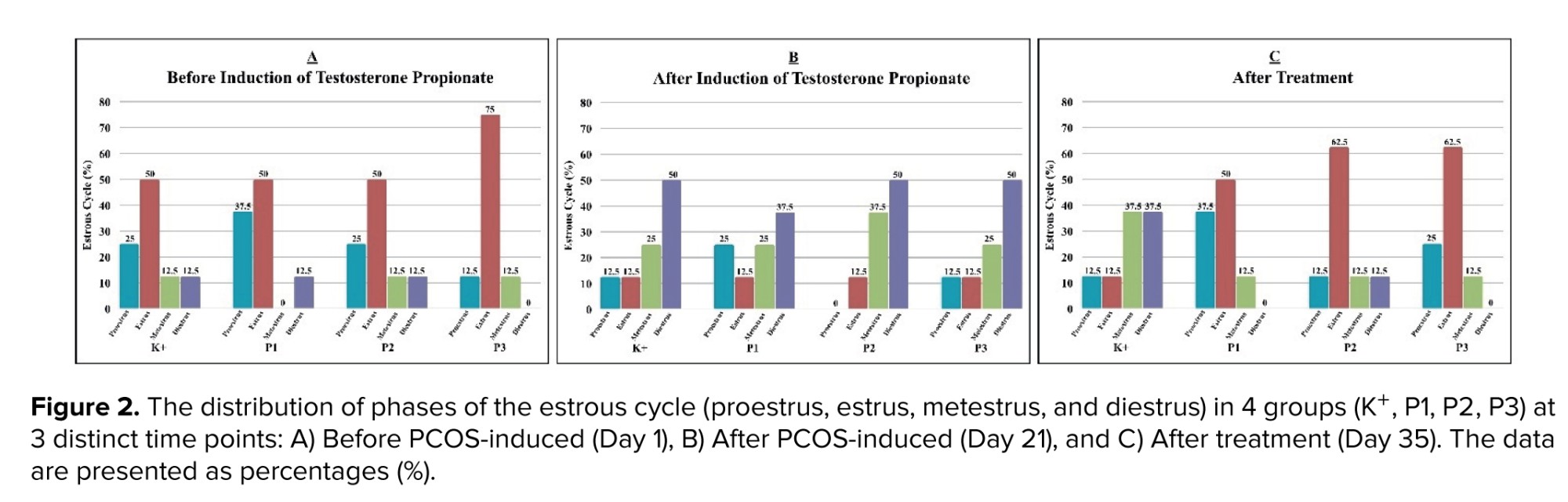
Figure 2 shows the distribution of estrous cycle phases across all experimental groups of PCOS rats, assessed at 3 time points. Before PCOS induction, the estrus cycle was dominated by the estrus phase (K+, P1, P2 = 50% and P3 = 75%), indicating that the majority of rats in each group were in the fertile period.
Based on the Friedman test, no significant differences were observed in estrous cycle distribution in the K+ (p = 0.311) or P1 (p = 0.135) groups. In contrast, significant differences were identified in the P2 (p = 0.048) and P3 (0.042) groups. Further analysis using the Wilcoxon test revealed that differences in both P2 and P3 occurred between the post-PCOS induction and post-treatment time points (p = 0.024 and p = 0.047). Specifically, the estrous cycle was predominantly in diestrus following PCOS induction, whereas after treatment, it was predominantly in estrus.
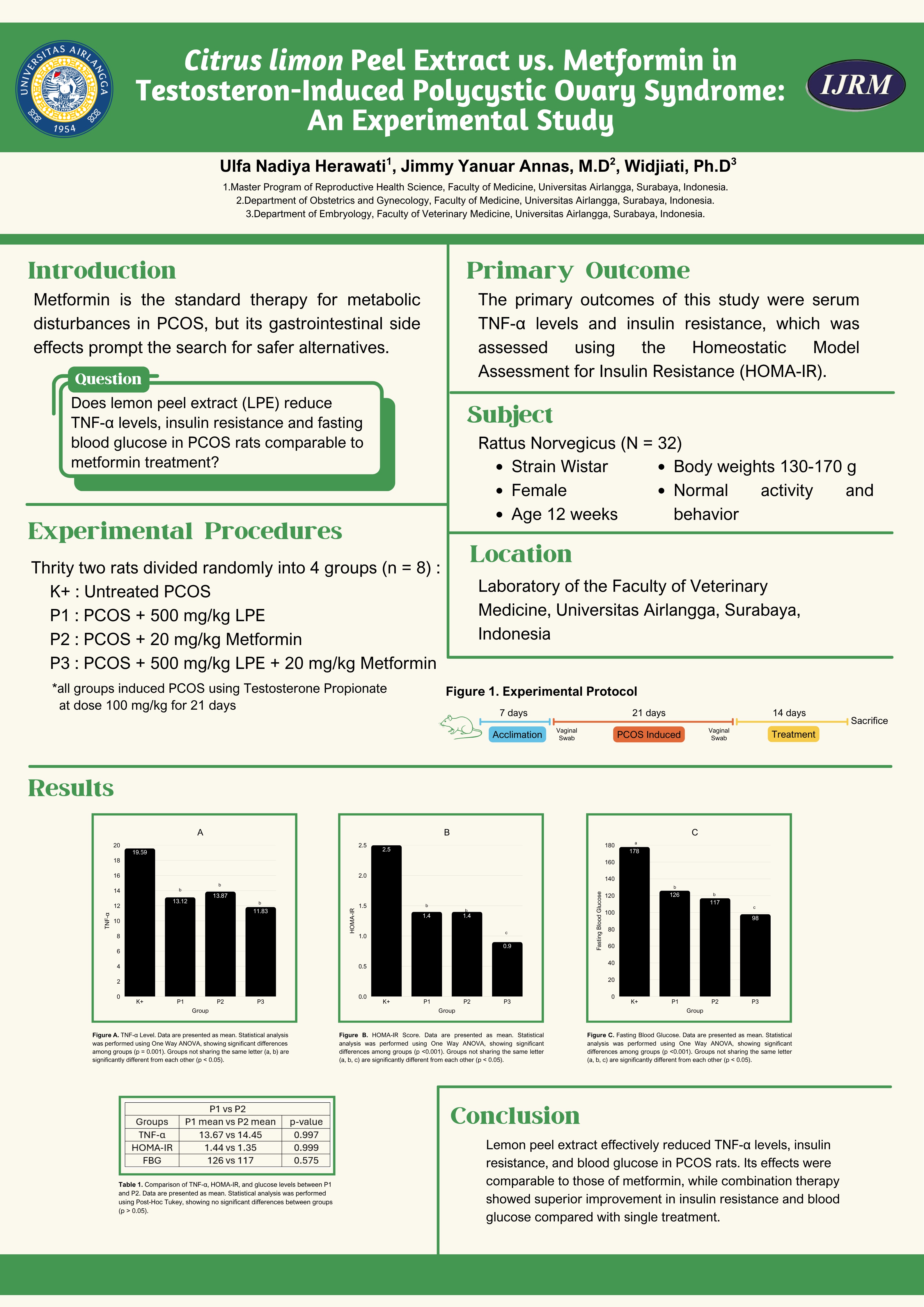
3.3. TNF-α levels
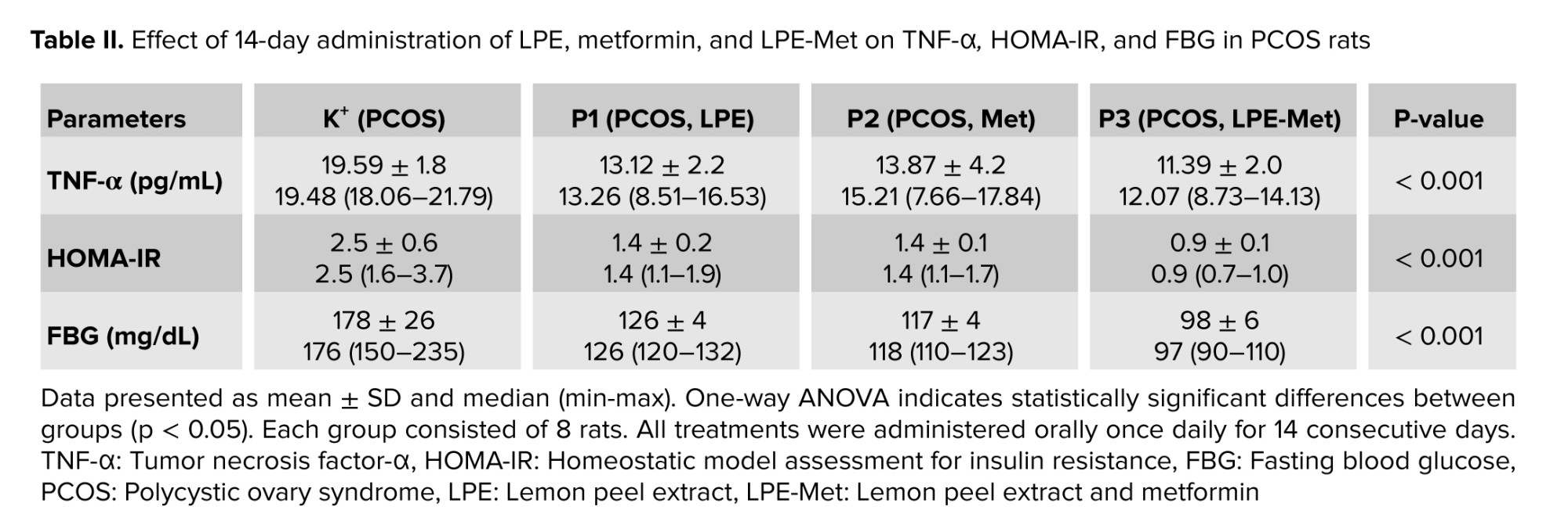
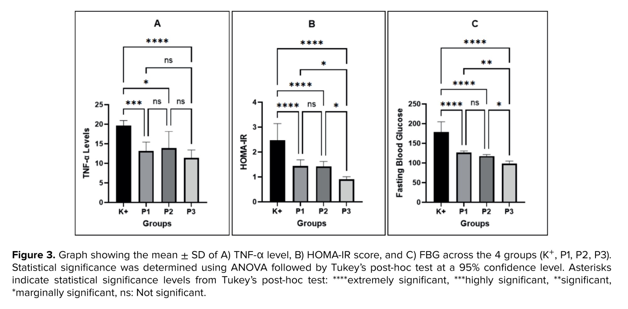
Table II shows significant differences in TNF-α levels among the groups (p < 0.001). Post-hoc analysis revealed that the mean of TNF-α level in the untreated PCOS group (19.59 ± 1.8 pg/mL) was significantly higher than in P1 (13.12 ± 2.2 pg/mL; p < 0.001), P2 (13.87 ± 4.2 pg/mL; p = 0.001), and P3 (11.39 ± 2.0 pg/mL; p < 0.001).
No significant difference was observed between the single treatment groups, LPE (P1) and metformin (P2) (p = 0.945). Although combination therapy of LPE-Met (P3) resulted in a greater reduction in TNF-α levels compared with single treatments, the differences were not statistically significant (P1 vs. P3, p = 0.584 and P2 vs. P3, p = 0.281) (Figure 3A).
3.4. HOMA-IR
In table II, the untreated PCOS rats had a higher average HOMA-IR score (2.5 ± 0.6) and were significantly different from the P1 (1.4 ± 0.2; p < 0.001), P2 (1.4 ± 0.1; p < 0.001), and P3 groups (0.9 ± 0.1; p < 0.001).
Figure 3B shows no significant differences in the HOMA-IR score after a single administration of LPE and metformin (P1 vs. P2, p = 0.554). The combination therapy of LPE-Met (P3) showed a lower reduction in HOMA-IR score and was significantly different from monotherapy (P1 vs. P3, p = 0.038 and P2 vs. P3, p = 0.044).
3.5. FBG
In table II, the untreated PCOS rats exhibited the highest average FBG at 178 ± 26 mg/dL and were significantly different from P1 (126 ± 4 mg/dL; p < 0.001), P2 (117 ± 4; p < 0.001), and P3 groups (98 ± 6 mg/dL; p < 0.001).
Among the 3 treatment groups, no significant difference was observed between single administration of LPE (P1) and metformin (P2), with p = 0.575 (Figure 3C). Meanwhile, the combination therapy of LPE-Met (P3) had the lowest average and was significantly different from monotherapy (P1 vs. P3, p = 0.002; P2 vs. P3, p = 0.048).
4. Discussion
Animal models of PCOS can be established using various validated approaches, one of which involves hyperandrogenism induced by TP. A previous study had demonstrated that TP administration in rats not only elicits PCOS-like phenotypes that closely resemble clinical manifestations in humans but also triggers systemic and local inflammatory responses, contributing to the pathophysiology of PCOS (10).
In the present study, the untreated PCOS rats exhibited the highest levels of TNF-α, HOMA-IR, and FBG, reflecting inflammation, insulin resistance, and metabolic disturbances in the TP-induced PCOS model. These findings are consistent with a previous study that TP induction not only produces polycystic ovarian morphology but also elevates TNF-α, insulin levels, and FBG in rodent models (17). Taken together, these results support the association between hyperandrogenism, inflammation, and metabolic disorder in the pathogenesis of PCOS.
In this study, the distribution of the diestrus phase increased following 21 days of TP administration. This finding is in line with a previous study demonstrating that TP exposure in female rats leads to irregular estrous cycle (21). Treatment with LPE, metformin, or their combination restored the distribution of the estrus phase, accompanied by an almost seven-fold reduction in the diestrus phase. Physiologically, the estrus phase is characterized by a peak in estrogen levels, which triggers a surge in luteinizing hormone and subsequently initiates ovulation (22). Therefore, the increased prevalence of the estrus phase observed in this study suggests the potential of LPE, metformin, and their combined administration to promote ovulatory function in PCOS rats. However, due to the limited evidence, we cannot confirm whether ovulatory function was fully restored, and further investigations incorporating ovarian histology are required.
The primary outcome of this study was that LPE administration reduced TNF-α levels compared to the untreated PCOS rats. Although a healthy control group was not included in this study, the observed reduction compared to the untreated PCOS rats suggests that LPE may attenuate inflammation in this experimental model. This finding aligns with prior work reporting reduced TNF-α following LPE treatment in rats with premature ovarian failure, and the anti-inflammatory activity of lemon peel has been suggested to involve bioactive compounds that modulate NF-κB signaling (23).
Our phytochemical analysis confirmed that LPE contains flavonoids, another study identified them as major bioactive constituents of lemon peel that function as anti-inflammatory through NF-κB inhibition (24). Eriocitrin is reported as the main flavonoid in lemon peel that can inhibit NF-κB activation, thereby limiting TNF-α production and dampening the inflammatory response (24, 25).
In addition to its anti-inflammatory effect, LPE administration reduced HOMA-IR scores and FBG compared with untreated PCOS rats, in line with a previous study reporting that LPE lowers FBG and improves glucose tolerance in diabetic rats (19). Flavonoids derived from herbal sources have been shown to exert anti-hyperglycemic effects by activating peroxisome proliferator-activated receptor gamma and glucose transporter type 1 receptors. This activation enhances glucose uptake and strengthens insulin-mediated glucose utilization, thereby improving glucose tolerance (26). Furthermore, flavonoids such as quercetin and naringenin in lemon peel have been reported to activate AMP-activated protein kinase (AMPK), which may contribute to improved insulin sensitivity in PCOS (27).
It is plausible that the reduction of TNF-α is linked to improvements in insulin resistance and glycemic control, as reflected by the decrease in TNF-α levels followed by a decrease in HOMA-IR score and FBG in PCOS rats treated with LPE. According to the literature, the decreased TNF-α production contributes to the recovery of insulin receptor substrate-1 (IRS-1) function in insulin signaling (28). Under physiological conditions, IRS-1 activates phosphoinositide 3-kinase or protein kinase B, leading to AMPK activation and the translocation of glucose transporter type 4 to the cell membrane. This enhances glucose uptake in muscle and adipose tissue and supports glucose homeostasis (29). However, this proposed mechanism remains speculative because insulin signaling components were not directly evaluated in this study. Further studies are needed to assess the effects of LPE on IRS-1, AMPK, and glucose transporter type 4 expression in insulin-sensitive tissues to confirm this mechanistic relationship.
Metformin administration reduced TNF-α levels, HOMA-IR scores, and FBG in the PCOS rats compared with the untreated PCOS rats. This observation is consistent with previous reports that metformin lowers TNF-α and improves insulin sensitivity in the PCOS model (17, 30). The primary mechanism of metformin involves AMPK, which may suppress TNF-α production through modulation of NF-κB signaling, reduce hepatic glucose production, and enhance glucose uptake in muscle (31, 32).
Our study shows that both LPE and metformin can reduce TNF-α levels, HOMA-IR score, and FBG compared to the untreated PCOS rats, with no statistically significant differences observed between the 2 treatments. These findings suggest that LPE may have potential anti-inflammatory and metabolic effects comparable to those of metformin in this experimental model. Interestingly, the combination therapy of LPE and metformin produced a greater reduction in TNF-α levels, HOMA-IR scores, and FBG than either treatment alone, suggesting a potential synergistic effect on inflammation suppression and metabolic regulation. This effect may be related to shared actions on AMPK activation and NF-κB inhibition.
5. Conclusion
We acknowledge the limitations of this study, particularly the absence of a healthy control group, which restricts the ability to evaluate whether the treatments restore parameters to normal physiological levels. Within these constraints, the results of this study suggest that LPE may have therapeutic potential comparable to metformin and could offer additional benefits when combined. Further studies are needed to confirm these findings and clarify the underlying mechanisms.
These findings contribute positively to the development of herbal-based PCOS management strategies. However, there are differences in dosage and metabolism of active compound between rats and humans, limiting the direct extrapolation of these results. Further analysis, including studies with healthy control and well-designed clinical trials, is required to substantiate the efficacy and safety of LPE in human populations.
Data Availability
The datasets generated and/or analyzed during the current study are available from the corresponding author on reasonable request.
Author Contributions
UN. Herawati is the principal author of this research, having developed the research topic, the study framework, prepared the manuscript, and conducted the data analysis. JY. Annas provided supervision and expert guidance in the areas of PCOS pathophysiology and therapeutic approaches. Widjiati contributed as a supervisor in the implementation of laboratory experiments.
Acknowledgments
The authors would like to express their sincere gratitude to the entire laboratory team at the Faculty of Veterinary Medicine, Airlangga University, Surabaya, Indonesia, for their technical assistance and support throughout this study. Appreciation is also extended to the UPT Materia Medica Herbal Laboratory in Batu, Indonesia, for their help in extracting lemon peel, and to Mulyant Farm in Kediri, Indonesia, for providing high-quality lemons used in this research. The authors also acknowledge the use of digital tools that facilitated the preparation of this manuscript, including GraphPad Prism, Grammarly, Canva, Mendeley, and DeepL. This research received no financial support from any institution or external party; the authors personally funded all expenses.
Conflict of Interest
The author declares that there is no conflict of interest.
A total of 15 kg of fresh lemons were peeled, resulting in 6.15 kg of fresh lemon peel. The lemon peel was dried and had a dry weight of 1.57 kg. The extraction was performed using the maceration method, producing 340.5 gr of thick extract.
Phytochemical screening shows that LPE contains active compounds, including flavonoid, alkaloid, tannin/phenol (Table I). Further analysis shows that the total flavonoid content in LPE is 58.4 mg equivalent quercetin (EQ)/gr, meaning that every gram of LPE contains 58.4 mg EQ.
3.2. Determining estrus cycle
Figure 2 shows the distribution of estrous cycle phases across all experimental groups of PCOS rats, assessed at 3 time points. Before PCOS induction, the estrus cycle was dominated by the estrus phase (K+, P1, P2 = 50% and P3 = 75%), indicating that the majority of rats in each group were in the fertile period.
Based on the Friedman test, no significant differences were observed in estrous cycle distribution in the K+ (p = 0.311) or P1 (p = 0.135) groups. In contrast, significant differences were identified in the P2 (p = 0.048) and P3 (0.042) groups. Further analysis using the Wilcoxon test revealed that differences in both P2 and P3 occurred between the post-PCOS induction and post-treatment time points (p = 0.024 and p = 0.047). Specifically, the estrous cycle was predominantly in diestrus following PCOS induction, whereas after treatment, it was predominantly in estrus.
3.3. TNF-α levels
Table II shows significant differences in TNF-α levels among the groups (p < 0.001). Post-hoc analysis revealed that the mean of TNF-α level in the untreated PCOS group (19.59 ± 1.8 pg/mL) was significantly higher than in P1 (13.12 ± 2.2 pg/mL; p < 0.001), P2 (13.87 ± 4.2 pg/mL; p = 0.001), and P3 (11.39 ± 2.0 pg/mL; p < 0.001).
No significant difference was observed between the single treatment groups, LPE (P1) and metformin (P2) (p = 0.945). Although combination therapy of LPE-Met (P3) resulted in a greater reduction in TNF-α levels compared with single treatments, the differences were not statistically significant (P1 vs. P3, p = 0.584 and P2 vs. P3, p = 0.281) (Figure 3A).
3.4. HOMA-IR
In table II, the untreated PCOS rats had a higher average HOMA-IR score (2.5 ± 0.6) and were significantly different from the P1 (1.4 ± 0.2; p < 0.001), P2 (1.4 ± 0.1; p < 0.001), and P3 groups (0.9 ± 0.1; p < 0.001).
Figure 3B shows no significant differences in the HOMA-IR score after a single administration of LPE and metformin (P1 vs. P2, p = 0.554). The combination therapy of LPE-Met (P3) showed a lower reduction in HOMA-IR score and was significantly different from monotherapy (P1 vs. P3, p = 0.038 and P2 vs. P3, p = 0.044).
3.5. FBG
In table II, the untreated PCOS rats exhibited the highest average FBG at 178 ± 26 mg/dL and were significantly different from P1 (126 ± 4 mg/dL; p < 0.001), P2 (117 ± 4; p < 0.001), and P3 groups (98 ± 6 mg/dL; p < 0.001).
Among the 3 treatment groups, no significant difference was observed between single administration of LPE (P1) and metformin (P2), with p = 0.575 (Figure 3C). Meanwhile, the combination therapy of LPE-Met (P3) had the lowest average and was significantly different from monotherapy (P1 vs. P3, p = 0.002; P2 vs. P3, p = 0.048).
4. Discussion
Animal models of PCOS can be established using various validated approaches, one of which involves hyperandrogenism induced by TP. A previous study had demonstrated that TP administration in rats not only elicits PCOS-like phenotypes that closely resemble clinical manifestations in humans but also triggers systemic and local inflammatory responses, contributing to the pathophysiology of PCOS (10).
In the present study, the untreated PCOS rats exhibited the highest levels of TNF-α, HOMA-IR, and FBG, reflecting inflammation, insulin resistance, and metabolic disturbances in the TP-induced PCOS model. These findings are consistent with a previous study that TP induction not only produces polycystic ovarian morphology but also elevates TNF-α, insulin levels, and FBG in rodent models (17). Taken together, these results support the association between hyperandrogenism, inflammation, and metabolic disorder in the pathogenesis of PCOS.
In this study, the distribution of the diestrus phase increased following 21 days of TP administration. This finding is in line with a previous study demonstrating that TP exposure in female rats leads to irregular estrous cycle (21). Treatment with LPE, metformin, or their combination restored the distribution of the estrus phase, accompanied by an almost seven-fold reduction in the diestrus phase. Physiologically, the estrus phase is characterized by a peak in estrogen levels, which triggers a surge in luteinizing hormone and subsequently initiates ovulation (22). Therefore, the increased prevalence of the estrus phase observed in this study suggests the potential of LPE, metformin, and their combined administration to promote ovulatory function in PCOS rats. However, due to the limited evidence, we cannot confirm whether ovulatory function was fully restored, and further investigations incorporating ovarian histology are required.
The primary outcome of this study was that LPE administration reduced TNF-α levels compared to the untreated PCOS rats. Although a healthy control group was not included in this study, the observed reduction compared to the untreated PCOS rats suggests that LPE may attenuate inflammation in this experimental model. This finding aligns with prior work reporting reduced TNF-α following LPE treatment in rats with premature ovarian failure, and the anti-inflammatory activity of lemon peel has been suggested to involve bioactive compounds that modulate NF-κB signaling (23).
Our phytochemical analysis confirmed that LPE contains flavonoids, another study identified them as major bioactive constituents of lemon peel that function as anti-inflammatory through NF-κB inhibition (24). Eriocitrin is reported as the main flavonoid in lemon peel that can inhibit NF-κB activation, thereby limiting TNF-α production and dampening the inflammatory response (24, 25).
In addition to its anti-inflammatory effect, LPE administration reduced HOMA-IR scores and FBG compared with untreated PCOS rats, in line with a previous study reporting that LPE lowers FBG and improves glucose tolerance in diabetic rats (19). Flavonoids derived from herbal sources have been shown to exert anti-hyperglycemic effects by activating peroxisome proliferator-activated receptor gamma and glucose transporter type 1 receptors. This activation enhances glucose uptake and strengthens insulin-mediated glucose utilization, thereby improving glucose tolerance (26). Furthermore, flavonoids such as quercetin and naringenin in lemon peel have been reported to activate AMP-activated protein kinase (AMPK), which may contribute to improved insulin sensitivity in PCOS (27).
It is plausible that the reduction of TNF-α is linked to improvements in insulin resistance and glycemic control, as reflected by the decrease in TNF-α levels followed by a decrease in HOMA-IR score and FBG in PCOS rats treated with LPE. According to the literature, the decreased TNF-α production contributes to the recovery of insulin receptor substrate-1 (IRS-1) function in insulin signaling (28). Under physiological conditions, IRS-1 activates phosphoinositide 3-kinase or protein kinase B, leading to AMPK activation and the translocation of glucose transporter type 4 to the cell membrane. This enhances glucose uptake in muscle and adipose tissue and supports glucose homeostasis (29). However, this proposed mechanism remains speculative because insulin signaling components were not directly evaluated in this study. Further studies are needed to assess the effects of LPE on IRS-1, AMPK, and glucose transporter type 4 expression in insulin-sensitive tissues to confirm this mechanistic relationship.
Metformin administration reduced TNF-α levels, HOMA-IR scores, and FBG in the PCOS rats compared with the untreated PCOS rats. This observation is consistent with previous reports that metformin lowers TNF-α and improves insulin sensitivity in the PCOS model (17, 30). The primary mechanism of metformin involves AMPK, which may suppress TNF-α production through modulation of NF-κB signaling, reduce hepatic glucose production, and enhance glucose uptake in muscle (31, 32).
Our study shows that both LPE and metformin can reduce TNF-α levels, HOMA-IR score, and FBG compared to the untreated PCOS rats, with no statistically significant differences observed between the 2 treatments. These findings suggest that LPE may have potential anti-inflammatory and metabolic effects comparable to those of metformin in this experimental model. Interestingly, the combination therapy of LPE and metformin produced a greater reduction in TNF-α levels, HOMA-IR scores, and FBG than either treatment alone, suggesting a potential synergistic effect on inflammation suppression and metabolic regulation. This effect may be related to shared actions on AMPK activation and NF-κB inhibition.
5. Conclusion
We acknowledge the limitations of this study, particularly the absence of a healthy control group, which restricts the ability to evaluate whether the treatments restore parameters to normal physiological levels. Within these constraints, the results of this study suggest that LPE may have therapeutic potential comparable to metformin and could offer additional benefits when combined. Further studies are needed to confirm these findings and clarify the underlying mechanisms.
These findings contribute positively to the development of herbal-based PCOS management strategies. However, there are differences in dosage and metabolism of active compound between rats and humans, limiting the direct extrapolation of these results. Further analysis, including studies with healthy control and well-designed clinical trials, is required to substantiate the efficacy and safety of LPE in human populations.
Data Availability
The datasets generated and/or analyzed during the current study are available from the corresponding author on reasonable request.
Author Contributions
UN. Herawati is the principal author of this research, having developed the research topic, the study framework, prepared the manuscript, and conducted the data analysis. JY. Annas provided supervision and expert guidance in the areas of PCOS pathophysiology and therapeutic approaches. Widjiati contributed as a supervisor in the implementation of laboratory experiments.
Acknowledgments
The authors would like to express their sincere gratitude to the entire laboratory team at the Faculty of Veterinary Medicine, Airlangga University, Surabaya, Indonesia, for their technical assistance and support throughout this study. Appreciation is also extended to the UPT Materia Medica Herbal Laboratory in Batu, Indonesia, for their help in extracting lemon peel, and to Mulyant Farm in Kediri, Indonesia, for providing high-quality lemons used in this research. The authors also acknowledge the use of digital tools that facilitated the preparation of this manuscript, including GraphPad Prism, Grammarly, Canva, Mendeley, and DeepL. This research received no financial support from any institution or external party; the authors personally funded all expenses.
Conflict of Interest
The author declares that there is no conflict of interest.
Type of Study: Original Article |
Subject:
Reproductive Endocrinology
Send email to the article author
| Rights and permissions | |
 |
This work is licensed under a Creative Commons Attribution-NonCommercial 4.0 International License. |
