
International Journal of
Reproductive Biomedicine
Thu, Oct 22, 2026
[Archive]
Volume 24, Issue 7 (July 2026)
IJRM 2026, 24(7): 571-584 |
Back to browse issues page




Download citation:
BibTeX | RIS | EndNote | Medlars | ProCite | Reference Manager | RefWorks
Send citation to:


BibTeX | RIS | EndNote | Medlars | ProCite | Reference Manager | RefWorks
Send citation to:
Kiaee F, Shahi H. The lactate-myeloid-derived suppressor cells axis in endometriosis from molecular reprogramming to translational therapies: A review article. IJRM 2026; 24 (7) :571-584
URL: http://ijrm.ir/article-1-3796-en.html
URL: http://ijrm.ir/article-1-3796-en.html


1- Blood Transfusion Research Center, High Institute for Research and Education in Transfusion Medicine, Tehran, Iran.
2- Department of Immunology, School of Medicine, Shahid Beheshti University of Medical Sciences, Tehran, Iran. ,Shahi@sbmu.ac.ir
2- Department of Immunology, School of Medicine, Shahid Beheshti University of Medical Sciences, Tehran, Iran. ,
Full-Text [PDF 464 kb]
(326 Downloads)
| Abstract (HTML) (414 Views)
Full-Text: (32 Views)
1. Introduction
Endometriosis is a chronic inflammatory gynecological disorder defined by the ectopic presence of endometrial-like tissue outside the uterine cavity, commonly affecting the ovaries, fallopian tubes, and peritoneal surfaces. This condition is estimated to impact 10-15% of women in their reproductive years and is prevalent in 35-50% of those experiencing pelvic pain and/or infertility (1). Some women frequently present with debilitating symptoms such as severe dysmenorrhea, chronic pelvic pain, dyspareunia, and infertility, all of which significantly impair their quality of life (2). Although the precise etiology and underlying pathogenic mechanisms of endometriosis remain incompletely elucidated, it is recognized as a multifactorial disease involving factors like retrograde menstruation, immune dysregulation, hormonal imbalances, genetic predispositions, and environmental influences (3).
The diagnostic and therapeutic management of endometriosis is frequently challenging and subject to delays (4) due to the absence of definitive and reliable biomarkers, its diverse and complex symptomology, and the suboptimal efficacy and safety profiles of current treatments. Presently, the gold-standard diagnostic approach involves laparoscopic surgery coupled with histological confirmation, procedures that are inherently expensive, invasive, and often inaccessible to many women (5). While treatment strategies for endometriosis aim to alleviate symptoms, reduce lesion burden, and restore fertility, they are frequently associated with adverse effects, recurrence, and treatment resistance (6, 7). Consequently, there is an urgent imperative for a deeper understanding of endometriosis pathophysiology and for the development of innovative and effective diagnostic and therapeutic strategies. Within this evolving landscape, the role of myeloid-derived suppressor cells (MDSCs) in endometriosis has emerged as a significant area of research (8). MDSCs are a heterogeneous population of immature myeloid cells known for their capacity to suppress immune responses and promote inflammation (9).
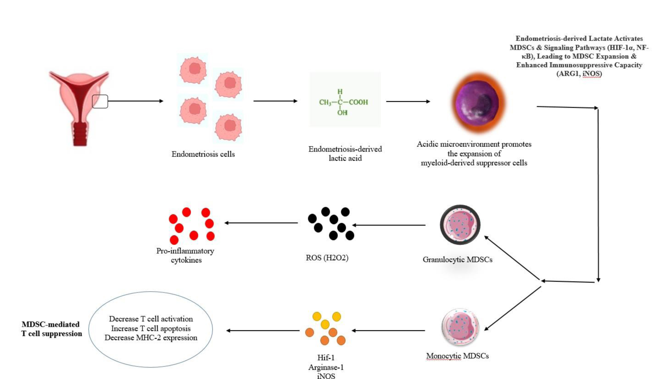
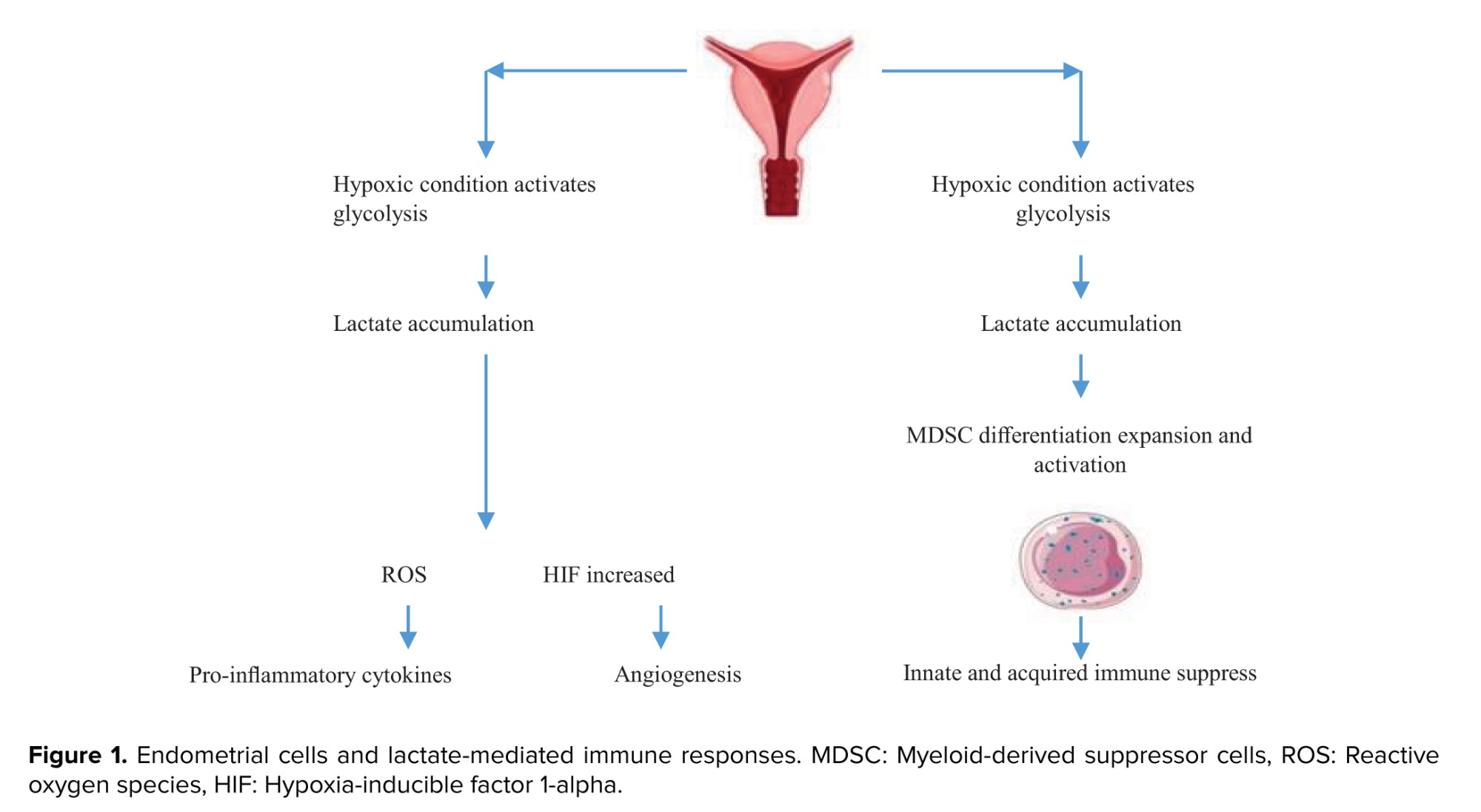
Recent investigations have revealed an increased abundance and heightened activity of MDSCs in both the peritoneal fluid and blood of individuals with endometriosis. These cells contribute to disease progression by inhibiting anti-endometrium associated immune responses and fostering angiogenesis and fibrosis within ectopic lesions (10, 11). Furthermore, the modulation of MDSCs by lactate originating from endometrium-associated lesions represent another burgeoning area of research in endometriosis (12). Endometrium cells, particularly under hypoxic conditions, predominantly utilize glycolysis for energy production, leading to the generation of lactate as a significant metabolic byproduct (13). The subsequent accumulation of lactate in the peritoneal fluid and systemic circulation of endometriosis women can profoundly influence the local cellular and molecular microenvironment of the disease (14). Recent studies have demonstrated that lactate can drive the differentiation and activation of MDSCs, mediated through the intricate regulation of various signaling pathways (15) (Figure 1). Therefore, the primary objective of this review is to comprehensively analyze the molecular and cellular mechanisms through which endometriosis-derived lactate modulates MDSC functions, and to critically evaluate the therapeutic implications of targeting this intricate interaction.
2. MDSCs' impact on endometriosis
MDSCs are a heterogeneous population of immature myeloid cells categorized into 2 main subsets: monocytic MDSCs (M-MDSCs) and granulocytic MDSCs (G-MDSCs). While both are elevated in women with endometriosis, G-MDSCs often represent the predominant phenotype in peripheral blood and peritoneal fluid, resembling neutrophils but exhibiting distinct immunosuppressive functions (11, 16). In contrast, M-MDSCs, more closely resemble monocytes and are potent producers of reactive oxygen species (ROS) and nitric oxide (NO) (9). The balance and specific functions of these subsets in the context of endometriosis pathogenesis, including their differential capacity to promote angiogenesis or fibrosis, warrant further investigation.
The activation and expansion of MDSCs in the endometrium-associated microenvironment are orchestrated by a complex network of factors. Pro-inflammatory cytokines such as tumor necrosis factor alpha (TNF-α), interleukin (IL)-6, and IL-1β, which are abundant in the peritoneal fluid of women with endometriosis, serve as potent activators. For instance, IL-6 and IL-1β can directly induce MDSC differentiation and activate the Janus kinase (JAK) /signal transducer and activator of transcription 3 (STAT3) pathway within these cells (17). Activation of STAT3 is crucial for MDSC survival, proliferation, and the upregulation of key immunosuppressive molecules like arginase-1 (ARG1) and inducible nitric oxide synthase (iNOS). Similarly, nuclear factor kappa-light-chain-enhancer of activated B cells (NF-κB) signaling is pivotal; activated by TNF-α and IL-1β, NF-κB drives the expression of genes essential for MDSC accumulation and function, including those involved in inflammatory responses and cell survival (18). Beyond T cells, MDSCs also impair B cell differentiation and antibody production through prostaglandin E2 (PGE2) and NO (19), and suppress natural killer (NK) cell cytotoxicity by downregulating activating receptors and producing inhibitory cytokines like transforming growth factor-β (20). In the context of macrophages, MDSCs drive M2 polarization, shifting the immune response toward a protumorigenic and profibrotic phenotype that supports lesion growth and survival (21).
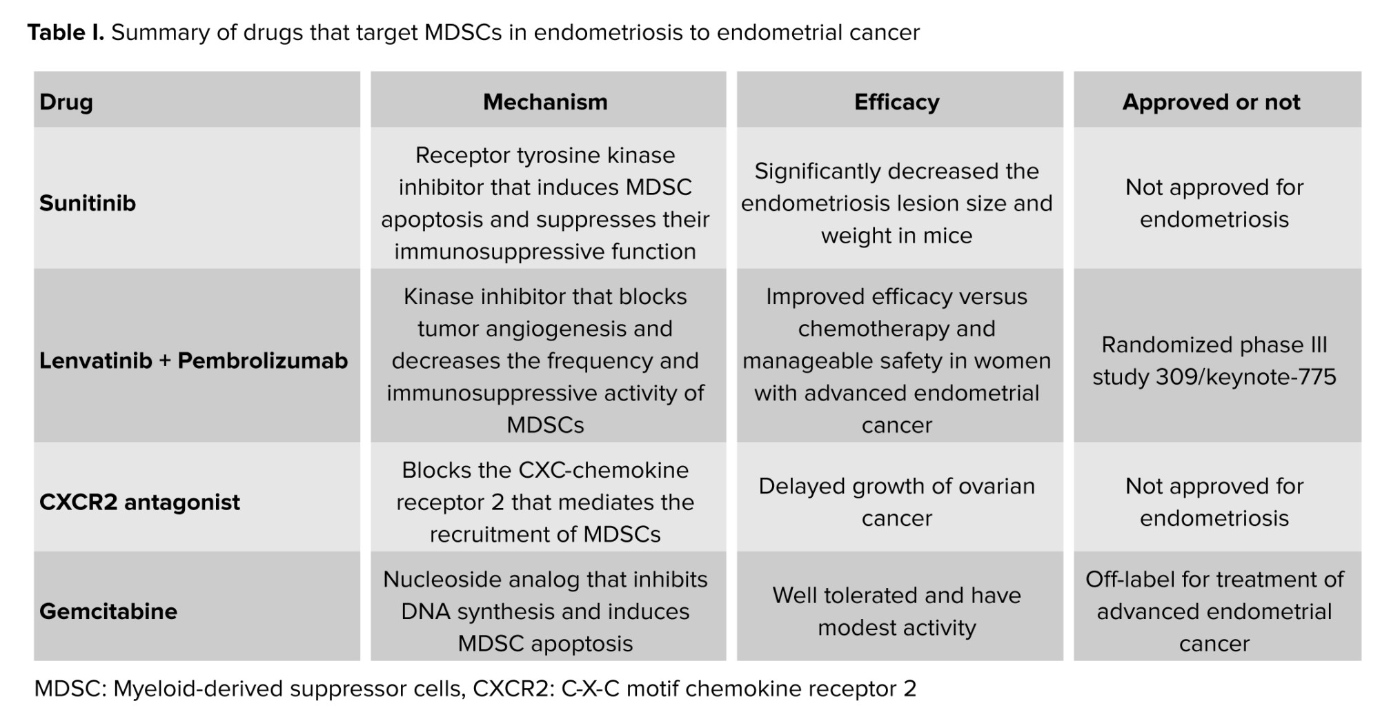
Moreover, MDSCs promote fibrosis by activating fibroblasts and inducing collagen deposition, leading to the characteristic adhesions and scar tissue observed in endometriosis (10, 11). The involvement of MDSCs in the malignant transformation of endometriosis into ovarian cancer further highlights their detrimental role (10, 22). This underscores MDSCs as a central orchestrator of the immune-inflammatory microenvironment in endometriosis, making them an attractive target for therapeutic interventions (Table I).
2.1. Factors driving MDSC accumulation and activation in endometriosis
Elevated levels of pro-inflammatory cytokines, such as TNF-α, IL-6, and IL-1β, are consistently observed in the peritoneal fluid of women with endometriosis, acting as potent inducers of MDSCs (10, 23). Estrogen, for instance, has been shown to enhance the immunosuppressive capacity of MDSCs through the upregulation of enzymes like ARG1 and by increasing the production of ROS. Highly oxidative environment serves as a potent inducer of MDSC differentiation and activation. MDSCs, in turn, can augment their survival and function in response to oxidative stress (21, 24). MDSCs play a central and decisive role in abrogating effective anti-endometriotic immune responses by directly suppressing various immune cell populations within the intricate peritoneal microenvironment (8, 10).
2.2. MDSC suppressive mechanisms
Arginine depletion: MDSCs characteristically express high levels of ARG1, an enzyme that catabolizes L-arginine, an amino acid critically essential for optimal T-cell activation and proliferation (25).
ROS and NO production: MDSCs actively generate abundant quantities of ROS and NO. While ROS can lead to oxidative modification of T-cell receptors and ultimately induce apoptosis, NO can inhibit T-cell signal transduction and impede proliferation (10, 26).
Programmed death-ligand 1 (PD-L1) expression: MDSCs frequently exhibit significant expression of PD-L1, which interacts with PD-1 on T cells. This interaction culminates in T cell exhaustion, anergy, and programmed cell death (27).
Induction of regulatory T cells (Tregs): beyond direct suppression, MDSCs actively promote the differentiation and expansion of immunosuppressive Tregs, thereby further dampening nascent anti-endometriotic immune responses (19).
B-cell inhibition: MDSCs also possess the capability to directly impair B-cell function via mechanisms involving ARG1, PGE2, and iNOS. This deleterious impact results in compromised B-cell differentiation, diminished antigen presentation capabilities, and a reduction in critical antibody production, all of which are vital for the efficient clearance of ectopic endometrial cells (19, 20).
NK cell suppression: MDSCs effectively inhibit the cytotoxic activity and cytokine production of NK cells, including critical effector molecules such as granzyme B and perforin, and key activating receptors like NKG2D. This observed reduction in NK cell efficacy is particularly relevant in the context of endometriosis, given that endometriotic cells often display low levels of major histocompatibility complex class I molecules (a strategy for evading T cells) and concurrently high levels of stress-induced ligands, making them prime targets for NK cell-mediated elimination (20).
Macrophage polarization: MDSCs exert a significant influence on the polarization and functional phenotype of macrophages, primarily through the judicious production of IL-10, transforming growth factor-β, and PGE2. This often profoundly skews macrophages toward an M2-like phenotype, which is distinctly characterized by profibrotic, proangiogenic, and anti-inflammatory properties, thereby significantly contributing to aberrant tissue remodeling and immune tolerance within the endometrium-associated lesions (21).
3. Lactate metabolism and micro environmental acidification in endometriosis
Lactate, frequently considered a mere metabolic byproduct, is predominantly generated through glycolysis, the fundamental pathway responsible for glucose breakdown and adenosine triphosphate (ATP) synthesis. While lactate can be efficiently recycled into glucose via hepatic gluconeogenesis or readily utilized as an energy substrate by metabolically active tissues such as the heart and brain, its excessive accumulation within tissues and biological fluids can precipitate significant physiological repercussions (22). In pathological contexts where glycolytic flux substantially surpasses the capacity for lactate clearance, localized lactate accumulation and consequent microenvironmental acidification are prone to occur (28-30). Endometriotic cells distinctly exhibit a profound metabolic reprogramming, characterized by a pronounced preference for aerobic glycolysis, a phenomenon widely recognized as the Warburg effect (13). This metabolic deviation involves sustained and often elevated utilization of glucose and concomitant lactate production, even in the presence of ample oxygen supply, a metabolic shift commonly observed in rapidly proliferating cells and various tumor types (13, 31). Consequently, substantial lactate accumulation has been consistently documented in both the peritoneal fluid and peripheral blood of women afflicted with endometriosis, reaching levels that profoundly impact the cellular and molecular landscape within ectopic lesions and the surrounding peritoneal cavity (14, 32).
3.1. Hypoxia in endometrium-associated lesions
Hypoxia, defined as a state of diminished oxygen availability, is a characteristic hallmark of endometrium-associated lesions. This localized micro-environmental hypoxia stems from a confluence of factors, including inadequate vascularization, rapid cellular proliferation leading to an escalated metabolic demand, and the inherently dense cellular composition of the ectopic implants (32, 33). Hypoxia is a potent physiological inducer of glycolysis, primarily by upregulating the expression of key glycolytic enzymes and glucose transporters. These include glucose transporter 1, hexokinase, phosphofructokinase and lactate dehydrogenase (LDH) A. Concurrently, hypoxia effectively suppresses mitochondrial oxidative phosphorylation (OXPHOS), the more energy-efficient pathway for ATP generation under normoxic conditions (33). A central orchestrator of the cellular hypoxic response is hypoxia-inducible factor 1-alpha (HIF-1α). HIF-1α acts as a master transcriptional regulator that, under hypoxic conditions, becomes stabilized and translocates to the nucleus. There, it actively induces the expression of numerous genes implicated in various cellular processes critical for endometriosis progression, including angiogenesis, inflammation, immune evasion, and metabolic adaptation. Specifically, HIF-1α significantly enhances the expression of GLUTs, LDHA, and monocarboxylate transporters (MCTs), thereby facilitating glucose uptake, lactate production, and lactate export from endometriotic cells, respectively (33, 34).
3.2. Enhanced glycolysis in the endometrium-associated microenvironment
Glycolysis represents the foundational metabolic pathway that converts glucose into pyruvate. In the absence of sufficient oxygen or within cells exhibiting the Warburg effect, pyruvate is predominantly converted to lactate-by-LDH rather than being channeled into the mitochondria for OXPHOS (34). Beyond hypoxia, other crucial factors contribute to the exacerbated glycolytic activity observed in endometriosis:
Inflammation: endometriosis is intrinsically an inflammatory disease. The peritoneal fluid, serum, and ectopic lesions of affected individuals are characterized by a robust immune response and elevated levels of various pro-inflammatory cytokines, including TNF-α, IL-1β, IL-6, and IL-17A (32, 35). These cytokines play a pivotal role in promoting glycolysis through several distinct mechanisms.
They can directly stimulate the expression and activity of HIF-1α, even under normoxic conditions, thereby establishing a critical link between inflammation and hypoxic-like metabolic responses (33, 34).
They elevate levels of ROS, which further impede OXPHOS and consequently divert pyruvate toward lactate production (35). Elevated ROS levels are also implicated in activating toll-like receptor signaling, NF-κB, and HIF-1α pathways, leading to enhanced angiogenesis and improved survival of endometriotic cells (31, 32).
Pro-inflammatory cytokines, alongside NO and HIF-1α, also stimulate the expression of cyclooxygenase-2 (COX-2) via intricate signaling cascades, such as the cAMP/PKA/CREB and JNK/Jun/ATF2 pathways (33). COX-2, in turn, activates mitogen-activated protein kinases (MAPKs). The sustained upregulation of COX-2 in endometriosis contributes to increased cell proliferation, reduced apoptosis, enhanced invasion, angiogenesis, and exacerbates the debilitating pain and infertility associated with the disease (36).
Estrogen signaling: estrogen serves as a primary driver of endometrial growth and function (34). Endometrium-associated lesions are notoriously characterized by elevated levels of estrogen receptors and robust local estrogen production, which is facilitated by high levels of aromatase, an enzyme responsible for converting androgens into estrogens (35). Estrogen signaling significantly enhances glycolysis in endometrium-associated cells through multiple intricate pathways (36, 37).
Estrogen can induce heat shock factor 1, a key transcription factor that upregulates the expression of phosphofructokinase-2/fructose-2, 6-bisphosphatase 3 a critical enzyme that profoundly boosts glycolytic flux. Heat shock factor also stabilizes HIF-1α and activates the PI3K/AKT/mTOR pathway, both of which are central regulators of glycolysis and are intimately implicated in endometriosis progression and cell survival (36).
Collectively, hypoxia, chronic inflammation, and aberrant estrogen signaling synergistically augment glycolysis within endometrium-associated lesions by influencing the expression and activity of pivotal metabolic enzymes, HIF-1α, and an extensive network of downstream signaling pathways. This enhanced glycolysis not only provides rapid ATP to meet demanding energy requirements but also generates essential biosynthetic building blocks, thereby supporting the survival, proliferation, invasion, and metastatic potential of endometriotic cells (38-42).
4. The modulatory role of lactate on MDSCs
The aberrant accumulation of lactate within the endometriotic microenvironment is not merely a passive metabolic consequence; it actively orchestrates the cellular landscape, profoundly impacting the biology of MDSCs. Endometriosis-derived lactate not only drives the differentiation, expansion, and activation of MDSCs but also significantly augments their potent immunosuppressive and pro-angiogenic capabilities (15, 43-46). Investigations have consistently demonstrated that elevated lactate concentrations can effectively induce the generation of MDSCs from murine bone marrow cells when co-cultured with granulocyte-macrophage colony-stimulating factor and IL-6 (44). While the precise molecular mechanisms underlying this direct induction remain subjects of ongoing inquiry, lactate's pivotal role in influencing MDSC differentiation pathways is becoming increasingly elucidated.
For instance, a recent study illuminated how lactate precisely steers the differentiation of M-MDSCs toward G-MDSCs rather than mature macrophages. This critical shift occurs via a robust MCT2/lactate/c-Jun/COX2 axis (46, 47). Specifically, lactate confers a protective role by maintaining the intracellular stability of the transcription factor c-Jun, which subsequently upregulates COX-2 expression. Conversely, the activation of notch signaling has been shown to inhibit MDSC differentiation by blocking MCT2, thereby curtailing intracellular lactate import (46). Furthermore, lactate possesses the capacity to modulate the activation of MDSCs through the G protein-coupled receptor 81 (GPR81), which functions as a lactate receptor, initiating the GPR81/mTOR/HIF-1α/STAT3 signaling pathway (47). This intricate pathway critically dictates MDSC function and survival.
4.1. Lactate-mediated cytokine production and MDSC accumulation
The pervasive presence of lactate within the endometrium-associated microenvironment actively fosters a pro-inflammatory milieu. Lactate significantly amplifies the production of key pro-inflammatory cytokines, notably IL-6 and IL-1β (48, 49). This phenomenon is primarily orchestrated through the activation of MAPK and NF-κB signaling cascades (48, 50). These lactate-induced cytokines, with IL-6 being particularly prominent, subsequently further stimulate the STAT3 pathway within MDSCs, culminating in their enhanced accumulation and prolonged maintenance (50-58). Moreover, IL-1β itself acts as a potent driver for the expansion of highly immunosuppressive MDSCs, operating through the established IL-6/STAT3 axis. Intriguingly, IL-1β can also activate the NOD-like receptor protein 3 inflammasome, which, in turn, promotes the phosphorylation of STAT3. This establishes a self-perpetuating autoinflammatory feedback loop, further amplifying IL-6 signaling and sustaining the MDSC-promoting environment (59).
5. Discussion
Our comprehensive literature review elucidates a critical role for endometriosis-derived lactate as a potent modulator of MDSCs within the complex microenvironment of ectopic lesions. We have highlighted that the distinct metabolic reprogramming of endometriotic cells, characterized by elevated aerobic glycolysis and subsequent lactate accumulation, actively orchestrates the expansion, differentiation, and functional potentiation of MDSCs. This multifaceted influence is mediated through several interconnected signaling pathways, including HIF-1α, STAT3, and NF-κB, as well as intricate interactions involving MCTs and the lactate receptor GPR81. The findings presented here underscore the profound implications of lactate-mediated MDSC modulation for the progression, prognosis, and potential therapeutic strategies in endometriosis. By augmenting MDSC immunosuppressive functions (through mechanisms involving ARG1, inducible iNOS, and ROS production) lactate contributes significantly to immune evasion within endometriotic lesions (17, 44, 53). This immune suppression can compromise the clearance of ectopic endometrial cells and facilitate lesion persistence and growth. Furthermore, lactate-activated MDSCs secrete a plethora of pro-angiogenic factors, such as vascular endothelial growth factor, basic fibroblast growth factor, and matrix metallo-proteinases, which are essential for the vascularization of new lesions and contribute to the pathological characteristics of endometriosis (51, 54).
The metabolic crosstalk between endometriotic cells and immune cells, particularly MDSCs, thus emerges as a central axis driving disease pathogenesis. This paradigm resonates strongly with observations in cancer biology, where tumor-derived lactate is a well-established orchestrator of MDSC activity, promoting tumor growth, metastasis, and immune escape (52, 55). This compelling analogy reinforces the potential significance of targeting this axis in endometriosis. The elucidation of these underlying mechanisms opens promising avenues for novel therapeutic interventions. Strategically targeting lactate metabolism, its transporters, or its receptors within the endometriotic microenvironment could offer a multi-pronged approach to disrupt MDSC function. For instance, inhibitors of LDHA could effectively reduce lactate production, while MCT inhibitors might impede lactate export, thereby altering the acidic microenvironment crucial for MDSC activation and maintenance (39, 46, 56).
6. Conclusion and therapeutic implications
In summary, endometriosis-derived lactate emerges as a pivotal and multifaceted factor in the intricate regulation of MDSC development and function within the unique endometriotic microenvironment. Its wide-ranging effects are meticulously mediated through a complex and interconnected network of signaling pathways and molecules, prominently including the MCT2/c-Jun/COX2, GPR81/mTOR/HIF-1α/STAT3, MAPK/NF-κB, and STAT1/STAT3 axes. These intricate mechanisms collectively drive MDSC accumulation, dramatically enhance their immunosuppressive capabilities (primarily via ARG1, iNOS, and ROS), and substantially bolster their critical contributions to angiogenesis.
These compelling findings resonate powerfully with observations from the field of oncology, where tumor-derived lactate is unequivocally known to profoundly influence the frequency of MDSC differentiation and their potent immunosuppressive activity, both in vitro and in vivo (44, 52). Consequently, therapeutically targeting lactate production or its critical signaling pathways presents a highly promising and novel strategy for endometriosis management. Strategically reducing lactate levels could offer a dual therapeutic benefit: it could substantially diminish the immune suppressive function by lowering both the number and intrinsic activity of MDSCs, and simultaneously significantly enhance the endogenous anti-endometrium associated immune response by preserving the vital activity of effector T and NK cells (44).
7. Targeting lactate: a novel approach to treat endometriosis
Given that lactate produced by endometriosis cells actively contributes to the accumulation of MDSCs, strategically targeting lactate offers a promising new avenue to abrogate the detrimental role of MDSCs in the initiation and progression of endometriosis. This therapeutic strategy holds the potential to effectively modulate the immune system, mitigate inflammation, inhibit pathological angiogenesis, and restore tissue homeostasis within the endometriotic microenvironment. Currently, several pharmacological agents are recognized for their ability to inhibit lactate production or transport. These include glycolytic inhibitors like 3-bromopyruvate and 2-deoxyglucose, which disrupt glucose-to-lactate conversion, though their clinical application is limited by toxicity concerns. Dichloroacetate (DCA) has shown promise in restoring normal glycolytic rates in human peritoneal mesothelial cells and reducing endometriosis lesion size in mice, leading to its exploration in an exploratory clinical trial for pain alleviation. Syrosingopine also presents a therapeutic option by inhibiting key lactate transporters MCT1 and MCT4, with enhanced effects when combined with metformin (57-61). Further targeted approaches include specific MCT1 inhibitors such as AZD3965, CHC, and AR-C155858, which are currently in various phases of clinical trials for cancer. Additionally, LDH inhibitors, comprising a range of natural compounds like polyphenols (e.g., quercetin, kaempferol, apigenin), terpenoids (e.g., oleanolic acid), and alkaloids (e.g., berberine, magnoflorine), demonstrate therapeutic potential by directly binding to the enzyme's active site or modulating its activity. Galloflavin, another LDH inhibitor, has shown efficacy in augmenting radiation therapy and suppressing endometrial cancer cell growth (61-63). However, the clinical translation of natural compounds faces hurdles due to issues, such as low bioavailability, poor solubility, and potential toxicity profiles. Researchers are exploring drug delivery systems and combinations with synthetic inhibitors to overcome these limitations. Despite promising preclinical results in inhibiting lactate production or transport, significant challenges persist regarding efficacy, safety, and the absence of robust validation and standardization for these therapeutic approaches (63-71). The limited number and quality of existing human and animal studies, coupled with the restricted availability and clinical approval status of these drugs, underscore the critical need for further rigorous research. Such investigations are essential to comprehensively assess the feasibility and effectiveness of these applications and to optimize methodologies for leveraging lactate as a multifaceted therapeutic target for endometriosis.
Author Contributions
F. Kiaee conducted the research and wrote the manuscript. H. Shahi designed the figure and reviewed the article. All authors approved the final manuscript and take responsibility for the integrity of the data.
Acknowledgments
There was no financial support from any center or institution. Artificial intelligence (Gemini 2.5) was used for grammar check.
Conflict of Interest
The authors declare that there is no conflict of interest.
Endometriosis is a chronic inflammatory gynecological disorder defined by the ectopic presence of endometrial-like tissue outside the uterine cavity, commonly affecting the ovaries, fallopian tubes, and peritoneal surfaces. This condition is estimated to impact 10-15% of women in their reproductive years and is prevalent in 35-50% of those experiencing pelvic pain and/or infertility (1). Some women frequently present with debilitating symptoms such as severe dysmenorrhea, chronic pelvic pain, dyspareunia, and infertility, all of which significantly impair their quality of life (2). Although the precise etiology and underlying pathogenic mechanisms of endometriosis remain incompletely elucidated, it is recognized as a multifactorial disease involving factors like retrograde menstruation, immune dysregulation, hormonal imbalances, genetic predispositions, and environmental influences (3).
The diagnostic and therapeutic management of endometriosis is frequently challenging and subject to delays (4) due to the absence of definitive and reliable biomarkers, its diverse and complex symptomology, and the suboptimal efficacy and safety profiles of current treatments. Presently, the gold-standard diagnostic approach involves laparoscopic surgery coupled with histological confirmation, procedures that are inherently expensive, invasive, and often inaccessible to many women (5). While treatment strategies for endometriosis aim to alleviate symptoms, reduce lesion burden, and restore fertility, they are frequently associated with adverse effects, recurrence, and treatment resistance (6, 7). Consequently, there is an urgent imperative for a deeper understanding of endometriosis pathophysiology and for the development of innovative and effective diagnostic and therapeutic strategies. Within this evolving landscape, the role of myeloid-derived suppressor cells (MDSCs) in endometriosis has emerged as a significant area of research (8). MDSCs are a heterogeneous population of immature myeloid cells known for their capacity to suppress immune responses and promote inflammation (9).
Recent investigations have revealed an increased abundance and heightened activity of MDSCs in both the peritoneal fluid and blood of individuals with endometriosis. These cells contribute to disease progression by inhibiting anti-endometrium associated immune responses and fostering angiogenesis and fibrosis within ectopic lesions (10, 11). Furthermore, the modulation of MDSCs by lactate originating from endometrium-associated lesions represent another burgeoning area of research in endometriosis (12). Endometrium cells, particularly under hypoxic conditions, predominantly utilize glycolysis for energy production, leading to the generation of lactate as a significant metabolic byproduct (13). The subsequent accumulation of lactate in the peritoneal fluid and systemic circulation of endometriosis women can profoundly influence the local cellular and molecular microenvironment of the disease (14). Recent studies have demonstrated that lactate can drive the differentiation and activation of MDSCs, mediated through the intricate regulation of various signaling pathways (15) (Figure 1). Therefore, the primary objective of this review is to comprehensively analyze the molecular and cellular mechanisms through which endometriosis-derived lactate modulates MDSC functions, and to critically evaluate the therapeutic implications of targeting this intricate interaction.
2. MDSCs' impact on endometriosis
MDSCs are a heterogeneous population of immature myeloid cells categorized into 2 main subsets: monocytic MDSCs (M-MDSCs) and granulocytic MDSCs (G-MDSCs). While both are elevated in women with endometriosis, G-MDSCs often represent the predominant phenotype in peripheral blood and peritoneal fluid, resembling neutrophils but exhibiting distinct immunosuppressive functions (11, 16). In contrast, M-MDSCs, more closely resemble monocytes and are potent producers of reactive oxygen species (ROS) and nitric oxide (NO) (9). The balance and specific functions of these subsets in the context of endometriosis pathogenesis, including their differential capacity to promote angiogenesis or fibrosis, warrant further investigation.
The activation and expansion of MDSCs in the endometrium-associated microenvironment are orchestrated by a complex network of factors. Pro-inflammatory cytokines such as tumor necrosis factor alpha (TNF-α), interleukin (IL)-6, and IL-1β, which are abundant in the peritoneal fluid of women with endometriosis, serve as potent activators. For instance, IL-6 and IL-1β can directly induce MDSC differentiation and activate the Janus kinase (JAK) /signal transducer and activator of transcription 3 (STAT3) pathway within these cells (17). Activation of STAT3 is crucial for MDSC survival, proliferation, and the upregulation of key immunosuppressive molecules like arginase-1 (ARG1) and inducible nitric oxide synthase (iNOS). Similarly, nuclear factor kappa-light-chain-enhancer of activated B cells (NF-κB) signaling is pivotal; activated by TNF-α and IL-1β, NF-κB drives the expression of genes essential for MDSC accumulation and function, including those involved in inflammatory responses and cell survival (18). Beyond T cells, MDSCs also impair B cell differentiation and antibody production through prostaglandin E2 (PGE2) and NO (19), and suppress natural killer (NK) cell cytotoxicity by downregulating activating receptors and producing inhibitory cytokines like transforming growth factor-β (20). In the context of macrophages, MDSCs drive M2 polarization, shifting the immune response toward a protumorigenic and profibrotic phenotype that supports lesion growth and survival (21).
Moreover, MDSCs promote fibrosis by activating fibroblasts and inducing collagen deposition, leading to the characteristic adhesions and scar tissue observed in endometriosis (10, 11). The involvement of MDSCs in the malignant transformation of endometriosis into ovarian cancer further highlights their detrimental role (10, 22). This underscores MDSCs as a central orchestrator of the immune-inflammatory microenvironment in endometriosis, making them an attractive target for therapeutic interventions (Table I).
2.1. Factors driving MDSC accumulation and activation in endometriosis
Elevated levels of pro-inflammatory cytokines, such as TNF-α, IL-6, and IL-1β, are consistently observed in the peritoneal fluid of women with endometriosis, acting as potent inducers of MDSCs (10, 23). Estrogen, for instance, has been shown to enhance the immunosuppressive capacity of MDSCs through the upregulation of enzymes like ARG1 and by increasing the production of ROS. Highly oxidative environment serves as a potent inducer of MDSC differentiation and activation. MDSCs, in turn, can augment their survival and function in response to oxidative stress (21, 24). MDSCs play a central and decisive role in abrogating effective anti-endometriotic immune responses by directly suppressing various immune cell populations within the intricate peritoneal microenvironment (8, 10).
2.2. MDSC suppressive mechanisms
Arginine depletion: MDSCs characteristically express high levels of ARG1, an enzyme that catabolizes L-arginine, an amino acid critically essential for optimal T-cell activation and proliferation (25).
ROS and NO production: MDSCs actively generate abundant quantities of ROS and NO. While ROS can lead to oxidative modification of T-cell receptors and ultimately induce apoptosis, NO can inhibit T-cell signal transduction and impede proliferation (10, 26).
Programmed death-ligand 1 (PD-L1) expression: MDSCs frequently exhibit significant expression of PD-L1, which interacts with PD-1 on T cells. This interaction culminates in T cell exhaustion, anergy, and programmed cell death (27).
Induction of regulatory T cells (Tregs): beyond direct suppression, MDSCs actively promote the differentiation and expansion of immunosuppressive Tregs, thereby further dampening nascent anti-endometriotic immune responses (19).
B-cell inhibition: MDSCs also possess the capability to directly impair B-cell function via mechanisms involving ARG1, PGE2, and iNOS. This deleterious impact results in compromised B-cell differentiation, diminished antigen presentation capabilities, and a reduction in critical antibody production, all of which are vital for the efficient clearance of ectopic endometrial cells (19, 20).
NK cell suppression: MDSCs effectively inhibit the cytotoxic activity and cytokine production of NK cells, including critical effector molecules such as granzyme B and perforin, and key activating receptors like NKG2D. This observed reduction in NK cell efficacy is particularly relevant in the context of endometriosis, given that endometriotic cells often display low levels of major histocompatibility complex class I molecules (a strategy for evading T cells) and concurrently high levels of stress-induced ligands, making them prime targets for NK cell-mediated elimination (20).
Macrophage polarization: MDSCs exert a significant influence on the polarization and functional phenotype of macrophages, primarily through the judicious production of IL-10, transforming growth factor-β, and PGE2. This often profoundly skews macrophages toward an M2-like phenotype, which is distinctly characterized by profibrotic, proangiogenic, and anti-inflammatory properties, thereby significantly contributing to aberrant tissue remodeling and immune tolerance within the endometrium-associated lesions (21).
3. Lactate metabolism and micro environmental acidification in endometriosis
Lactate, frequently considered a mere metabolic byproduct, is predominantly generated through glycolysis, the fundamental pathway responsible for glucose breakdown and adenosine triphosphate (ATP) synthesis. While lactate can be efficiently recycled into glucose via hepatic gluconeogenesis or readily utilized as an energy substrate by metabolically active tissues such as the heart and brain, its excessive accumulation within tissues and biological fluids can precipitate significant physiological repercussions (22). In pathological contexts where glycolytic flux substantially surpasses the capacity for lactate clearance, localized lactate accumulation and consequent microenvironmental acidification are prone to occur (28-30). Endometriotic cells distinctly exhibit a profound metabolic reprogramming, characterized by a pronounced preference for aerobic glycolysis, a phenomenon widely recognized as the Warburg effect (13). This metabolic deviation involves sustained and often elevated utilization of glucose and concomitant lactate production, even in the presence of ample oxygen supply, a metabolic shift commonly observed in rapidly proliferating cells and various tumor types (13, 31). Consequently, substantial lactate accumulation has been consistently documented in both the peritoneal fluid and peripheral blood of women afflicted with endometriosis, reaching levels that profoundly impact the cellular and molecular landscape within ectopic lesions and the surrounding peritoneal cavity (14, 32).
3.1. Hypoxia in endometrium-associated lesions
Hypoxia, defined as a state of diminished oxygen availability, is a characteristic hallmark of endometrium-associated lesions. This localized micro-environmental hypoxia stems from a confluence of factors, including inadequate vascularization, rapid cellular proliferation leading to an escalated metabolic demand, and the inherently dense cellular composition of the ectopic implants (32, 33). Hypoxia is a potent physiological inducer of glycolysis, primarily by upregulating the expression of key glycolytic enzymes and glucose transporters. These include glucose transporter 1, hexokinase, phosphofructokinase and lactate dehydrogenase (LDH) A. Concurrently, hypoxia effectively suppresses mitochondrial oxidative phosphorylation (OXPHOS), the more energy-efficient pathway for ATP generation under normoxic conditions (33). A central orchestrator of the cellular hypoxic response is hypoxia-inducible factor 1-alpha (HIF-1α). HIF-1α acts as a master transcriptional regulator that, under hypoxic conditions, becomes stabilized and translocates to the nucleus. There, it actively induces the expression of numerous genes implicated in various cellular processes critical for endometriosis progression, including angiogenesis, inflammation, immune evasion, and metabolic adaptation. Specifically, HIF-1α significantly enhances the expression of GLUTs, LDHA, and monocarboxylate transporters (MCTs), thereby facilitating glucose uptake, lactate production, and lactate export from endometriotic cells, respectively (33, 34).
3.2. Enhanced glycolysis in the endometrium-associated microenvironment
Glycolysis represents the foundational metabolic pathway that converts glucose into pyruvate. In the absence of sufficient oxygen or within cells exhibiting the Warburg effect, pyruvate is predominantly converted to lactate-by-LDH rather than being channeled into the mitochondria for OXPHOS (34). Beyond hypoxia, other crucial factors contribute to the exacerbated glycolytic activity observed in endometriosis:
Inflammation: endometriosis is intrinsically an inflammatory disease. The peritoneal fluid, serum, and ectopic lesions of affected individuals are characterized by a robust immune response and elevated levels of various pro-inflammatory cytokines, including TNF-α, IL-1β, IL-6, and IL-17A (32, 35). These cytokines play a pivotal role in promoting glycolysis through several distinct mechanisms.
They can directly stimulate the expression and activity of HIF-1α, even under normoxic conditions, thereby establishing a critical link between inflammation and hypoxic-like metabolic responses (33, 34).
They elevate levels of ROS, which further impede OXPHOS and consequently divert pyruvate toward lactate production (35). Elevated ROS levels are also implicated in activating toll-like receptor signaling, NF-κB, and HIF-1α pathways, leading to enhanced angiogenesis and improved survival of endometriotic cells (31, 32).
Pro-inflammatory cytokines, alongside NO and HIF-1α, also stimulate the expression of cyclooxygenase-2 (COX-2) via intricate signaling cascades, such as the cAMP/PKA/CREB and JNK/Jun/ATF2 pathways (33). COX-2, in turn, activates mitogen-activated protein kinases (MAPKs). The sustained upregulation of COX-2 in endometriosis contributes to increased cell proliferation, reduced apoptosis, enhanced invasion, angiogenesis, and exacerbates the debilitating pain and infertility associated with the disease (36).
Estrogen signaling: estrogen serves as a primary driver of endometrial growth and function (34). Endometrium-associated lesions are notoriously characterized by elevated levels of estrogen receptors and robust local estrogen production, which is facilitated by high levels of aromatase, an enzyme responsible for converting androgens into estrogens (35). Estrogen signaling significantly enhances glycolysis in endometrium-associated cells through multiple intricate pathways (36, 37).
Estrogen can induce heat shock factor 1, a key transcription factor that upregulates the expression of phosphofructokinase-2/fructose-2, 6-bisphosphatase 3 a critical enzyme that profoundly boosts glycolytic flux. Heat shock factor also stabilizes HIF-1α and activates the PI3K/AKT/mTOR pathway, both of which are central regulators of glycolysis and are intimately implicated in endometriosis progression and cell survival (36).
Collectively, hypoxia, chronic inflammation, and aberrant estrogen signaling synergistically augment glycolysis within endometrium-associated lesions by influencing the expression and activity of pivotal metabolic enzymes, HIF-1α, and an extensive network of downstream signaling pathways. This enhanced glycolysis not only provides rapid ATP to meet demanding energy requirements but also generates essential biosynthetic building blocks, thereby supporting the survival, proliferation, invasion, and metastatic potential of endometriotic cells (38-42).
4. The modulatory role of lactate on MDSCs
The aberrant accumulation of lactate within the endometriotic microenvironment is not merely a passive metabolic consequence; it actively orchestrates the cellular landscape, profoundly impacting the biology of MDSCs. Endometriosis-derived lactate not only drives the differentiation, expansion, and activation of MDSCs but also significantly augments their potent immunosuppressive and pro-angiogenic capabilities (15, 43-46). Investigations have consistently demonstrated that elevated lactate concentrations can effectively induce the generation of MDSCs from murine bone marrow cells when co-cultured with granulocyte-macrophage colony-stimulating factor and IL-6 (44). While the precise molecular mechanisms underlying this direct induction remain subjects of ongoing inquiry, lactate's pivotal role in influencing MDSC differentiation pathways is becoming increasingly elucidated.
For instance, a recent study illuminated how lactate precisely steers the differentiation of M-MDSCs toward G-MDSCs rather than mature macrophages. This critical shift occurs via a robust MCT2/lactate/c-Jun/COX2 axis (46, 47). Specifically, lactate confers a protective role by maintaining the intracellular stability of the transcription factor c-Jun, which subsequently upregulates COX-2 expression. Conversely, the activation of notch signaling has been shown to inhibit MDSC differentiation by blocking MCT2, thereby curtailing intracellular lactate import (46). Furthermore, lactate possesses the capacity to modulate the activation of MDSCs through the G protein-coupled receptor 81 (GPR81), which functions as a lactate receptor, initiating the GPR81/mTOR/HIF-1α/STAT3 signaling pathway (47). This intricate pathway critically dictates MDSC function and survival.
4.1. Lactate-mediated cytokine production and MDSC accumulation
The pervasive presence of lactate within the endometrium-associated microenvironment actively fosters a pro-inflammatory milieu. Lactate significantly amplifies the production of key pro-inflammatory cytokines, notably IL-6 and IL-1β (48, 49). This phenomenon is primarily orchestrated through the activation of MAPK and NF-κB signaling cascades (48, 50). These lactate-induced cytokines, with IL-6 being particularly prominent, subsequently further stimulate the STAT3 pathway within MDSCs, culminating in their enhanced accumulation and prolonged maintenance (50-58). Moreover, IL-1β itself acts as a potent driver for the expansion of highly immunosuppressive MDSCs, operating through the established IL-6/STAT3 axis. Intriguingly, IL-1β can also activate the NOD-like receptor protein 3 inflammasome, which, in turn, promotes the phosphorylation of STAT3. This establishes a self-perpetuating autoinflammatory feedback loop, further amplifying IL-6 signaling and sustaining the MDSC-promoting environment (59).
5. Discussion
Our comprehensive literature review elucidates a critical role for endometriosis-derived lactate as a potent modulator of MDSCs within the complex microenvironment of ectopic lesions. We have highlighted that the distinct metabolic reprogramming of endometriotic cells, characterized by elevated aerobic glycolysis and subsequent lactate accumulation, actively orchestrates the expansion, differentiation, and functional potentiation of MDSCs. This multifaceted influence is mediated through several interconnected signaling pathways, including HIF-1α, STAT3, and NF-κB, as well as intricate interactions involving MCTs and the lactate receptor GPR81. The findings presented here underscore the profound implications of lactate-mediated MDSC modulation for the progression, prognosis, and potential therapeutic strategies in endometriosis. By augmenting MDSC immunosuppressive functions (through mechanisms involving ARG1, inducible iNOS, and ROS production) lactate contributes significantly to immune evasion within endometriotic lesions (17, 44, 53). This immune suppression can compromise the clearance of ectopic endometrial cells and facilitate lesion persistence and growth. Furthermore, lactate-activated MDSCs secrete a plethora of pro-angiogenic factors, such as vascular endothelial growth factor, basic fibroblast growth factor, and matrix metallo-proteinases, which are essential for the vascularization of new lesions and contribute to the pathological characteristics of endometriosis (51, 54).
The metabolic crosstalk between endometriotic cells and immune cells, particularly MDSCs, thus emerges as a central axis driving disease pathogenesis. This paradigm resonates strongly with observations in cancer biology, where tumor-derived lactate is a well-established orchestrator of MDSC activity, promoting tumor growth, metastasis, and immune escape (52, 55). This compelling analogy reinforces the potential significance of targeting this axis in endometriosis. The elucidation of these underlying mechanisms opens promising avenues for novel therapeutic interventions. Strategically targeting lactate metabolism, its transporters, or its receptors within the endometriotic microenvironment could offer a multi-pronged approach to disrupt MDSC function. For instance, inhibitors of LDHA could effectively reduce lactate production, while MCT inhibitors might impede lactate export, thereby altering the acidic microenvironment crucial for MDSC activation and maintenance (39, 46, 56).
6. Conclusion and therapeutic implications
In summary, endometriosis-derived lactate emerges as a pivotal and multifaceted factor in the intricate regulation of MDSC development and function within the unique endometriotic microenvironment. Its wide-ranging effects are meticulously mediated through a complex and interconnected network of signaling pathways and molecules, prominently including the MCT2/c-Jun/COX2, GPR81/mTOR/HIF-1α/STAT3, MAPK/NF-κB, and STAT1/STAT3 axes. These intricate mechanisms collectively drive MDSC accumulation, dramatically enhance their immunosuppressive capabilities (primarily via ARG1, iNOS, and ROS), and substantially bolster their critical contributions to angiogenesis.
These compelling findings resonate powerfully with observations from the field of oncology, where tumor-derived lactate is unequivocally known to profoundly influence the frequency of MDSC differentiation and their potent immunosuppressive activity, both in vitro and in vivo (44, 52). Consequently, therapeutically targeting lactate production or its critical signaling pathways presents a highly promising and novel strategy for endometriosis management. Strategically reducing lactate levels could offer a dual therapeutic benefit: it could substantially diminish the immune suppressive function by lowering both the number and intrinsic activity of MDSCs, and simultaneously significantly enhance the endogenous anti-endometrium associated immune response by preserving the vital activity of effector T and NK cells (44).
7. Targeting lactate: a novel approach to treat endometriosis
Given that lactate produced by endometriosis cells actively contributes to the accumulation of MDSCs, strategically targeting lactate offers a promising new avenue to abrogate the detrimental role of MDSCs in the initiation and progression of endometriosis. This therapeutic strategy holds the potential to effectively modulate the immune system, mitigate inflammation, inhibit pathological angiogenesis, and restore tissue homeostasis within the endometriotic microenvironment. Currently, several pharmacological agents are recognized for their ability to inhibit lactate production or transport. These include glycolytic inhibitors like 3-bromopyruvate and 2-deoxyglucose, which disrupt glucose-to-lactate conversion, though their clinical application is limited by toxicity concerns. Dichloroacetate (DCA) has shown promise in restoring normal glycolytic rates in human peritoneal mesothelial cells and reducing endometriosis lesion size in mice, leading to its exploration in an exploratory clinical trial for pain alleviation. Syrosingopine also presents a therapeutic option by inhibiting key lactate transporters MCT1 and MCT4, with enhanced effects when combined with metformin (57-61). Further targeted approaches include specific MCT1 inhibitors such as AZD3965, CHC, and AR-C155858, which are currently in various phases of clinical trials for cancer. Additionally, LDH inhibitors, comprising a range of natural compounds like polyphenols (e.g., quercetin, kaempferol, apigenin), terpenoids (e.g., oleanolic acid), and alkaloids (e.g., berberine, magnoflorine), demonstrate therapeutic potential by directly binding to the enzyme's active site or modulating its activity. Galloflavin, another LDH inhibitor, has shown efficacy in augmenting radiation therapy and suppressing endometrial cancer cell growth (61-63). However, the clinical translation of natural compounds faces hurdles due to issues, such as low bioavailability, poor solubility, and potential toxicity profiles. Researchers are exploring drug delivery systems and combinations with synthetic inhibitors to overcome these limitations. Despite promising preclinical results in inhibiting lactate production or transport, significant challenges persist regarding efficacy, safety, and the absence of robust validation and standardization for these therapeutic approaches (63-71). The limited number and quality of existing human and animal studies, coupled with the restricted availability and clinical approval status of these drugs, underscore the critical need for further rigorous research. Such investigations are essential to comprehensively assess the feasibility and effectiveness of these applications and to optimize methodologies for leveraging lactate as a multifaceted therapeutic target for endometriosis.
Author Contributions
F. Kiaee conducted the research and wrote the manuscript. H. Shahi designed the figure and reviewed the article. All authors approved the final manuscript and take responsibility for the integrity of the data.
Acknowledgments
There was no financial support from any center or institution. Artificial intelligence (Gemini 2.5) was used for grammar check.
Conflict of Interest
The authors declare that there is no conflict of interest.
Type of Study: Review Article |
Subject:
Reproductive Biology
Send email to the article author
| Rights and permissions | |
 |
This work is licensed under a Creative Commons Attribution-NonCommercial 4.0 International License. |