
International Journal of
Reproductive Biomedicine
Mon, Oct 12, 2026
[Archive]
Volume 24, Issue 6 (June 2026)
IJRM 2026, 24(6): 499-516 |
Back to browse issues page



Ethics code: IR.SSU.RSI.REC.1399.036


Download citation:
BibTeX | RIS | EndNote | Medlars | ProCite | Reference Manager | RefWorks
Send citation to:


BibTeX | RIS | EndNote | Medlars | ProCite | Reference Manager | RefWorks
Send citation to:
Jahanara M, Sharifiyazdi H, Kalantar S M, Ghasemi N, Moshrefi M, Ghaemi M et al . Non-invasive embryo assessment: Cell-free DNA-based genetic testing and amino acid metabolomics in relation to morphology: A case-control study. IJRM 2026; 24 (6) :499-516
URL: http://ijrm.ir/article-1-3825-en.html
URL: http://ijrm.ir/article-1-3825-en.html
Maliheh Jahanara1 

 , Hassan Sharifiyazdi2 , Seyed Mehdi Kalantar3 , Nasrin Ghasemi3 , Mojgan Moshrefi4 , Mehran Ghaemi5 , Fateme Montazeri *6
, Hassan Sharifiyazdi2 , Seyed Mehdi Kalantar3 , Nasrin Ghasemi3 , Mojgan Moshrefi4 , Mehran Ghaemi5 , Fateme Montazeri *6
1- Department of Biotechnology, School of Veterinary Medicine, Shiraz University, Shiraz, Iran.
2- Department of Clinical Sciences, School of Veterinary Medicine, Shiraz University, Shiraz, Iran.
3- Abortion Research Center, Yazd Reproductive Sciences Institute, Shahid Sadoghi University of Medical Sciences, Yazd, Iran.
4- Biotechnology Research Center, Yazd Reproductive Sciences Institute, Shahid Sadoughi University of Medical Sciences, Yazd, Iran.
5- Department of Pathobiology, Faculty of Veterinary Medicine, Ferdowsi University of Mashhad, Mashhad, Iran.
6- Abortion Research Center, Yazd Reproductive Sciences Institute, Shahid Sadoghi University of Medical Sciences, Yazd, Iran. ,f.montazeri@ssu.ac.ir
2- Department of Clinical Sciences, School of Veterinary Medicine, Shiraz University, Shiraz, Iran.
3- Abortion Research Center, Yazd Reproductive Sciences Institute, Shahid Sadoghi University of Medical Sciences, Yazd, Iran.
4- Biotechnology Research Center, Yazd Reproductive Sciences Institute, Shahid Sadoughi University of Medical Sciences, Yazd, Iran.
5- Department of Pathobiology, Faculty of Veterinary Medicine, Ferdowsi University of Mashhad, Mashhad, Iran.
6- Abortion Research Center, Yazd Reproductive Sciences Institute, Shahid Sadoghi University of Medical Sciences, Yazd, Iran. ,
Keywords: Cell-free nucleic acids, Noninvasive prenatal testing, Embryo culture techniques, Amino acid metabolism, Blastocyst.
Full-Text [PDF 1483 kb]
(348 Downloads)
| Abstract (HTML) (382 Views)
1. Introduction
Multiple embryo transfers have historically been used to improve the success of in vitro fertilization (IVF). Still, they significantly increase the risk of adverse outcomes such as perinatal mortality, preterm birth, and maternal complications. To reduce these risks, many countries have limited the number of embryos transferred per cycle, particularly in older women (1). However, advanced maternal age itself is strongly associated with aneuploidy, which compromises embryo competence and IVF success. As embryonic aneuploidy is a significant cause of implantation failure, accurate identification of euploid embryos is critical to improve live birth outcomes (2).
Morphological grading remains the most common non-invasive tool, but it is unreliable because chromosomally abnormal embryos may still appear morphologically standard (3). Invasive preimplantation genetic testing for aneuploidy (PGT-A) overcomes this limitation, with the first successful pregnancy achieved in 1995 using fluorescence in situ hybridization (FISH). FISH has largely been replaced by more comprehensive approaches such as array comparative genomic hybridization (CGH), single-nucleotide polymorphism microarray, quantitative polymerase chain reaction (qPCR), and next-generation sequencing (NGS) (4). Yet invasive biopsy has drawbacks: it samples only a few cells that may not represent the whole embryo, especially in mosaic cases (5), and it requires costly equipment and specialized expertise, with evidence suggesting that biopsy itself can impair implantation (6).
These limitations have prompted growing interest in non-invasive alternatives. Time-lapse imaging, proteomics, metabolomics, and analysis of embryo culture media have all been investigated, though their diagnostic accuracy remains insufficient (7). Cell-free DNA (cfDNA) has emerged as a particularly promising biomarker. Genomic DNA has been detected in blastocoele fluid (8, 9), but aspiration remains semi-invasive and technically demanding. More recently, cfDNA has been isolated from spent blastocyst culture medium (SBM), a truly non-invasive approach (10). cfDNA levels are typically higher in SBM than in blastocoele fluid (11) and likely originate from both trophectoderm and inner cell mass (12), potentially providing a more complete representation of the embryo's chromosomal status (13). Still, concordance between cfDNA results and trophectoderm biopsy has varied widely (14).
In parallel, metabolic profiling of embryo culture media, particularly amino acid turnover, has been proposed as another non-invasive indicator of embryo viability and developmental competence (15). This study, therefore, aims to evaluate 2 complementary approaches: i) cfDNA-based assays (real-time polymerase chain reaction, quantitative fluorescent polymerase chain reaction [QF-PCR], and IRFiling) applied to SBM, and ii) amino acid profiling using liquid chromatography-tandem mass spectrometry (LC-MS/MS). These strategies are considered less invasive and potentially cost-effective alternatives to conventional invasive PGT-A and to the often unreliable morphological assessment (16).
2. Materials and Methods
2.1. Participants and study group
In this case-control study, 48 couples undergoing assisted reproduction treatment at the Yazd Institute of Reproductive Sciences in Yazd, Iran, between February 2021 and March 2023 were enrolled. The inclusion criteria were a normal parental karyotype, maternal age below 40 yr, and absence of Y-chromosome microdeletions in the male partner. Couples with a history of recurrent miscarriage or a child affected by chromosomal abnormalities were excluded. Furthermore, couples with fewer than 5 embryos were not eligible for inclusion, and a maximum of 4 embryos per couple were analyzed to ensure methodological consistency and minimize sampling bias.
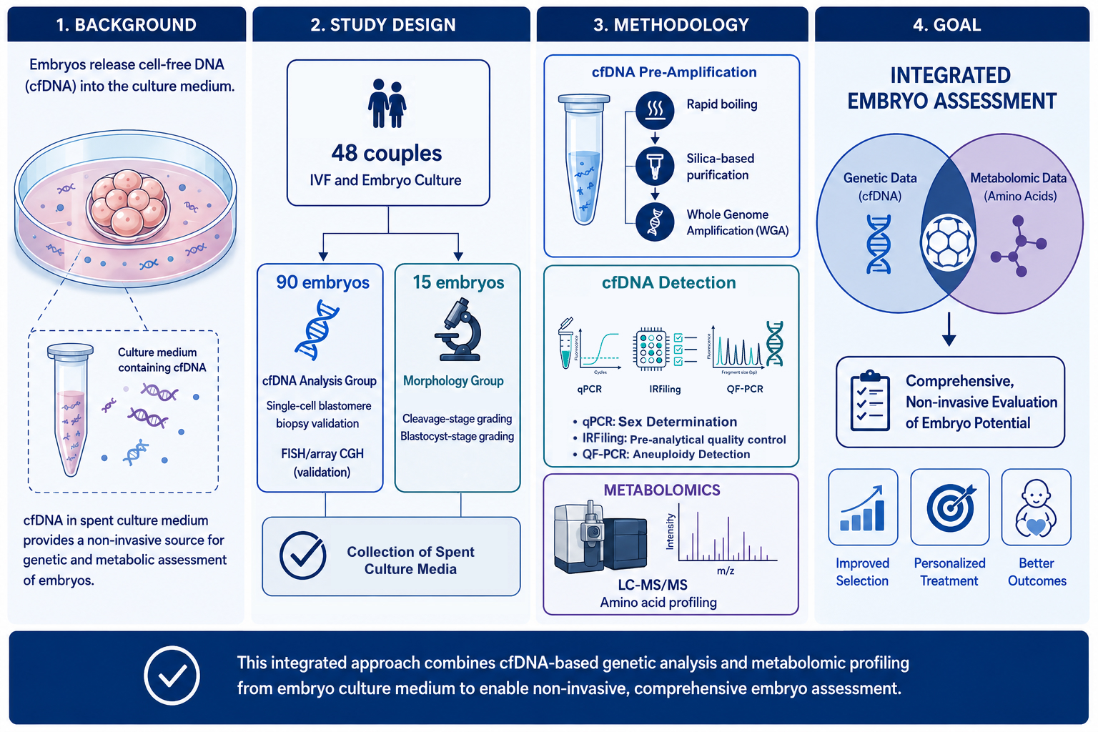
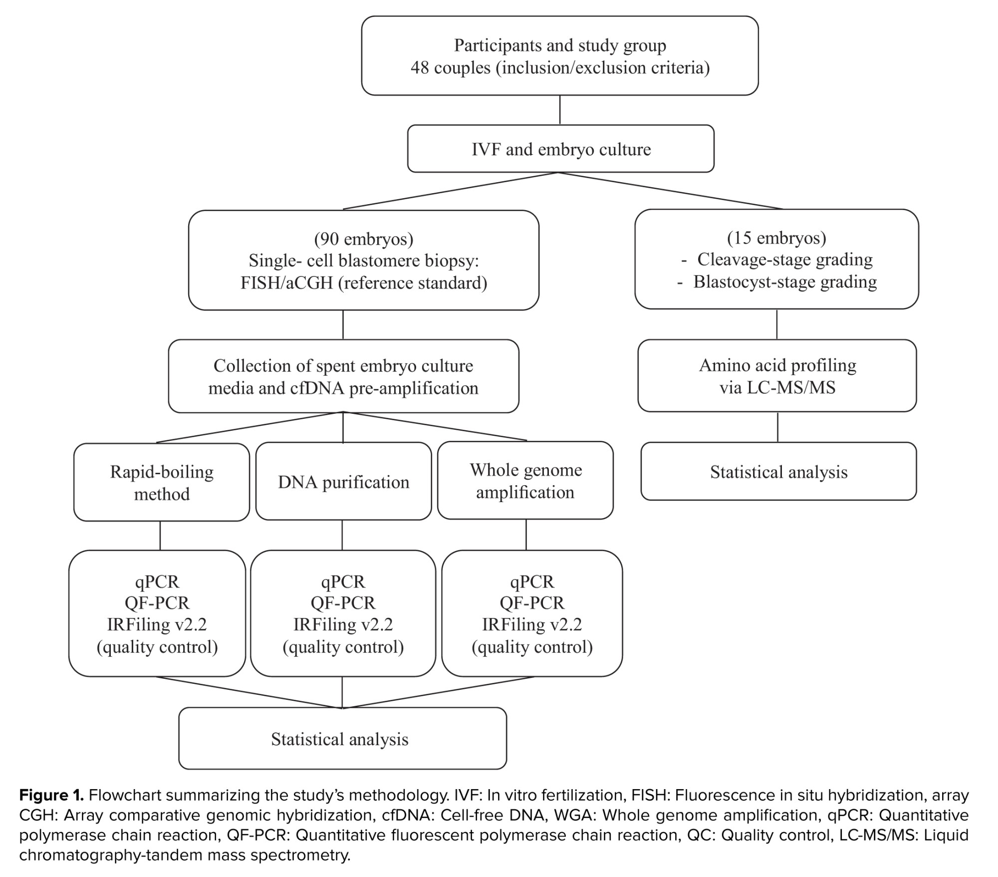
A total of 90 embryos were included in the genetic analysis phase. In addition, a separate set of 30 embryos were evaluated, from which 15 embryos with the desired characteristics were selected for the amino acid profiling phase. Figure 1 provides a schematic overview of the study methodology.
Full-Text: (54 Views)
1. Introduction
Multiple embryo transfers have historically been used to improve the success of in vitro fertilization (IVF). Still, they significantly increase the risk of adverse outcomes such as perinatal mortality, preterm birth, and maternal complications. To reduce these risks, many countries have limited the number of embryos transferred per cycle, particularly in older women (1). However, advanced maternal age itself is strongly associated with aneuploidy, which compromises embryo competence and IVF success. As embryonic aneuploidy is a significant cause of implantation failure, accurate identification of euploid embryos is critical to improve live birth outcomes (2).
Morphological grading remains the most common non-invasive tool, but it is unreliable because chromosomally abnormal embryos may still appear morphologically standard (3). Invasive preimplantation genetic testing for aneuploidy (PGT-A) overcomes this limitation, with the first successful pregnancy achieved in 1995 using fluorescence in situ hybridization (FISH). FISH has largely been replaced by more comprehensive approaches such as array comparative genomic hybridization (CGH), single-nucleotide polymorphism microarray, quantitative polymerase chain reaction (qPCR), and next-generation sequencing (NGS) (4). Yet invasive biopsy has drawbacks: it samples only a few cells that may not represent the whole embryo, especially in mosaic cases (5), and it requires costly equipment and specialized expertise, with evidence suggesting that biopsy itself can impair implantation (6).
These limitations have prompted growing interest in non-invasive alternatives. Time-lapse imaging, proteomics, metabolomics, and analysis of embryo culture media have all been investigated, though their diagnostic accuracy remains insufficient (7). Cell-free DNA (cfDNA) has emerged as a particularly promising biomarker. Genomic DNA has been detected in blastocoele fluid (8, 9), but aspiration remains semi-invasive and technically demanding. More recently, cfDNA has been isolated from spent blastocyst culture medium (SBM), a truly non-invasive approach (10). cfDNA levels are typically higher in SBM than in blastocoele fluid (11) and likely originate from both trophectoderm and inner cell mass (12), potentially providing a more complete representation of the embryo's chromosomal status (13). Still, concordance between cfDNA results and trophectoderm biopsy has varied widely (14).
In parallel, metabolic profiling of embryo culture media, particularly amino acid turnover, has been proposed as another non-invasive indicator of embryo viability and developmental competence (15). This study, therefore, aims to evaluate 2 complementary approaches: i) cfDNA-based assays (real-time polymerase chain reaction, quantitative fluorescent polymerase chain reaction [QF-PCR], and IRFiling) applied to SBM, and ii) amino acid profiling using liquid chromatography-tandem mass spectrometry (LC-MS/MS). These strategies are considered less invasive and potentially cost-effective alternatives to conventional invasive PGT-A and to the often unreliable morphological assessment (16).
2. Materials and Methods
2.1. Participants and study group
In this case-control study, 48 couples undergoing assisted reproduction treatment at the Yazd Institute of Reproductive Sciences in Yazd, Iran, between February 2021 and March 2023 were enrolled. The inclusion criteria were a normal parental karyotype, maternal age below 40 yr, and absence of Y-chromosome microdeletions in the male partner. Couples with a history of recurrent miscarriage or a child affected by chromosomal abnormalities were excluded. Furthermore, couples with fewer than 5 embryos were not eligible for inclusion, and a maximum of 4 embryos per couple were analyzed to ensure methodological consistency and minimize sampling bias.
A total of 90 embryos were included in the genetic analysis phase. In addition, a separate set of 30 embryos were evaluated, from which 15 embryos with the desired characteristics were selected for the amino acid profiling phase. Figure 1 provides a schematic overview of the study methodology.
2.2. IVF procedure and embryo culture
Controlled ovarian stimulation was performed using a gonadotropin-releasing hormone antagonist protocol tailored to individual patient responses. Oocyte retrieval was conducted 36 hr after human chorionic gonadotropin administration. To eliminate and minimize maternal contamination, cumulus-oocyte complexes were isolated using a sterile pipette. The oocytes were washed in G-IVF medium (Vitrolife, Sweden) and evaluated for nuclear maturity. Mature oocytes were inseminated 2-3 hr later via intracytoplasmic sperm injection (ICSI) (17). ICSI was used to eliminate paternal contamination due to sperm adhesion to the zona pellucida.
Following fertilization, embryos were cultured individually in 20 μL microdrops of ONESTEP medium (VITROMEDia, Germany) under 2.5 mL of mineral oil (Vitrolife, Sweden) at 37°C in an atmosphere of 6% CO₂, 5% O₂, and 89% N₂. Fertilization was assessed 16-18 hr post-ICSI. At the cleavage stage, all embryos were morphologically graded, and a single-cell biopsy was performed. The embryos were subsequently cultured until the blastocyst stage (day 5/6), at which point the culture medium was collected, and the embryos were graded again (18). As a negative control, 6 drops of culture medium without embryos were incubated under the same conditions. A positive control was prepared from the skin of a healthy male donor with a normal karyotype.
2.3. Blastomere biopsy
At the cleavage stage (2-8 cells) on the morning of day 3, approximately 68-72 hr after microinjection, embryo biopsy was performed using a micromanipulation system (Narashige, Japan) fitted on an inverted microscope (Diaphot 300, Nikon, Japan). A 1.48 µm diode laser (drilling method), in accordance with ESHRE guidelines, was used to dissect the zona pellucida before biopsy. A single blastomere was carefully removed under controlled laboratory conditions to minimize impact on embryo viability and evaluated under a ×400 inverted microscope for its integrity, presence of a nucleus, and freedom from other cells/debris (19). The samples were subsequently analyzed using CGH (n = 45) and FISH (n = 45).
2.4. Array CGH
On the third day, 45 embryos underwent blastomere biopsy followed by whole-genome amplification using the PicoPLEX Single Cell WGA kit (Rubicon Genomics, USA). Amplified DNA was hybridized on Genetisure Pre-Screen 8x60K custom microarrays (Agilent Technologies, USA). Hybridization signals were analyzed using Agilent Cytogenomics Software v4 against male and female reference standards (20). This procedure was performed at the Kariminejad-Najmabadi Pathology & Genetics Center, Tehran, Iran.
2.5. FISH
Another set of 45 embryos was analyzed using FISH technique. Following zona pellucida drilling and blastomere aspiration in Ca²⁺/Mg2+ free biopsy medium (LifeGlobal), isolated nuclei were fixed on pre-labeled slides. 2 sequential FISH protocols were performed to assess chromosomal abnormalities involving chromosomes 13, 18, 21, X (round 1), and 15, 16, 22, Y (round 2) using commercial probes (MetaSystems GmbH, Germany). Slide evaluation and scoring were performed by 2 technicians using an Olympus BX61 fluorescence microscope (21).
2.6. Collection of spent culture media
Spent blastocyst culture media were collected individually on day 5 or 6 and transferred into sterile 0.2 mL PCR-grade microtubes (Eppendorf, Germany). Samples were stored at -80°C. Blank media incubated under identical conditions served as controls. To enhance cfDNA yield, 3 pre-amplification strategies were employed.
2.7. cfDNA pre-amplification methods
Rapid-boiling method: the 30 samples (15 from the CGH group and 15 from the FISH group) were incubated at 56°C for 30 min, then at 100°C for 20 min to reduce the volume by ~50% and concentrate cfDNA (22). Concentration was assessed using a nanodrop spectrophotometer (Thermo Scientific).
DNA purification: a second set of 30 (15 from the CGH group and 15 from the FISH group) cfDNA samples was extracted using the High Pure Viral Nucleic Acid kit (Roche, Germany, Cat. No. 11858874001) and eluted in 15 µL of buffer, with concentration quantified using a nanodrop.
Whole genome amplification (WGA): the REPLI-g Single Cell Mini kit (QIAGEN, Cat. No. 150023) (23) was used for multiple displacement amplification. The amplification success of 30 SBMs (15 from the CGH group and 15 from the FISH group) was verified via nanodrop and 2% agarose gel electrophoresis.
2.8. Cell-free genomic DNA detection
2.8.1. Real-time PCR
qPCR amplification of the ZFX (495 bp) and SRY (472 bp) genes was performed using SYBR green chemistry on an applied biosystems real-time PCR system (24, 25). The primer sequences used were as follows: ZFX (forward: 5′-ACC RCT GTA CTG ACT GTG ATT ACA C-3′; reverse: 5′-GCA CYT CTT TGG TAT CYG AGA AAG T-3′) and SRY (forward: 5’-GAA TAT TCC CGC TCT CCG GA-3’; reverse: 5’-GCT GGT GCT CCA TTC TTG AG-3’). Each 20-µL reaction contained 10 µL SYBR green master mix (Applied Biosystems), 0.8 µL of each primer (10 µM), 2 µL of template DNA, and 6.4 µL nuclease-free water; it was run on an Applied Biosystems thermocycler. Thermal cycling consisted of 95°C for 5 min, followed by 40 cycles of 95°C for 15 s, 58°C for 30 s, and 72°C for 20 s, and a melt-curve analysis from 65-95°C, at 0.5°C/sec increments. Samples positive for both ZFX and SRY were classified as male, whereas samples positive only for ZFX were considered female. Each run included male, female, and no-template controls (24, 25).
2.8.2. IRFiling™ kit v2.2 (Kawsar Biotech): pre-analytical quality control (QC)
IRFiling v2.2 (Kawsar Biotech) was employed as a pre-analytical QC assay to assess the integrity and amplifiability of SBM-derived cfDNA prior to QF-PCR. The multiplex STR panel includes 17 autosomal and sex chromosome markers, analyzed by capillary electrophoresis with 5-dye resolution. For each sample, IRFiling profiles were visually inspected and classified based on signal quality, peak height, and allele-pattern consistency.
Importantly, IRFiling results were not used as exclusion criteria. To avoid potential data loss and minimize selection bias associated with low-quantity and fragmented cfDNA, all samples underwent QF-PCR analysis regardless of IRFiling performance. IRFiling status (adequate vs. suboptimal profile) was recorded and later used in stratified analyses to explore its association with QF-PCR success rate and interpretability of results, rather than as a stand-alone diagnostic tool for aneuploidy in SBM.
2.8.3. QF-PCR (AneuQuick™ v3.2, Kawsar Biotech)
Amplified 26 STR markers across chromosomes 13, 18, 21, X, and Y. PCR products were analyzed on an ABI Genetic Analyzer and interpreted using GeneMapper v6.
2.9. Amino acid profiling via LC-MS/MS
15 embryos were selected based on their morphology and developmental stage. The day 3 group included 9 embryos: three 2 pronuclear (PN) (normal), three 1PN, and three 3PN embryos. The day 5 group consisted of 6 blastocysts: 3 with high morphological quality and 3 with poor morphological quality. High-quality blastocysts were defined as having an expanded blastocoel, cohesive inner cell mass, and well-organized trophectoderm (18).
For amino acid profiling, spent culture media from each embryo were collected and sent to Farzaneh Clinical Laboratory (Shiraz, Iran) for LC-MS/MS analysis. Measurements were performed on a triple quadrupole LC-MS/MS system (AB Sciex, Framingham, MA, USA) equipped with a TurboIonSpray electrospray ionization source, operated in positive-ion mode under multiple reaction monitoring conditions. 3 drops of blank culture medium were used as negative controls to confirm the absence of background contamination.
2.10. Ethical Considerations
This study was approved by the Ethics Committee of the Yazd Institute of Reproductive Sciences, Yazd, Iran (Code: IR.SSU.RSI.REC.1399.036). Written informed consent was obtained from all participants prior to enrollment. All couples underwent pre-treatment counseling and provided explicit permission for the use of their clinical data and treatment outcomes for research purposes, with strict confidentiality guaranteed.
2.11. Statistical Analysis
Statistical analyses for cfDNA-based assays were performed using IBM SPSS Statistics (v26.0; IBM Corp., Armonk, NY, USA). Because qPCR and QF-PCR yielded categorical call outcomes and the sample size was limited, results were summarized descriptively. Diagnostic performance metrics, including concordance and misclassification rate, were calculated using FISH/array CGH as the reference standard. Incorrect outcomes were defined as any discordant call relative to the reference standard or a failed result (no-detect), and no-detect samples were therefore classified as incorrect calls in all performance calculations. No formal inferential statistical testing was performed to compare cfDNA workflows.
IRFiling results were not used for diagnostic performance calculations, as the assay served only as a pre-analytical QC step. Instead, IRFiling status (adequate vs. suboptimal profile) was recorded and descriptively compared with QF-PCR outcomes to explore its potential association with the QF-PCR success rate and the interpretability of the results.
For LC-MS/MS amino-acid quantification, between-group differences were evaluated using one-way ANOVA for each amino acid. When the ANOVA indicated a significant group effect (p < 0.05), Tukey’s HSD test was used for post-hoc pairwise comparisons to control the family-wise error rate. Data were reported as mean ± SD from 3 biological replicates per group (n = 3). Assumptions of normality and homogeneity of variances were assessed using the Shapiro-Wilk and Levene’s tests, respectively. All statistical analyses were performed using SPSS (version 26.0; IBM Corp., Armonk, NY, USA).
3. Results
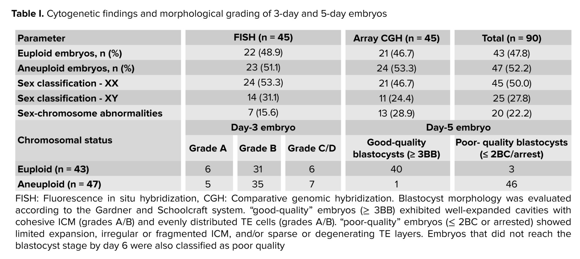
90 embryos were analyzed via FISH (n = 45) and array CGH (n = 45) to validate cfDNA-based assays. FISH identified 22 (48.9%) euploid and 23 (51.1%) aneuploid embryos; similarly, array CGH classified 21 (46.7%) as euploid and 24 (53.3%) as aneuploid (Table I). Sex-chromosome abnormalities (XO, XXY, XXX, XXYY) occurred in 22.2% of embryos across both methods, while trisomies 13, 18, and 21 were the most frequent autosomal anomalies.
Morphological assessment at cleavage (day 3) and blastocyst (day 5/6) stages revealed that most day-3 embryos were grade B (Table I). By day 5/6, euploid embryos typically progressed to high-quality blastocysts, whereas aneuploid embryos frequently arrested or displayed poor quality, establishing a robust baseline for cfDNA analyses (Table I).
3.1. Detection performance of cfDNA-based methods
3.1.1. Rapid-boiling group
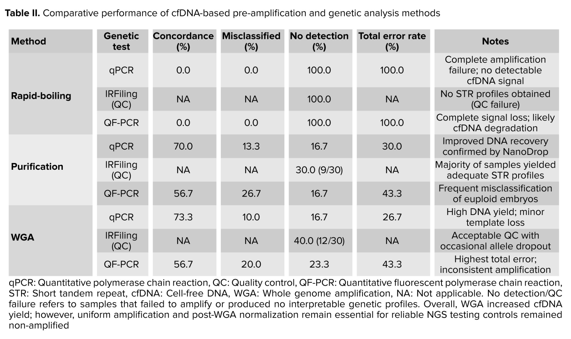
30 heat-concentrated SBM samples failed to amplify ZFX or SRY genes, resulting in 100% failure in sex determination. Likewise, both IRFiling™ and QF-PCR assays yielded no detectable STR profiles, resulting in 0% concordance with reference methods (Table II). Although reference testing identified 14 euploid and 16 aneuploid embryos, heat-processed samples generated no informative data, suggesting cfDNA degradation or poor recovery. Nanodrop spectrophotometry showed no significant differences in DNA concentration or purity compared with untreated samples, indicating that despite volume loss, thermal exposure did not intrinsically compromise cfDNA integrity.
3.1.2. Purification group
30 cfDNA samples from spent blastocyst media were purified before qPCR, IRFiling, and QF-PCR analysis. NanoDrop confirmed successful DNA recovery with higher purity than heat-treated samples. qPCR-based sex determination showed 70.0% concordance, with 13.3% misclassified and 16.7% undetectable samples (total error: 30.0%), mainly due to low template yield or partial degradation. IRFiling, used exclusively as a pre-analytical fluorescence QC tool, showed 9 of 30 failures (30.0%). Subsequent QF-PCR analysis demonstrated 56.7% overall concordance and a 16.7% undetectable rate (Table II).
3.1.3. WGA group
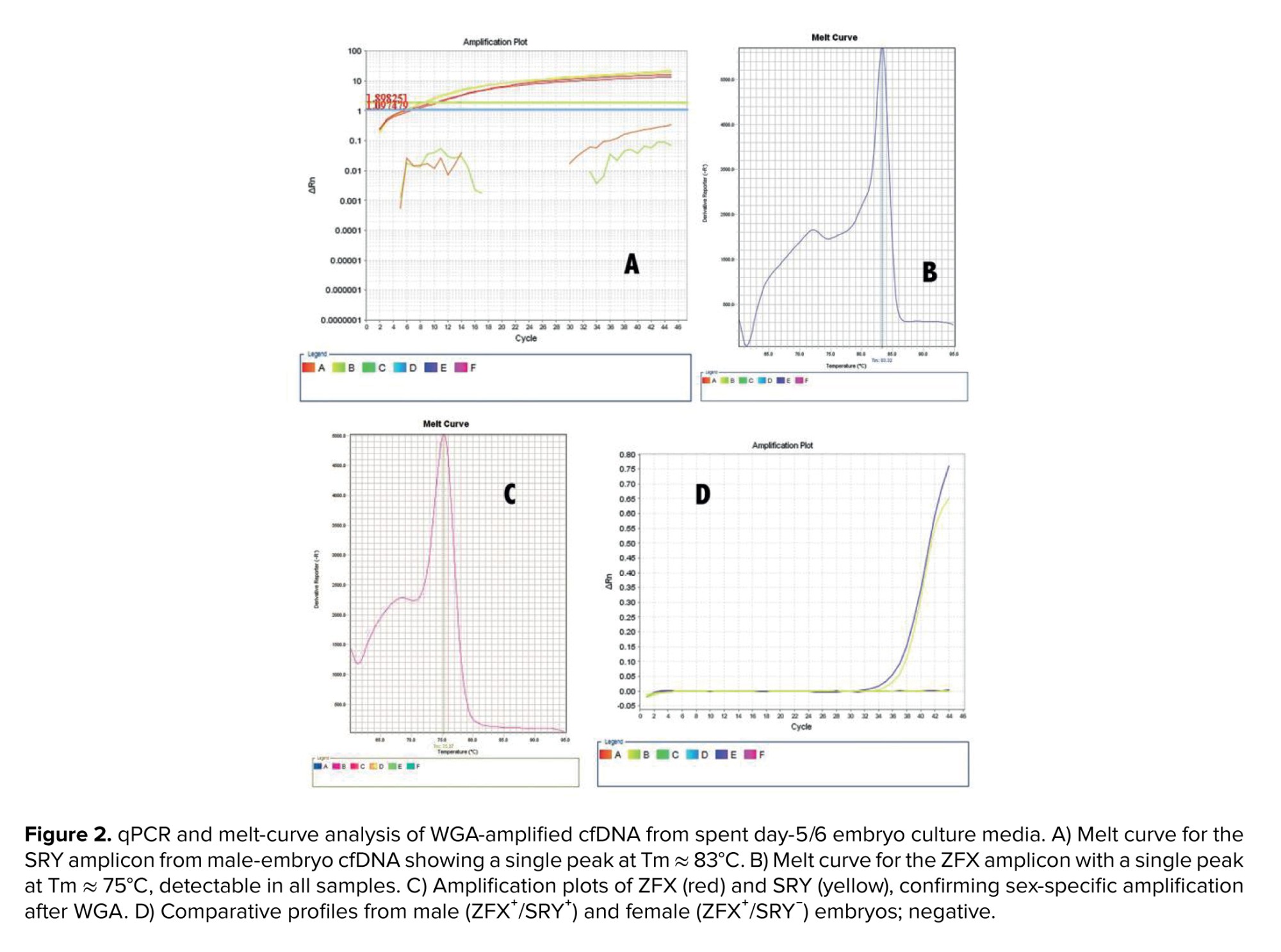
All 30 cfDNA samples underwent successful WGA, confirmed via NanoDrop and agarose gel electrophoresis. Sex-specific qPCR targeting ZFX and SRY confirmed cfDNA origin and assay performance: ZFX was detected in all samples, whereas SRY amplification was observed only in media from male embryos (Figure 2). qPCR-based sex determination achieved 73.3% concordance, with 10.0% misclassified and 16.7% undetectable samples (26.7% total error), likely due to template loss during pre-amplification. IRFiling QC failure was 40.0% (12/30 samples). QF-PCR achieved 56.7% concordance, with 20.0% misclassified and 23.3% undetectable outcomes (43.3% error; Table II). Assay performance was assessed via descriptive diagnostic measures, given the categorical data and limited sample size.
3.2. Amino acid profiling of embryo culture media
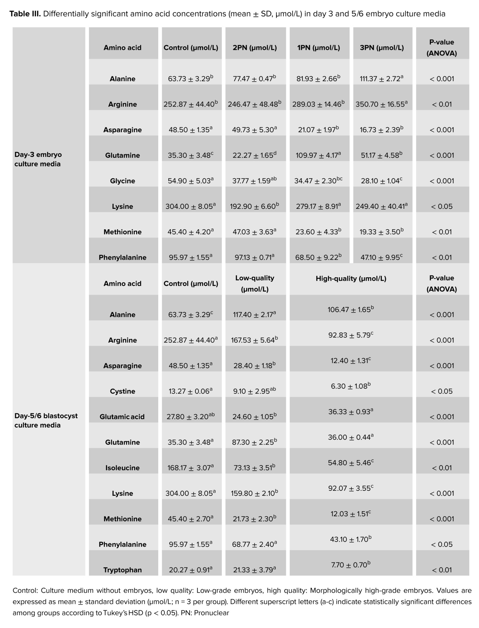
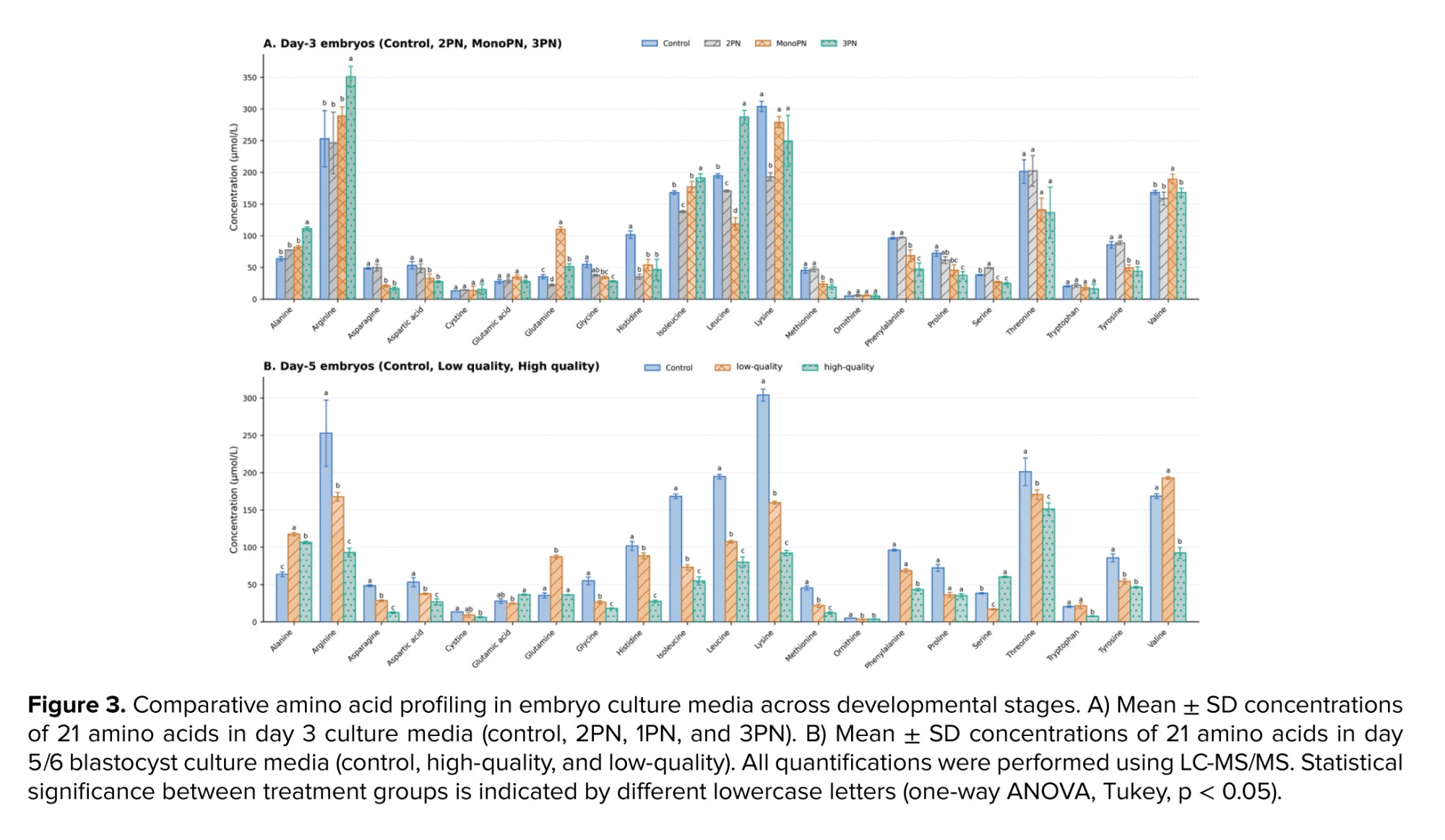
A targeted LC-MS/MS workflow was employed to quantify amino acids in spent culture media from day-3 embryos (control, 2PN, 1PN, 3PN) and day-5/6 blastocysts (control, high-quality, low-quality). Data were presented as mean ± SD and analyzed by one-way ANOVA followed by Tukey's post hoc test, with p < 0.05 considered statistically significant (Table III; Figure 3).
3.2.1. Amino-acid profiling at the cleavage stage (day 3)
Amino acid profiling of day-3 culture media revealed distinct metabolic patterns across PN groups. The most pronounced deviations were observed in 3PN embryos, characterized by marked elevations in alanine (111.37 ± 2.72 µmol/L vs. control 63.73 ± 3.29 and 2PN 77.47 ± 0.47), arginine (350.70 ± 16.55 vs. control 252.87 ± 44.40 and 2PN 246.47 ± 48.48), and leucine (287.40 ± 10.42), together with reductions in methionine (19.33 ± 3.50), phenylalanine (47.10 ± 9.95), glycine (28.10 ± 1.04), and asparagine (16.73 ± 2.39). This pattern indicates widespread metabolic disturbance associated with tripronuclear development (Figure 3).
MonoPN embryos exhibited a distinct glutamine-enriched profile, with glutamine reaching 109.97 ± 4.17 µmol/L compared with control (35.30 ± 3.48), 2PN (22.27 ± 1.65), and 3PN (51.17 ± 4.58). Both MonoPN and 3PN embryos showed markedly reduced asparagine levels compared with the control and 2PN groups, suggesting a shared disruption of amino acid metabolism.
In contrast, 2PN embryos displayed a comparatively less perturbed metabolic pattern than the abnormal PN groups, although several amino acids still differed from control levels. Overall, these findings demonstrate that PN status is strongly associated with amino-acid metabolic variation at the cleavage stage, with 3PN embryos showing the broadest disturbance and MonoPN embryos exhibiting a selective glutamine-dominant shift (Table III).
3.2.2. Amino-acid profiling at the blastocyst stage (day 5/6)
Amino-acid profiling of day-5/6 blastocyst media revealed clear metabolic differences between low and high-quality embryos. Low-quality blastocysts showed selective increases in a limited number of amino acids, most notably alanine (117.40 ± 2.17 µmol/L vs. high-quality 106.47 ± 1.65 and control 63.73 ± 3.29), glutamine (87.30 ± 2.25 vs. control 35.30 ± 3.48), and valine (192.70 ± 1.91 vs. control 168.40 ± 3.10). In contrast, many other amino acids (including glycine, histidine, isoleucine, leucine, lysine, methionine, phenylalanine, and asparagine) were reduced in low-quality embryos, indicating impaired metabolic turnover.
High-quality blastocysts exhibited a distinct pattern of pronounced depletion across multiple amino acids. Arginine fell to 92.83 ± 5.79 µmol/L (low-quality 167.53 ± 5.64; control 252.87 ± 44.40), asparagine to 12.40 ± 1.31 µmol/L (low-quality 28.40 ± 1.18; control 48.50 ± 1.35), and methionine to 12.03 ± 1.51 µmol/L. Broad reductions across several essential and aromatic amino acids further reflected enhanced metabolic utilization.
A notable exception was serine, which was markedly elevated in the spent medium of high-quality embryos (60.17 ± 1.12 µmol/L vs. low-quality 16.83 ± 0.90), consistent with upregulated one-carbon metabolism necessary for blastocyst expansion. Overall, the day-5/6 metabolic profile indicates that the spent medium of low-quality embryos exhibit mixed disruptions involving selective accumulation and widespread depletion, whereas high-quality blastocysts demonstrate coordinated amino-acid consumption and pathway-specific metabolic activity, underscoring the strong association between metabolic regulation and developmental competence.
3.2.3. Cross-stage metabolic comparison (day 3 → day 5/6)
Integration of day-3 and day-5/6 amino-acid profiles revealed consistent metabolic signatures associated with embryo quality. The disturbances observed in 3PN embryos at day 3 notably elevated alanine (111.4 ± 2.7 µmol/L) and reduced glycine (28.1 ± 1.0 µmol/L) persisted into the blastocyst stage in low-quality embryos, where alanine remained elevated (117.4 ± 2.2 µmol/L), and glycine remained low (26.5 ± 2.1 µmol/L). This parallel pattern suggests that impaired uptake and mitochondrial inefficiency are persistent features of metabolically compromised embryos.
Conversely, the balanced profile of 2PN embryos at day 3 aligned with high-quality blastocysts, characterized by pronounced depletion of arginine (92.8 ± 5.8 µmol/L), asparagine (12.4 ± 1.3 µmol/L), and methionine (12.0 ± 1.9 µmol/L), indicating efficient intracellular utilization and biosynthetic competence. While 1PN embryos showed transient glutamine enrichment (110.0 ± 4.2 µmol/L) at day 3, this phenotype did not persist. Instead, high-quality blastocysts uniquely exhibited elevated serine (60.2 ± 1.1 µmol/L), supporting one-carbon metabolism required for expansion. Overall, cleavage-stage metabolic status predicts blastocyst quality: 3PN embryos show persistent accumulation, 2PN embryos maintain efficient utilization, and 1PN embryos display a transient, non-beneficial metabolic phenotype.
4. Discussion
This study demonstrated that WGA and DNA purification significantly enhance cfDNA recovery compared to rapid boiling (which yielded no detectable signal) achieving 70% concordance for sex determination and 56.7% for aneuploidy detection. Concurrently, LC-MS/MS profiling identified distinct quality-dependent metabolic signatures in culture media, supporting their potential as complementary markers for embryo viability. Nonetheless, both cfDNA and metabolomics-based strategies require substantial technical optimization before clinical implementation.
4.1. Inefficacy of rapid boiling for cfDNA recovery
Rapid boiling failed to detect cfDNA, with both real-time PCR and QF-PCR showing 0% concordance with FISH/array CGH data. Despite reports of successful heat-based extraction (22), its performance remains inconsistent-likely due to thermal degradation, poor DNA release, or co-extracted inhibitors. Consequently, rapid boiling lacks the requisite sensitivity for reliable cfDNA-based PGT.
4.2. DNA purification improves cfDNA yield but still suffers from low-copy limitations
While DNA purification enabled downstream PCR, real-time PCR yielded only ~70% concordance for sex determination (13% misclassified, 17% undetectable). This reflects the low abundance and biological variability of embryonic cfDNA in culture media (23, 26). Nonetheless, combining spent media with whole-genome amplification and NGS can yield sufficient DNA for PGT-A (27).
Multiplex STR genotyping via QF-PCR showed moderate concordance, limited by allele dropout, contamination, mosaicism, and low template copy numbers (15, 28). Here, IRFiling served as a pre-analytical fluorescence-based QC to assess cfDNA signal adequacy before STR analysis.
Spent culture media analysis remains partially feasible; multicopy loci (e.g., TBC1D3) amplify in > 90% of samples, and Y-linked markers show higher reliability at the blastocyst stage. Similarly, qPCR-based single-gene diagnostic rates are high, despite occasional allele dropout (29). Semi-invasive blastocoel fluid aspiration also recovers cfDNA in most samples (8). Overall, the low-copy, fragmented nature of embryonic cfDNA remains the primary bottleneck for clinical reliability.
4.3. WGA delivers the strongest analytical performance among cfDNA workflows
4.3.1. Real-time PCR outcomes
WGA increased sex-determination concordance to ~73% and reduced the number of undetectable samples compared with purification alone. However, occasional misclassifications persisted, highlighting the challenge of amplifying low-copy cfDNA (26). While combining WGA with NGS has achieved full FISH concordance (27), our findings confirm that template stabilization and workflow optimization remain essential to maximize diagnostic reliability.
4.3.2. QF-PCR and IRFiling outcomes
In multiplex STR analysis, WGA achieved ~56.7% concordance, limited by undetectable signals and allele dropout. This aligns with reported cfDNA-based embryo testing concordances of 50-70%, where low abundance and contamination remain major constraints (30, 31). Here, IRFiling served as a pre-analytical QC; the lower failure rate observed after WGA indicates improved signal detectability via template pre-amplification.
Although WGA enhances downstream detectability by amplifying low-copy templates, stochastic amplification bias and allelic imbalance can introduce false positives. Consequently, refining primer design, cycle control, and post-WGA normalization remains essential (32).
4.4. Amino-acid profiling reveals functional metabolic signatures of embryo viability
Amino acid profiling complemented cfDNA analysis by capturing development-associated metabolic activity. LC-MS/MS quantification revealed distinct stage- and quality-dependent differences in amino acid utilization between day 3 embryos and day 5/6 blastocysts, reflecting developmental shifts in carbon-nitrogen processing. Furthermore, abnormal PN embryos (1PN, 3PN) and low-grade blastocysts exhibited altered amino acid turnover, suggesting disrupted redox balance and impaired mTOR-related metabolic regulation.
4.4.1. Amino-acid changes in embryo-free control media
In control droplets, amino acid concentrations either remained stable or declined relative to fresh media (33). These reductions likely stem from spontaneous chemical degradation rather than biological uptake. Key mechanisms include non-enzymatic glutamine hydrolysis, oxidative instability of sulfur-containing amino acids, oil adsorption, and the absence of embryo-driven metabolic homeostasis (34).
4.4.2. Day-3 embryos display distinct PN-dependent metabolic patterns (1PN, 2PN, 3PN)
2PN embryos exhibited a balanced profile, consistent with a “quiet but competent” phenotype and optimal anabolic flux (35). Conversely, 1PN embryos showed marked metabolic disruption: glutamine accumulation (212% vs. control) and substantial asparagine depletion (-58%). This pattern likely reflects impaired zygotic activation and altered amide group diversion, rather than enhanced glutaminolysis (36). 3PN embryos showed the most pronounced deviations. Compared to 2PN, alanine increased by 45%, whereas asparagine, methionine, and phenylalanine dropped by over 50%, and leucine rose 1.5-fold. These abnormalities signal redox imbalance and mitochondrial dysfunction; specifically, elevated alanine levels suggest enhanced pyruvate transamination under oxidative stress (37), while increased arginine levels indicate reduced utilization in the NO and polyamine pathways (38). The concurrent depletion of methionine and phenylalanine suggests reduced methylation capacity and weakened antioxidant defense.
Across all groups, alanine consistently differentiated fertilization outcomes, increasing by 70% in 3PN vs. 2PN embryos. Given that elevated alanine in spent media is strongly correlated with reduced implantation potential (35, 37), it emerges as a robust, non-invasive marker that distinguishes 2PN from abnormal fertilization and serves as a potential indicator of developmental competence.
4.4.3. High-quality blastocysts show selective amino-acid uptake and efficient nitrogen metabolism
High-quality blastocysts displayed selective amino acid uptake, characterized by significant extracellular depletion of arginine (63%), asparagine (74%), glycine (67%), leucine (59%), methionine (74%), phenylalanine (65%), and tryptophan (62%) relative to controls. Conversely, glutamic acid levels increased by 31% over controls and 48% versus low-quality embryos. This metabolic profile indicates active transamination, coordinated one-carbon metabolism, and mTORC1 activation-hallmarks of metabolically competent, implantation-ready embryos (37).
Although the literature suggests that amino acid flux is influenced by oxygen tension, protein supplementation, and analytical platform variability (34, 39, 40), our results confirm robust depletion trends. Contextual factors may modulate absolute flux values; however, under physiological culture conditions (5 percent oxygen, optimized media), high-quality blastocysts consistently exhibit depletion patterns indicative of active uptake and oxidation.
4.4.4. Low-quality blastocysts release excess amino acids and exhibit metabolic stress signatures
Low-quality blastocysts showed a release-dominant amino acid profile consistent with inefficient utilization and metabolic stress. Alanine increased by 84% relative to controls and by 10% relative to high-quality embryos, suggesting enhanced pyruvate transamination under mitochondrial stress (35). Glutamine exhibited the largest rise (147% vs. control; 2.4-fold vs. high-quality), indicating reduced uptake rather than effective glutaminolysis (36). Arginine and asparagine displayed intermediate depletion patterns (Arg 34% below control but 80% higher than high-quality; Asn 41% below control but 129% higher than high-quality), reflecting incomplete engagement of nucleotide synthesis and NO/polyamine pathways (38). Aromatic amino acids were markedly elevated (phenylalanine, 265%; tryptophan, 177%), consistent with reduced uptake and diminished antioxidant capacity (37). External conditions and analytical factors may influence amino-acid flux measurements. Oxygen tension, protein supplementation, and medium composition can alter metabolic turnover, and in some cases even competent embryos may appear to release amino acids. Additionally, different metabolomic platforms, including LC-MS/MS and Raman profiling, report partially distinct discriminant metabolites (34, 37, 39). Despite these methodological differences, several studies consistently identify elevated alanine, glutamine, and aromatic amino acids as indicators of metabolic inefficiency under physiological culture conditions.
Among all metabolites, phenylalanine showed the strongest discrimination between embryo qualities, with concentrations 3.65-fold higher in low-quality embryos. Lower levels in high-quality blastocysts likely reflect active uptake for protein synthesis and redox regulation, whereas accumulation in low-quality embryos suggests impaired utilization (37). Overall, the day-5/6 metabolic profile indicates that efficient amino acid uptake and coordinated transamination characterize high-quality blastocysts, whereas extracellular accumulation of alanine, glutamine, and aromatic amino acids reflects metabolic dysfunction in low-quality counterparts. Early amino-acid flux may also contribute to long-term metabolic programming and epigenetic regulation during embryonic development (41).
Overall, the findings show that WGA and DNA purification improved cfDNA yield and analytical quality compared with the rapid boiling method, although concordance with embryo biopsy remained moderate. Amino-acid profiling also revealed metabolic signatures associated with embryonic developmental competence, supporting the potential value of integrating genetic and metabolomic approaches for non-invasive embryo assessment. Consistent with recent systematic reviews, noninvasive preimplantation genetic testing shows high specificity for detecting euploid embryos but limited sensitivity for mosaicism (42), with cfDNA quantity and timing of media collection identified as key factors affecting diagnostic performance (43). Other studies likewise report very low false-negative rates for euploid embryos while confirming moderate but reproducible concordance across laboratories using cfDNA from spent culture media (44).
5. Conclusion
Non-invasive analysis of embryo culture media may provide complementary genetic and metabolic information for embryo assessment without biopsy. In this study, WGA achieved the highest sensitivity for cfDNA analysis, while LC-MS/MS amino acid profiling identified metabolic patterns associated with embryonic competence. However, low cfDNA yield, amplification bias, and metabolite instability continue to limit diagnostic reliability. Future studies should focus on standardized sampling, improved cfDNA capture, and integration of genomic and metabolomic approaches. Large multicenter validation studies will be required before these non-invasive strategies can be reliably implemented in clinical practice.
Data Availability
Data supporting the findings of this study are available upon reasonable request from the corresponding author.
Author Contributions
SM. Kalantar, H. Sharifiyazdi, and F. Montazeri designed the study. M. Jahanara, H. Sharifiyazdi, F. Montazeri, and M. Moshrefi conducted the research, evaluated, and analyzed the results of the study. Further, SM. Kalantar, N. Ghasemi, and M. Ghaemi reviewed the article. All authors approved the final manuscript and take responsibility for the integrity of the data.
This work was conducted under joint senior supervision, with 2 authors assuming complementary corresponding-level responsibilities. One author was primarily responsible for the clinical and genetic analysis components of the study, while the other oversaw the metabolomic analyses, manuscript development, and scientific correspondence. Given the interdisciplinary nature of the work, both authors made essential and continuous contributions to the conception of the study, its supervision, the interpretation of the findings, and the reporting of the results.
Accordingly, listing both individuals as corresponding authors accurately reflects the leadership structure of the study and helps ensure that editorial and scientific queries are addressed appropriately and efficiently. To facilitate streamlined communication, Dr. Fateme Montazeri can serve as the primary contact for administrative correspondence, if preferred by the journal.
Acknowledgments
The authors thank all contributors, with special gratitude to Professor Abbas Aflatoonian for his ongoing guidance. Grammarly was used solely for English language editing.
Conflict of Interest
The authors declare that there is no conflict of interest.
Following fertilization, embryos were cultured individually in 20 μL microdrops of ONESTEP medium (VITROMEDia, Germany) under 2.5 mL of mineral oil (Vitrolife, Sweden) at 37°C in an atmosphere of 6% CO₂, 5% O₂, and 89% N₂. Fertilization was assessed 16-18 hr post-ICSI. At the cleavage stage, all embryos were morphologically graded, and a single-cell biopsy was performed. The embryos were subsequently cultured until the blastocyst stage (day 5/6), at which point the culture medium was collected, and the embryos were graded again (18). As a negative control, 6 drops of culture medium without embryos were incubated under the same conditions. A positive control was prepared from the skin of a healthy male donor with a normal karyotype.
2.3. Blastomere biopsy
At the cleavage stage (2-8 cells) on the morning of day 3, approximately 68-72 hr after microinjection, embryo biopsy was performed using a micromanipulation system (Narashige, Japan) fitted on an inverted microscope (Diaphot 300, Nikon, Japan). A 1.48 µm diode laser (drilling method), in accordance with ESHRE guidelines, was used to dissect the zona pellucida before biopsy. A single blastomere was carefully removed under controlled laboratory conditions to minimize impact on embryo viability and evaluated under a ×400 inverted microscope for its integrity, presence of a nucleus, and freedom from other cells/debris (19). The samples were subsequently analyzed using CGH (n = 45) and FISH (n = 45).
2.4. Array CGH
On the third day, 45 embryos underwent blastomere biopsy followed by whole-genome amplification using the PicoPLEX Single Cell WGA kit (Rubicon Genomics, USA). Amplified DNA was hybridized on Genetisure Pre-Screen 8x60K custom microarrays (Agilent Technologies, USA). Hybridization signals were analyzed using Agilent Cytogenomics Software v4 against male and female reference standards (20). This procedure was performed at the Kariminejad-Najmabadi Pathology & Genetics Center, Tehran, Iran.
2.5. FISH
Another set of 45 embryos was analyzed using FISH technique. Following zona pellucida drilling and blastomere aspiration in Ca²⁺/Mg2+ free biopsy medium (LifeGlobal), isolated nuclei were fixed on pre-labeled slides. 2 sequential FISH protocols were performed to assess chromosomal abnormalities involving chromosomes 13, 18, 21, X (round 1), and 15, 16, 22, Y (round 2) using commercial probes (MetaSystems GmbH, Germany). Slide evaluation and scoring were performed by 2 technicians using an Olympus BX61 fluorescence microscope (21).
2.6. Collection of spent culture media
Spent blastocyst culture media were collected individually on day 5 or 6 and transferred into sterile 0.2 mL PCR-grade microtubes (Eppendorf, Germany). Samples were stored at -80°C. Blank media incubated under identical conditions served as controls. To enhance cfDNA yield, 3 pre-amplification strategies were employed.
2.7. cfDNA pre-amplification methods
Rapid-boiling method: the 30 samples (15 from the CGH group and 15 from the FISH group) were incubated at 56°C for 30 min, then at 100°C for 20 min to reduce the volume by ~50% and concentrate cfDNA (22). Concentration was assessed using a nanodrop spectrophotometer (Thermo Scientific).
DNA purification: a second set of 30 (15 from the CGH group and 15 from the FISH group) cfDNA samples was extracted using the High Pure Viral Nucleic Acid kit (Roche, Germany, Cat. No. 11858874001) and eluted in 15 µL of buffer, with concentration quantified using a nanodrop.
Whole genome amplification (WGA): the REPLI-g Single Cell Mini kit (QIAGEN, Cat. No. 150023) (23) was used for multiple displacement amplification. The amplification success of 30 SBMs (15 from the CGH group and 15 from the FISH group) was verified via nanodrop and 2% agarose gel electrophoresis.
2.8. Cell-free genomic DNA detection
2.8.1. Real-time PCR
qPCR amplification of the ZFX (495 bp) and SRY (472 bp) genes was performed using SYBR green chemistry on an applied biosystems real-time PCR system (24, 25). The primer sequences used were as follows: ZFX (forward: 5′-ACC RCT GTA CTG ACT GTG ATT ACA C-3′; reverse: 5′-GCA CYT CTT TGG TAT CYG AGA AAG T-3′) and SRY (forward: 5’-GAA TAT TCC CGC TCT CCG GA-3’; reverse: 5’-GCT GGT GCT CCA TTC TTG AG-3’). Each 20-µL reaction contained 10 µL SYBR green master mix (Applied Biosystems), 0.8 µL of each primer (10 µM), 2 µL of template DNA, and 6.4 µL nuclease-free water; it was run on an Applied Biosystems thermocycler. Thermal cycling consisted of 95°C for 5 min, followed by 40 cycles of 95°C for 15 s, 58°C for 30 s, and 72°C for 20 s, and a melt-curve analysis from 65-95°C, at 0.5°C/sec increments. Samples positive for both ZFX and SRY were classified as male, whereas samples positive only for ZFX were considered female. Each run included male, female, and no-template controls (24, 25).
2.8.2. IRFiling™ kit v2.2 (Kawsar Biotech): pre-analytical quality control (QC)
IRFiling v2.2 (Kawsar Biotech) was employed as a pre-analytical QC assay to assess the integrity and amplifiability of SBM-derived cfDNA prior to QF-PCR. The multiplex STR panel includes 17 autosomal and sex chromosome markers, analyzed by capillary electrophoresis with 5-dye resolution. For each sample, IRFiling profiles were visually inspected and classified based on signal quality, peak height, and allele-pattern consistency.
Importantly, IRFiling results were not used as exclusion criteria. To avoid potential data loss and minimize selection bias associated with low-quantity and fragmented cfDNA, all samples underwent QF-PCR analysis regardless of IRFiling performance. IRFiling status (adequate vs. suboptimal profile) was recorded and later used in stratified analyses to explore its association with QF-PCR success rate and interpretability of results, rather than as a stand-alone diagnostic tool for aneuploidy in SBM.
2.8.3. QF-PCR (AneuQuick™ v3.2, Kawsar Biotech)
Amplified 26 STR markers across chromosomes 13, 18, 21, X, and Y. PCR products were analyzed on an ABI Genetic Analyzer and interpreted using GeneMapper v6.
2.9. Amino acid profiling via LC-MS/MS
15 embryos were selected based on their morphology and developmental stage. The day 3 group included 9 embryos: three 2 pronuclear (PN) (normal), three 1PN, and three 3PN embryos. The day 5 group consisted of 6 blastocysts: 3 with high morphological quality and 3 with poor morphological quality. High-quality blastocysts were defined as having an expanded blastocoel, cohesive inner cell mass, and well-organized trophectoderm (18).
For amino acid profiling, spent culture media from each embryo were collected and sent to Farzaneh Clinical Laboratory (Shiraz, Iran) for LC-MS/MS analysis. Measurements were performed on a triple quadrupole LC-MS/MS system (AB Sciex, Framingham, MA, USA) equipped with a TurboIonSpray electrospray ionization source, operated in positive-ion mode under multiple reaction monitoring conditions. 3 drops of blank culture medium were used as negative controls to confirm the absence of background contamination.
2.10. Ethical Considerations
This study was approved by the Ethics Committee of the Yazd Institute of Reproductive Sciences, Yazd, Iran (Code: IR.SSU.RSI.REC.1399.036). Written informed consent was obtained from all participants prior to enrollment. All couples underwent pre-treatment counseling and provided explicit permission for the use of their clinical data and treatment outcomes for research purposes, with strict confidentiality guaranteed.
2.11. Statistical Analysis
Statistical analyses for cfDNA-based assays were performed using IBM SPSS Statistics (v26.0; IBM Corp., Armonk, NY, USA). Because qPCR and QF-PCR yielded categorical call outcomes and the sample size was limited, results were summarized descriptively. Diagnostic performance metrics, including concordance and misclassification rate, were calculated using FISH/array CGH as the reference standard. Incorrect outcomes were defined as any discordant call relative to the reference standard or a failed result (no-detect), and no-detect samples were therefore classified as incorrect calls in all performance calculations. No formal inferential statistical testing was performed to compare cfDNA workflows.
IRFiling results were not used for diagnostic performance calculations, as the assay served only as a pre-analytical QC step. Instead, IRFiling status (adequate vs. suboptimal profile) was recorded and descriptively compared with QF-PCR outcomes to explore its potential association with the QF-PCR success rate and the interpretability of the results.
For LC-MS/MS amino-acid quantification, between-group differences were evaluated using one-way ANOVA for each amino acid. When the ANOVA indicated a significant group effect (p < 0.05), Tukey’s HSD test was used for post-hoc pairwise comparisons to control the family-wise error rate. Data were reported as mean ± SD from 3 biological replicates per group (n = 3). Assumptions of normality and homogeneity of variances were assessed using the Shapiro-Wilk and Levene’s tests, respectively. All statistical analyses were performed using SPSS (version 26.0; IBM Corp., Armonk, NY, USA).
3. Results
90 embryos were analyzed via FISH (n = 45) and array CGH (n = 45) to validate cfDNA-based assays. FISH identified 22 (48.9%) euploid and 23 (51.1%) aneuploid embryos; similarly, array CGH classified 21 (46.7%) as euploid and 24 (53.3%) as aneuploid (Table I). Sex-chromosome abnormalities (XO, XXY, XXX, XXYY) occurred in 22.2% of embryos across both methods, while trisomies 13, 18, and 21 were the most frequent autosomal anomalies.
Morphological assessment at cleavage (day 3) and blastocyst (day 5/6) stages revealed that most day-3 embryos were grade B (Table I). By day 5/6, euploid embryos typically progressed to high-quality blastocysts, whereas aneuploid embryos frequently arrested or displayed poor quality, establishing a robust baseline for cfDNA analyses (Table I).
3.1. Detection performance of cfDNA-based methods
3.1.1. Rapid-boiling group
30 heat-concentrated SBM samples failed to amplify ZFX or SRY genes, resulting in 100% failure in sex determination. Likewise, both IRFiling™ and QF-PCR assays yielded no detectable STR profiles, resulting in 0% concordance with reference methods (Table II). Although reference testing identified 14 euploid and 16 aneuploid embryos, heat-processed samples generated no informative data, suggesting cfDNA degradation or poor recovery. Nanodrop spectrophotometry showed no significant differences in DNA concentration or purity compared with untreated samples, indicating that despite volume loss, thermal exposure did not intrinsically compromise cfDNA integrity.
3.1.2. Purification group
30 cfDNA samples from spent blastocyst media were purified before qPCR, IRFiling, and QF-PCR analysis. NanoDrop confirmed successful DNA recovery with higher purity than heat-treated samples. qPCR-based sex determination showed 70.0% concordance, with 13.3% misclassified and 16.7% undetectable samples (total error: 30.0%), mainly due to low template yield or partial degradation. IRFiling, used exclusively as a pre-analytical fluorescence QC tool, showed 9 of 30 failures (30.0%). Subsequent QF-PCR analysis demonstrated 56.7% overall concordance and a 16.7% undetectable rate (Table II).
3.1.3. WGA group
All 30 cfDNA samples underwent successful WGA, confirmed via NanoDrop and agarose gel electrophoresis. Sex-specific qPCR targeting ZFX and SRY confirmed cfDNA origin and assay performance: ZFX was detected in all samples, whereas SRY amplification was observed only in media from male embryos (Figure 2). qPCR-based sex determination achieved 73.3% concordance, with 10.0% misclassified and 16.7% undetectable samples (26.7% total error), likely due to template loss during pre-amplification. IRFiling QC failure was 40.0% (12/30 samples). QF-PCR achieved 56.7% concordance, with 20.0% misclassified and 23.3% undetectable outcomes (43.3% error; Table II). Assay performance was assessed via descriptive diagnostic measures, given the categorical data and limited sample size.
3.2. Amino acid profiling of embryo culture media
A targeted LC-MS/MS workflow was employed to quantify amino acids in spent culture media from day-3 embryos (control, 2PN, 1PN, 3PN) and day-5/6 blastocysts (control, high-quality, low-quality). Data were presented as mean ± SD and analyzed by one-way ANOVA followed by Tukey's post hoc test, with p < 0.05 considered statistically significant (Table III; Figure 3).
3.2.1. Amino-acid profiling at the cleavage stage (day 3)
Amino acid profiling of day-3 culture media revealed distinct metabolic patterns across PN groups. The most pronounced deviations were observed in 3PN embryos, characterized by marked elevations in alanine (111.37 ± 2.72 µmol/L vs. control 63.73 ± 3.29 and 2PN 77.47 ± 0.47), arginine (350.70 ± 16.55 vs. control 252.87 ± 44.40 and 2PN 246.47 ± 48.48), and leucine (287.40 ± 10.42), together with reductions in methionine (19.33 ± 3.50), phenylalanine (47.10 ± 9.95), glycine (28.10 ± 1.04), and asparagine (16.73 ± 2.39). This pattern indicates widespread metabolic disturbance associated with tripronuclear development (Figure 3).
MonoPN embryos exhibited a distinct glutamine-enriched profile, with glutamine reaching 109.97 ± 4.17 µmol/L compared with control (35.30 ± 3.48), 2PN (22.27 ± 1.65), and 3PN (51.17 ± 4.58). Both MonoPN and 3PN embryos showed markedly reduced asparagine levels compared with the control and 2PN groups, suggesting a shared disruption of amino acid metabolism.
In contrast, 2PN embryos displayed a comparatively less perturbed metabolic pattern than the abnormal PN groups, although several amino acids still differed from control levels. Overall, these findings demonstrate that PN status is strongly associated with amino-acid metabolic variation at the cleavage stage, with 3PN embryos showing the broadest disturbance and MonoPN embryos exhibiting a selective glutamine-dominant shift (Table III).
3.2.2. Amino-acid profiling at the blastocyst stage (day 5/6)
Amino-acid profiling of day-5/6 blastocyst media revealed clear metabolic differences between low and high-quality embryos. Low-quality blastocysts showed selective increases in a limited number of amino acids, most notably alanine (117.40 ± 2.17 µmol/L vs. high-quality 106.47 ± 1.65 and control 63.73 ± 3.29), glutamine (87.30 ± 2.25 vs. control 35.30 ± 3.48), and valine (192.70 ± 1.91 vs. control 168.40 ± 3.10). In contrast, many other amino acids (including glycine, histidine, isoleucine, leucine, lysine, methionine, phenylalanine, and asparagine) were reduced in low-quality embryos, indicating impaired metabolic turnover.
High-quality blastocysts exhibited a distinct pattern of pronounced depletion across multiple amino acids. Arginine fell to 92.83 ± 5.79 µmol/L (low-quality 167.53 ± 5.64; control 252.87 ± 44.40), asparagine to 12.40 ± 1.31 µmol/L (low-quality 28.40 ± 1.18; control 48.50 ± 1.35), and methionine to 12.03 ± 1.51 µmol/L. Broad reductions across several essential and aromatic amino acids further reflected enhanced metabolic utilization.
A notable exception was serine, which was markedly elevated in the spent medium of high-quality embryos (60.17 ± 1.12 µmol/L vs. low-quality 16.83 ± 0.90), consistent with upregulated one-carbon metabolism necessary for blastocyst expansion. Overall, the day-5/6 metabolic profile indicates that the spent medium of low-quality embryos exhibit mixed disruptions involving selective accumulation and widespread depletion, whereas high-quality blastocysts demonstrate coordinated amino-acid consumption and pathway-specific metabolic activity, underscoring the strong association between metabolic regulation and developmental competence.
3.2.3. Cross-stage metabolic comparison (day 3 → day 5/6)
Integration of day-3 and day-5/6 amino-acid profiles revealed consistent metabolic signatures associated with embryo quality. The disturbances observed in 3PN embryos at day 3 notably elevated alanine (111.4 ± 2.7 µmol/L) and reduced glycine (28.1 ± 1.0 µmol/L) persisted into the blastocyst stage in low-quality embryos, where alanine remained elevated (117.4 ± 2.2 µmol/L), and glycine remained low (26.5 ± 2.1 µmol/L). This parallel pattern suggests that impaired uptake and mitochondrial inefficiency are persistent features of metabolically compromised embryos.
Conversely, the balanced profile of 2PN embryos at day 3 aligned with high-quality blastocysts, characterized by pronounced depletion of arginine (92.8 ± 5.8 µmol/L), asparagine (12.4 ± 1.3 µmol/L), and methionine (12.0 ± 1.9 µmol/L), indicating efficient intracellular utilization and biosynthetic competence. While 1PN embryos showed transient glutamine enrichment (110.0 ± 4.2 µmol/L) at day 3, this phenotype did not persist. Instead, high-quality blastocysts uniquely exhibited elevated serine (60.2 ± 1.1 µmol/L), supporting one-carbon metabolism required for expansion. Overall, cleavage-stage metabolic status predicts blastocyst quality: 3PN embryos show persistent accumulation, 2PN embryos maintain efficient utilization, and 1PN embryos display a transient, non-beneficial metabolic phenotype.
4. Discussion
This study demonstrated that WGA and DNA purification significantly enhance cfDNA recovery compared to rapid boiling (which yielded no detectable signal) achieving 70% concordance for sex determination and 56.7% for aneuploidy detection. Concurrently, LC-MS/MS profiling identified distinct quality-dependent metabolic signatures in culture media, supporting their potential as complementary markers for embryo viability. Nonetheless, both cfDNA and metabolomics-based strategies require substantial technical optimization before clinical implementation.
4.1. Inefficacy of rapid boiling for cfDNA recovery
Rapid boiling failed to detect cfDNA, with both real-time PCR and QF-PCR showing 0% concordance with FISH/array CGH data. Despite reports of successful heat-based extraction (22), its performance remains inconsistent-likely due to thermal degradation, poor DNA release, or co-extracted inhibitors. Consequently, rapid boiling lacks the requisite sensitivity for reliable cfDNA-based PGT.
4.2. DNA purification improves cfDNA yield but still suffers from low-copy limitations
While DNA purification enabled downstream PCR, real-time PCR yielded only ~70% concordance for sex determination (13% misclassified, 17% undetectable). This reflects the low abundance and biological variability of embryonic cfDNA in culture media (23, 26). Nonetheless, combining spent media with whole-genome amplification and NGS can yield sufficient DNA for PGT-A (27).
Multiplex STR genotyping via QF-PCR showed moderate concordance, limited by allele dropout, contamination, mosaicism, and low template copy numbers (15, 28). Here, IRFiling served as a pre-analytical fluorescence-based QC to assess cfDNA signal adequacy before STR analysis.
Spent culture media analysis remains partially feasible; multicopy loci (e.g., TBC1D3) amplify in > 90% of samples, and Y-linked markers show higher reliability at the blastocyst stage. Similarly, qPCR-based single-gene diagnostic rates are high, despite occasional allele dropout (29). Semi-invasive blastocoel fluid aspiration also recovers cfDNA in most samples (8). Overall, the low-copy, fragmented nature of embryonic cfDNA remains the primary bottleneck for clinical reliability.
4.3. WGA delivers the strongest analytical performance among cfDNA workflows
4.3.1. Real-time PCR outcomes
WGA increased sex-determination concordance to ~73% and reduced the number of undetectable samples compared with purification alone. However, occasional misclassifications persisted, highlighting the challenge of amplifying low-copy cfDNA (26). While combining WGA with NGS has achieved full FISH concordance (27), our findings confirm that template stabilization and workflow optimization remain essential to maximize diagnostic reliability.
4.3.2. QF-PCR and IRFiling outcomes
In multiplex STR analysis, WGA achieved ~56.7% concordance, limited by undetectable signals and allele dropout. This aligns with reported cfDNA-based embryo testing concordances of 50-70%, where low abundance and contamination remain major constraints (30, 31). Here, IRFiling served as a pre-analytical QC; the lower failure rate observed after WGA indicates improved signal detectability via template pre-amplification.
Although WGA enhances downstream detectability by amplifying low-copy templates, stochastic amplification bias and allelic imbalance can introduce false positives. Consequently, refining primer design, cycle control, and post-WGA normalization remains essential (32).
4.4. Amino-acid profiling reveals functional metabolic signatures of embryo viability
Amino acid profiling complemented cfDNA analysis by capturing development-associated metabolic activity. LC-MS/MS quantification revealed distinct stage- and quality-dependent differences in amino acid utilization between day 3 embryos and day 5/6 blastocysts, reflecting developmental shifts in carbon-nitrogen processing. Furthermore, abnormal PN embryos (1PN, 3PN) and low-grade blastocysts exhibited altered amino acid turnover, suggesting disrupted redox balance and impaired mTOR-related metabolic regulation.
4.4.1. Amino-acid changes in embryo-free control media
In control droplets, amino acid concentrations either remained stable or declined relative to fresh media (33). These reductions likely stem from spontaneous chemical degradation rather than biological uptake. Key mechanisms include non-enzymatic glutamine hydrolysis, oxidative instability of sulfur-containing amino acids, oil adsorption, and the absence of embryo-driven metabolic homeostasis (34).
4.4.2. Day-3 embryos display distinct PN-dependent metabolic patterns (1PN, 2PN, 3PN)
2PN embryos exhibited a balanced profile, consistent with a “quiet but competent” phenotype and optimal anabolic flux (35). Conversely, 1PN embryos showed marked metabolic disruption: glutamine accumulation (212% vs. control) and substantial asparagine depletion (-58%). This pattern likely reflects impaired zygotic activation and altered amide group diversion, rather than enhanced glutaminolysis (36). 3PN embryos showed the most pronounced deviations. Compared to 2PN, alanine increased by 45%, whereas asparagine, methionine, and phenylalanine dropped by over 50%, and leucine rose 1.5-fold. These abnormalities signal redox imbalance and mitochondrial dysfunction; specifically, elevated alanine levels suggest enhanced pyruvate transamination under oxidative stress (37), while increased arginine levels indicate reduced utilization in the NO and polyamine pathways (38). The concurrent depletion of methionine and phenylalanine suggests reduced methylation capacity and weakened antioxidant defense.
Across all groups, alanine consistently differentiated fertilization outcomes, increasing by 70% in 3PN vs. 2PN embryos. Given that elevated alanine in spent media is strongly correlated with reduced implantation potential (35, 37), it emerges as a robust, non-invasive marker that distinguishes 2PN from abnormal fertilization and serves as a potential indicator of developmental competence.
4.4.3. High-quality blastocysts show selective amino-acid uptake and efficient nitrogen metabolism
High-quality blastocysts displayed selective amino acid uptake, characterized by significant extracellular depletion of arginine (63%), asparagine (74%), glycine (67%), leucine (59%), methionine (74%), phenylalanine (65%), and tryptophan (62%) relative to controls. Conversely, glutamic acid levels increased by 31% over controls and 48% versus low-quality embryos. This metabolic profile indicates active transamination, coordinated one-carbon metabolism, and mTORC1 activation-hallmarks of metabolically competent, implantation-ready embryos (37).
Although the literature suggests that amino acid flux is influenced by oxygen tension, protein supplementation, and analytical platform variability (34, 39, 40), our results confirm robust depletion trends. Contextual factors may modulate absolute flux values; however, under physiological culture conditions (5 percent oxygen, optimized media), high-quality blastocysts consistently exhibit depletion patterns indicative of active uptake and oxidation.
4.4.4. Low-quality blastocysts release excess amino acids and exhibit metabolic stress signatures
Low-quality blastocysts showed a release-dominant amino acid profile consistent with inefficient utilization and metabolic stress. Alanine increased by 84% relative to controls and by 10% relative to high-quality embryos, suggesting enhanced pyruvate transamination under mitochondrial stress (35). Glutamine exhibited the largest rise (147% vs. control; 2.4-fold vs. high-quality), indicating reduced uptake rather than effective glutaminolysis (36). Arginine and asparagine displayed intermediate depletion patterns (Arg 34% below control but 80% higher than high-quality; Asn 41% below control but 129% higher than high-quality), reflecting incomplete engagement of nucleotide synthesis and NO/polyamine pathways (38). Aromatic amino acids were markedly elevated (phenylalanine, 265%; tryptophan, 177%), consistent with reduced uptake and diminished antioxidant capacity (37). External conditions and analytical factors may influence amino-acid flux measurements. Oxygen tension, protein supplementation, and medium composition can alter metabolic turnover, and in some cases even competent embryos may appear to release amino acids. Additionally, different metabolomic platforms, including LC-MS/MS and Raman profiling, report partially distinct discriminant metabolites (34, 37, 39). Despite these methodological differences, several studies consistently identify elevated alanine, glutamine, and aromatic amino acids as indicators of metabolic inefficiency under physiological culture conditions.
Among all metabolites, phenylalanine showed the strongest discrimination between embryo qualities, with concentrations 3.65-fold higher in low-quality embryos. Lower levels in high-quality blastocysts likely reflect active uptake for protein synthesis and redox regulation, whereas accumulation in low-quality embryos suggests impaired utilization (37). Overall, the day-5/6 metabolic profile indicates that efficient amino acid uptake and coordinated transamination characterize high-quality blastocysts, whereas extracellular accumulation of alanine, glutamine, and aromatic amino acids reflects metabolic dysfunction in low-quality counterparts. Early amino-acid flux may also contribute to long-term metabolic programming and epigenetic regulation during embryonic development (41).
Overall, the findings show that WGA and DNA purification improved cfDNA yield and analytical quality compared with the rapid boiling method, although concordance with embryo biopsy remained moderate. Amino-acid profiling also revealed metabolic signatures associated with embryonic developmental competence, supporting the potential value of integrating genetic and metabolomic approaches for non-invasive embryo assessment. Consistent with recent systematic reviews, noninvasive preimplantation genetic testing shows high specificity for detecting euploid embryos but limited sensitivity for mosaicism (42), with cfDNA quantity and timing of media collection identified as key factors affecting diagnostic performance (43). Other studies likewise report very low false-negative rates for euploid embryos while confirming moderate but reproducible concordance across laboratories using cfDNA from spent culture media (44).
5. Conclusion
Non-invasive analysis of embryo culture media may provide complementary genetic and metabolic information for embryo assessment without biopsy. In this study, WGA achieved the highest sensitivity for cfDNA analysis, while LC-MS/MS amino acid profiling identified metabolic patterns associated with embryonic competence. However, low cfDNA yield, amplification bias, and metabolite instability continue to limit diagnostic reliability. Future studies should focus on standardized sampling, improved cfDNA capture, and integration of genomic and metabolomic approaches. Large multicenter validation studies will be required before these non-invasive strategies can be reliably implemented in clinical practice.
Data Availability
Data supporting the findings of this study are available upon reasonable request from the corresponding author.
Author Contributions
SM. Kalantar, H. Sharifiyazdi, and F. Montazeri designed the study. M. Jahanara, H. Sharifiyazdi, F. Montazeri, and M. Moshrefi conducted the research, evaluated, and analyzed the results of the study. Further, SM. Kalantar, N. Ghasemi, and M. Ghaemi reviewed the article. All authors approved the final manuscript and take responsibility for the integrity of the data.
This work was conducted under joint senior supervision, with 2 authors assuming complementary corresponding-level responsibilities. One author was primarily responsible for the clinical and genetic analysis components of the study, while the other oversaw the metabolomic analyses, manuscript development, and scientific correspondence. Given the interdisciplinary nature of the work, both authors made essential and continuous contributions to the conception of the study, its supervision, the interpretation of the findings, and the reporting of the results.
Accordingly, listing both individuals as corresponding authors accurately reflects the leadership structure of the study and helps ensure that editorial and scientific queries are addressed appropriately and efficiently. To facilitate streamlined communication, Dr. Fateme Montazeri can serve as the primary contact for administrative correspondence, if preferred by the journal.
Acknowledgments
The authors thank all contributors, with special gratitude to Professor Abbas Aflatoonian for his ongoing guidance. Grammarly was used solely for English language editing.
Conflict of Interest
The authors declare that there is no conflict of interest.
Type of Study: Original Article |
Subject:
Reproductive Genetics
Send email to the article author
| Rights and permissions | |
 |
This work is licensed under a Creative Commons Attribution-NonCommercial 4.0 International License. |