
International Journal of
Reproductive Biomedicine
Mon, Oct 19, 2026
[Archive]
Volume 24, Issue 8 (August 2026)
IJRM 2026, 24(8): 687-696 |
Back to browse issues page



Ethics code: IR.TUMU.SHARIATI.REC.1404.051


Download citation:
BibTeX | RIS | EndNote | Medlars | ProCite | Reference Manager | RefWorks
Send citation to:


BibTeX | RIS | EndNote | Medlars | ProCite | Reference Manager | RefWorks
Send citation to:
Mokhtari S, Eslamian L, Marsoosi V, Golbabaei A, Saedi N, Boustani P. Comparison of indomethacin and nifedipine regarding ductus arteriosus Doppler findings after laser therapy for monochorionic diamniotic twin-to-twin transfusion syndrome: A retrospective cohort study. IJRM 2026; 24 (8) :687-696
URL: http://ijrm.ir/article-1-3882-en.html
URL: http://ijrm.ir/article-1-3882-en.html
Sareh Mokhtari1 

 , Laleh Eslamian *2 , Vajiheh Marsoosi1 , Alireza Golbabaei3 , Nafiseh Saedi4 , Paria Boustani5
1- Department of Obstetrics and Gynecology, Tehran University of Medical Sciences, Tehran, Iran.
2- Department of Obstetrics and Gynecology, Tehran University of Medical Sciences, Tehran, Iran. ,leslamian@tums.ac.ir
3- Department of Perinatology and Fetal Cardiology, Shariati Hospital, Tehran University of Medical Sciences, Tehran, Iran.
4- Department of Obstetrics and Gynecology, Yas Hospital, Tehran University of Medical Sciences, Tehran, Iran.
5- Digestive Disease Research Institute, Tehran University of Medical Sciences, Tehran, Iran.
2- Department of Obstetrics and Gynecology, Tehran University of Medical Sciences, Tehran, Iran. ,
3- Department of Perinatology and Fetal Cardiology, Shariati Hospital, Tehran University of Medical Sciences, Tehran, Iran.
4- Department of Obstetrics and Gynecology, Yas Hospital, Tehran University of Medical Sciences, Tehran, Iran.
5- Digestive Disease Research Institute, Tehran University of Medical Sciences, Tehran, Iran.
Full-Text [PDF 400 kb]
(202 Downloads)
| Abstract (HTML) (276 Views)
2.7. Monitoring during pregnancy
In addition to DA monitoring, routine fetal growth monitoring (such as biophysical profile and Doppler studies) were performed. Any complications such as heart failure, pulmonary artery pressure, or abnormal Doppler patterns in the umbilical artery were managed according to clinical protocols. Doppler findings included open, narrow, or closed status and quantitative Doppler flow indices (PSV, EDV, and PI).
2.8. Data collection instruments
Maternal demographics, TTTS stage, fetal echocardiographic findings, and pregnancy outcomes were recorded. All ultrasound images were reviewed by an experienced pediatric cardiologist to ensure accuracy and consistency of data. Follow-up data were recorded at routine clinical visits and supplemented by telephone calls as needed.
2.9. Ethical Considerations
This study was approved by the ethics committee of Shariati hospital of Tehran University of Medical Sciences, Tehran, Iran (Code: IR.TUMU.SHARIATI.REC.1404.051). The confidentiality of participants' data was ensured by anonymizing all patient information prior to analysis and restricting data access to the research team.
2.10. Statistical Analysis
Number (percentage) and mean (standard deviation) were used to describe qualitative and quantitative data, respectively. The Shapiro-Wilk test was used to examine the normal distribution of quantitative variables, and if the data were normal, parametric tests (one-way analysis of variance) were used; otherwise, nonparametric tests (Kruskal-Wallis test) were used. A likelihood ratio Chi-square test was used to compare qualitative variables between groups. All analyses were performed using Stata software version 13 and at a significance level of 0.05.
3. Results
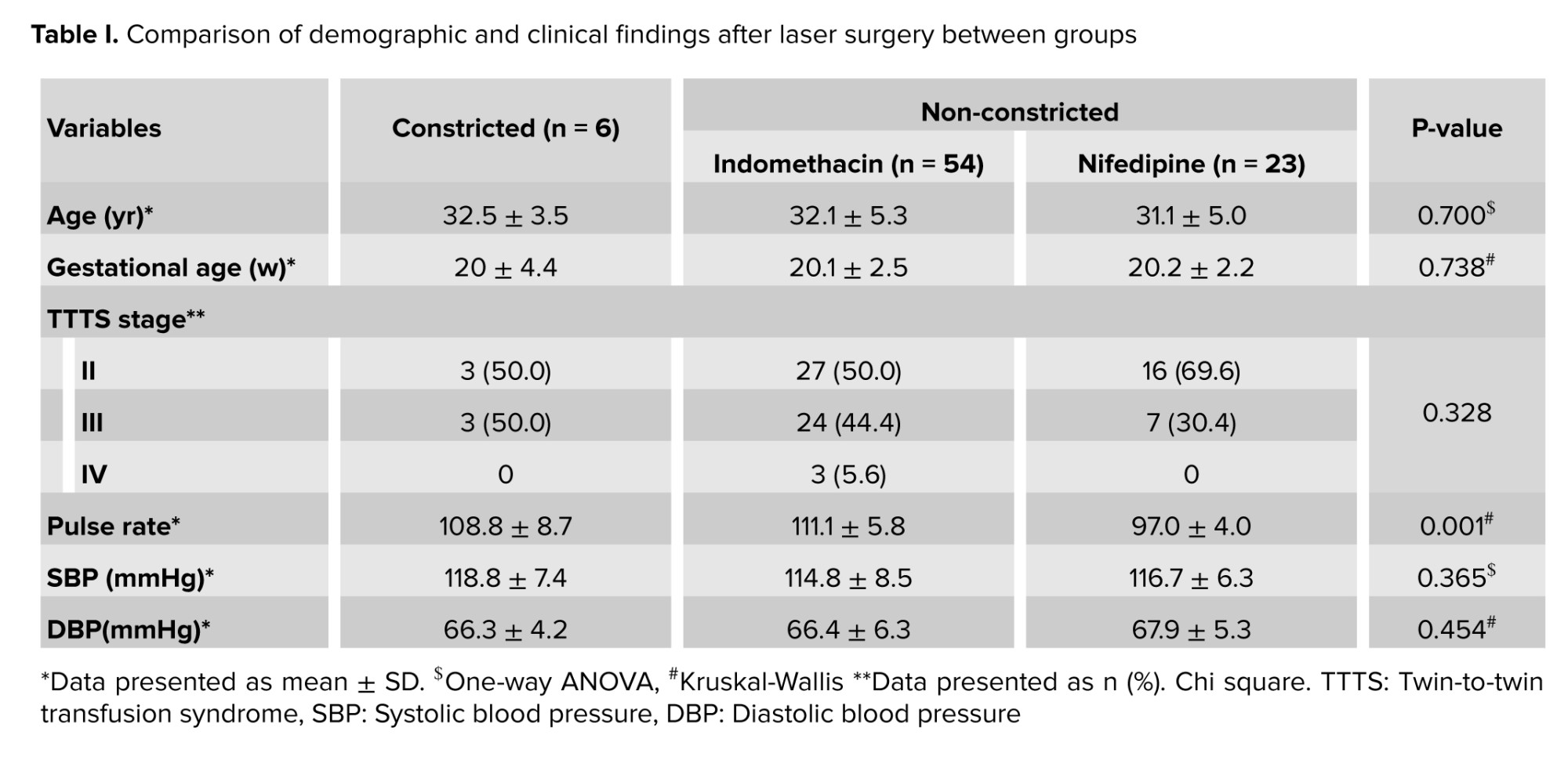
A total of 83 cases were included in the study, of which 6 cases had DA constriction and 77 cases had non-constriction, and were divided into 2 groups receiving indomethacin and nifedipine. Table I shows the demographic and clinical variables after laser surgery between these groups. Based on the information presented in this table, no statistically significant difference was observed between the 3 groups in terms of the mean age of the mothers, nor was there a significant difference between the groups in terms of the stage of TTTS and gestational age. In addition, the mean systolic blood pressure (p = 0.365) and diastolic blood pressure (p = 0.454) were uniformly distributed between groups. However, the results revealed that a significant difference (p = 0.001) between pulse rates of the 3 groups and the lowest mean pulse rate was observed in the nifedipine group (97.0 ± 4.0).
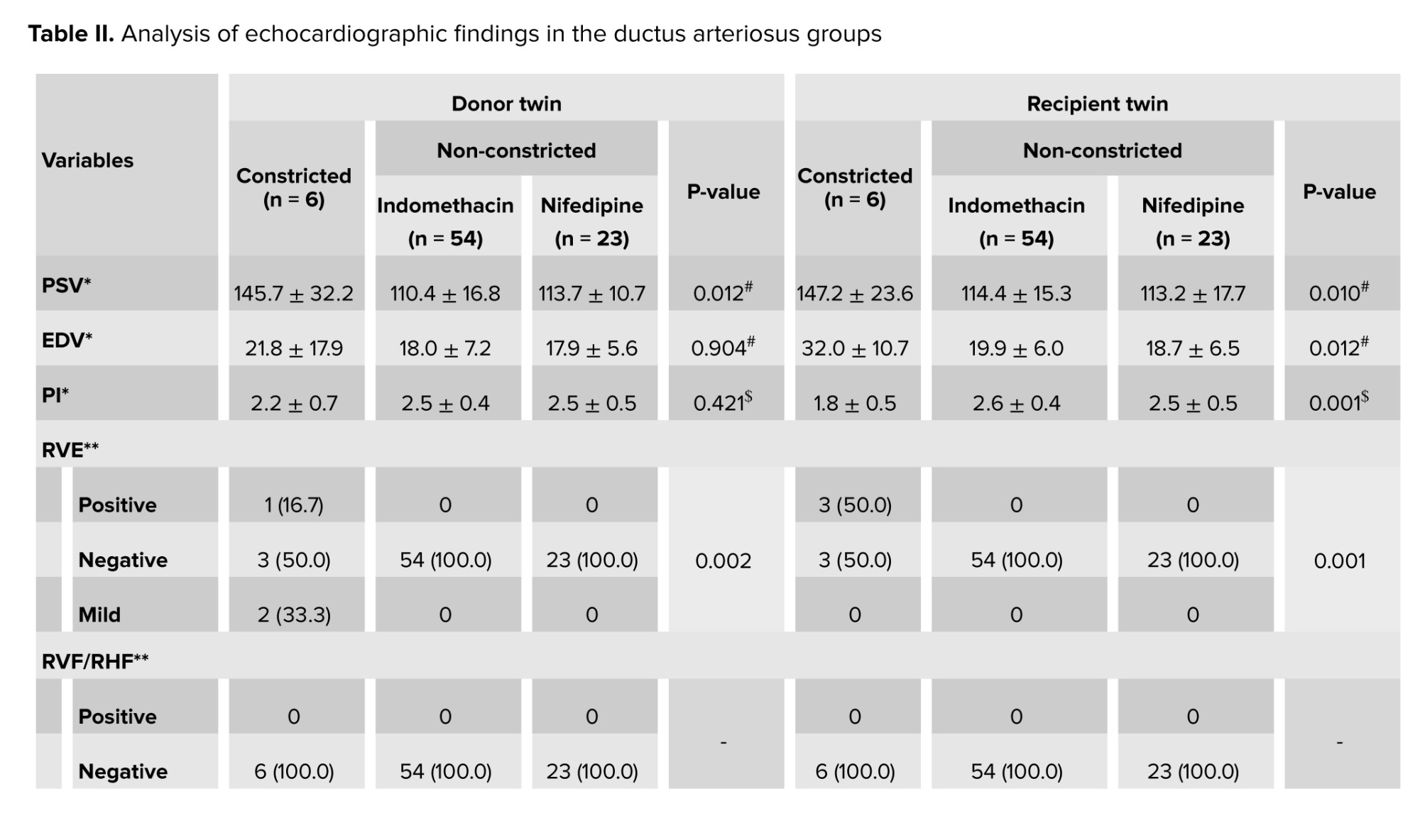
According to the results presented in table II, Doppler findings of the DA were compared among 3 groups of participants with TTTS after FLP surgery: cases who developed DA constriction and 2 groups without constriction who received indomethacin or nifedipine 48 hr after the procedure. Based on the results, PSV was significantly higher in both donor (p = 0.012) and recipient twins (p = 0.010) in the ductal constriction group compared with the 2 non-constriction groups.
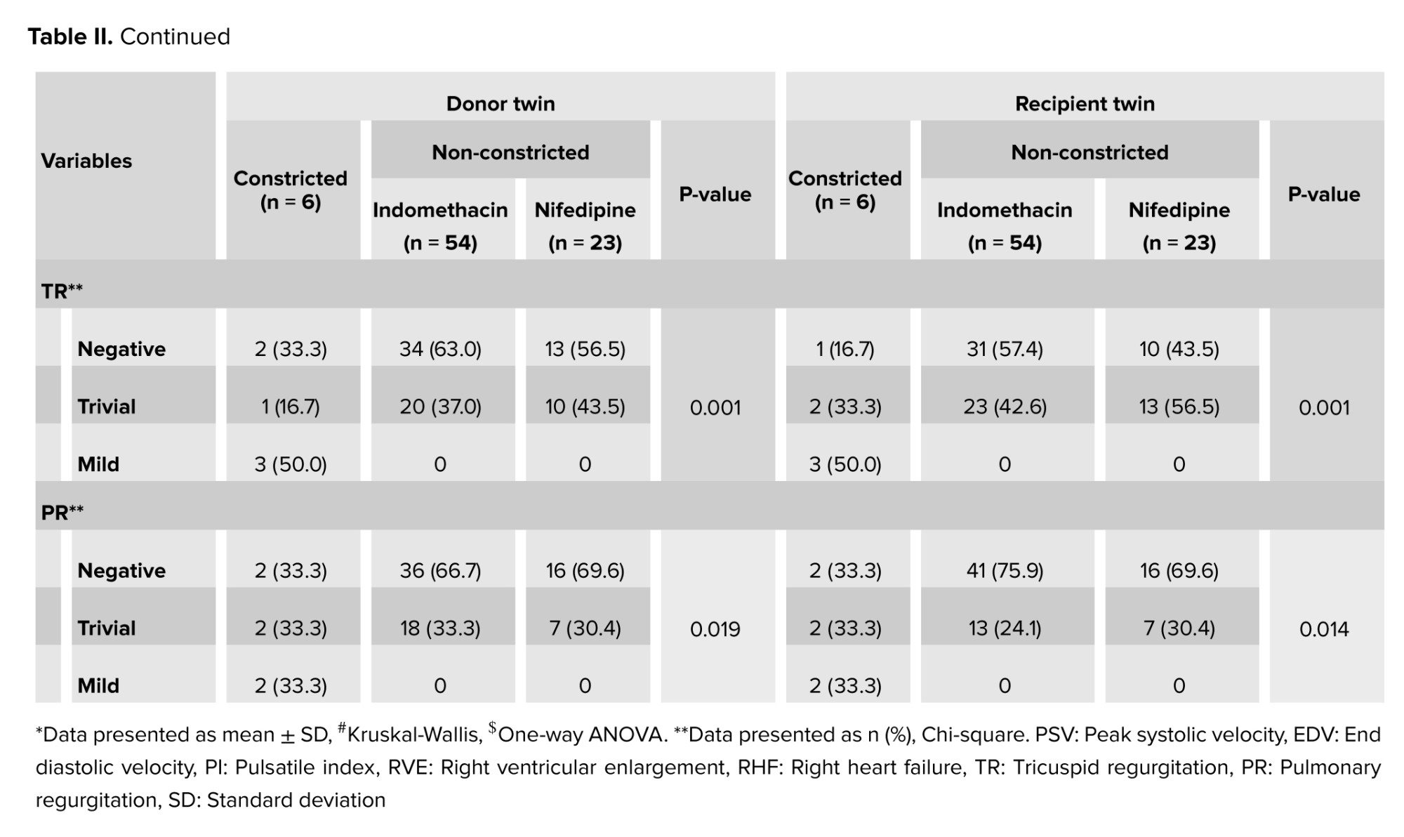
In recipient twins, EDV was also significantly increased (p = 0.012), while the PI was significantly lower in the constriction group (p = 0.001), indicating a distinct Doppler flow pattern compared with participants without ductal constriction. Right ventricular enlargement was observed exclusively in cases with DA constriction and was significantly more frequent among DA cases of donor (p = 0.002) and recipient twin (p = 0.001), whereas no cases of right ventricular enlargement were detected among participants without constriction who received either indomethacin or nifedipine. No cases of right ventricular failure or right heart failure were observed in any of the study groups. In addition, tricuspid regurgitation (p = 0.001) and pulmonary regurgitation (among donor twin: p = 0.019, among recipient twin: p = 0.014) were significantly more prevalent in the ductal constriction group, predominantly at trivial to mild levels. In contrast, participants without ductal constriction who received indomethacin or nifedipine demonstrated similar and relatively stable Doppler and valvular findings.
1 wk after FLP, the 6 cases with ductal constriction were re-evaluated, and all showed normal findings.
4. Discussion
The present study offers valuable insight into Doppler alterations of the DA following fetoscopic laser surgery in MCDA pregnancies complicated by TTTS in cases where DA constriction developed following the administration of tocolytic agents (indomethacin or nifedipine). The findings indicate that post-laser DA constriction is associated with significant changes in Doppler indices, as well as a higher prevalence of right-sided cardiac abnormalities compared with cases without constriction. Furthermore, the results suggest that the administration of indomethacin or nifedipine after laser therapy may be beneficial in TTTS cases with normal Doppler findings. Although DA constriction was identified in a relatively small subset of participants, its presence was clearly linked to both Doppler alterations and cardiac changes.
In cases complicated by ductal constriction, PSV was significantly elevated in both donor and recipient twins, reflecting increased flow velocity across the narrowed ductus. Among recipient twins, the simultaneous rise in EDV and reduction in PI further indicate altered hemodynamic patterns secondary to ductal constriction. These observations are in line with previous studies demonstrating that ductal narrowing can result in measurable and clinically relevant Doppler modifications (11). The lack of clinically meaningful differences in Doppler and cardiac parameters between the indomethacin and nifedipine groups in the absence of ductal constriction suggests that, when ductal narrowing is not present, the short-term hemodynamic effects of these 2 medications after laser surgery are largely comparable. Overall, these findings underscore the value of routine Doppler assessment of the DA following laser therapy for TTTS, particularly in the early postoperative phase. Future investigations involving larger cohorts and extended follow-up periods are needed to establish the incidence, associated risk factors, and clinical consequences of DA constriction more clearly in this context.
Indomethacin exerts its pharmacological effects by inhibiting cyclooxygenase enzymes responsible for prostaglandin synthesis. Prostaglandins, particularly prostaglandin estradiol, play a crucial role in maintaining patency of the fetal DA by facilitating blood flow from the pulmonary artery to the aorta and bypassing the nonfunctional lungs (12, 13). By reducing prostaglandin estradiol levels, indomethacin may induce constriction or premature closure of the DA. This effect is dose-dependent and is more likely to occur with prolonged use or in pregnancies beyond 30 wk of gestation, when the ductus becomes increasingly sensitive to prostaglandin inhibition (14, 15).
Premature ductal constriction can lead to significant hemodynamic alterations. Closure of the ductus redirects blood flow toward the pulmonary circulation, thereby increasing pulmonary vascular resistance and right ventricular afterload (16). Elevated pulmonary vascular resistance may result in pulmonary hypertension, compromising fetal oxygenation and overall well-being. Persistent ductal constriction may also lead to right ventricular hypertrophy and, ultimately, cardiac failure. Furthermore, reduced placental perfusion secondary to altered fetal circulation may impair oxygen delivery, resulting in fetal hypoxia and metabolic acidosis (17-20).
More than 95% of MCDA twins share placental circulation between the twins, which can lead to TTTS. Hypovolemia seen in the donor twin leads to renal hypoperfusion, which can stimulate the renin-angiotensin-aldosterone system of the donor twin. This effect results in oliguria and oligohydramnios. In addition, hypervolemia in the recipient twin can cause cardiac dilation, which leads to increased atrial and brain natriuretic peptide in the recipient twin. These changes ultimately inhibit the renin-angiotensin-aldosterone system, leading to polyuria and, in some cases, polyhydramnios.
Similar to the results of the current study, another study investigated the susceptibility of complicated monochorionic twin pregnancies to indomethacin-induced constriction of the fetal DA. They reported that 2 participants with TTTS underwent placental laser surgery and concluded that the constrictive effect of indomethacin on the DA was detectable even after a single dose and from as early as 20 wk of gestation in complicated monochorionic twin pregnancies (21). Moreover, a study conducted a meta-analysis to evaluate the association between NSAID use during the third trimester of pregnancy and the risk of premature ductal closure.
Based on data from 217 indomethacin-exposed participants and 221 placebo-exposed participants, the risk of ductal closure among women exposed to NSAIDs was 15 times higher than among those receiving a placebo. No statistically significant difference was observed in ductal closure risk between neonates exposed to indomethacin and those exposed to other NSAIDs (22). In a study, the researchers examined the effectiveness of prolonged indomethacin therapy in prolonging gestation following fetoscopic laser surgery for TTTS. Long-term indomethacin treatment was defined as administration for at least 48 hr. Their results suggested that the risk of preterm birth before 34, 32, 28, and 26 wk of gestation was significantly lower in the long-term indomethacin therapy group than in the non-long-term indomethacin therapy group (9).
4.1. Strengths and Limitations
This study has several strengths and limitations. A notable strength is the comparative evaluation of outcomes between groups using Doppler parameters, which enhances the robustness and internal validity of the findings.
The relatively small sample size may have reduced the statistical power to detect more subtle differences, and the cohort design limits the ability to establish causal relationships. Accordingly, future research incorporating larger study populations and preferably interventional designs would help generate more definitive and generalizable conclusions. One of the limitations of the present study is its retrospective non-randomized design, which introduces a substantial risk of selection bias and confounding. The administration of indomethacin or nifedipine was determined according to maternal heart rate (cutoff ≥ 100 beats/min), which may have introduced selection bias and potential confounding. Therefore, confounding by indication cannot be excluded, and the observed differences between treatment groups should be interpreted with caution, and future randomized controlled studies are recommended. Another limitation of this study was that clinical variables such as pulmonary hypertension, neonatal cardiac dysfunction, neonatal intensive care unit admission, and neonatal mortality were not examined, which is why it is recommended that in future studies, in addition to Doppler findings, other relevant clinical variables be compared to draw more comprehensive conclusions. In addition, although a formal twin-pair analysis was not performed, fetal observations were analyzed as independent units. Because both fetuses originated from the same pregnancy, their measurements are inherently correlated rather than statistically independent. Consequently, the use of conventional statistical methods that assume independent observations may have underestimated the standard errors and affected the validity of the statistical inferences.
5. Conclusion
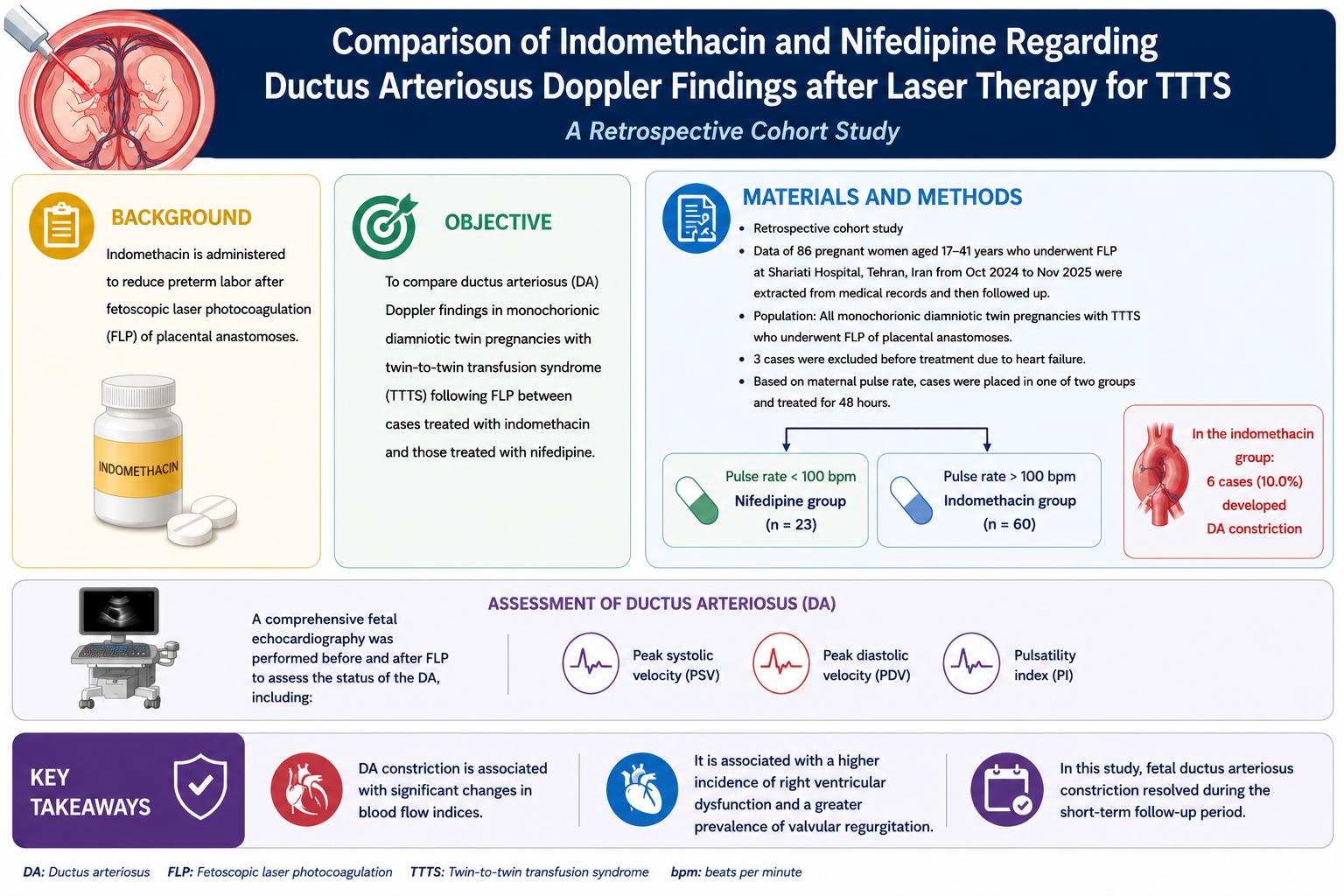
DA constriction following laser surgery in MCDA pregnancies complicated by TTTS is associated with significant alterations in Doppler indices and a greater incidence of right-sided cardiac changes compared with cases without constriction. Treatment with either indomethacin or nifedipine was associated with favorable short-term outcomes in TTTS cases in which Doppler parameters remain within normal limits. Although DA constriction was identified in only a small proportion of participants, its presence was clearly linked to measurable Doppler abnormalities and associated cardiac changes. In this study, fetal DA constriction was resolved during the short-term follow-up period.
Data Availability
The data that support the findings of this study are available from the corresponding author upon reasonable request.
Author Contributions
S. Mokhtari, L. Eslamian, V. Marsoosi, and A. Golbabaei contributed to conceptualization, methodology, supervision, and writing, review and editing. N. Saedi and P. Boustani contributed to data curation, formal analysis, investigation, and writing, original draft. All authors reviewed and approved the final manuscript and agreed to be accountable for all aspects of the work.
Acknowledgments
We would like to thank the vice chancellor for the research of Tehran University of Medical Sciences, Tehran, Iran for their financial and scientific support (Grant no.: 97112900). The authors used ChatGPT (OpenAI, USA) to assist with language editing and improvement of grammar. All scientific content, interpretation, and final approval of the manuscript were performed by the authors.
Conflict of Interest
All authors declare that there is no conflict of interest.
Full-Text: (34 Views)
1. Introduction
Twin-to-twin transfusion syndrome (TTTS) is a serious condition that occurs exclusively in monochorionic twin pregnancies, in which both fetuses share a single placenta. This syndrome arises from abnormal vascular connections within the shared placenta, leading to an unequal distribution of blood flow between the 2 fetuses. In this condition, one fetus, referred to as the “donor”, experiences reduced blood volume and decreased amniotic fluid levels (oligohydramnios), whereas the other fetus, known as the “recipient”, receives excessive blood flow, resulting in increased amniotic fluid volume (polyhydramnios) and increased strain on the cardiovascular system (1).
Laser ablation, particularly fetoscopic laser photocoagulation (FLP), is currently regarded as the standard treatment for TTTS. This minimally invasive procedure directly targets the underlying pathophysiology by eliminating abnormal vascular anastomoses within the shared placenta. Under fetoscopic guidance, laser energy is applied to coagulate the communicating vessels, thereby interrupting pathological intertwin blood flow. By functionally separating the placental circulations of each fetus, laser ablation reduces abnormal transfusion, stabilizes the intrauterine environment, and decreases the risk of adverse outcomes for both twins (2). Despite its effectiveness, laser ablation is associated with an increased risk of preterm birth. This complication may arise from several factors, including the invasive nature of the procedure, intrauterine inflammation, and pre-existing uterine conditions that may be exacerbated by intervention. Studies indicate that preterm delivery occurs in approximately 60% of cases following laser ablation, with most deliveries taking place before 34 wk of gestation (3). Instrumentation during the procedure may induce localized inflammatory responses, thereby increasing the risk of premature labor. In addition, perforation of the amniotic sac during fetoscope insertion may result in premature rupture of membranes (4, 5).
Indomethacin is a nonsteroidal anti-inflammatory drug (NSAID) that primarily exerts its effects through inhibition of cyclooxygenase enzymes. These enzymes are key mediators in the synthesis of prostaglandins, which play a central role in the initiation and maintenance of labor. The pharmacokinetic profile of indomethacin is characterized by rapid absorption, extensive plasma protein binding, and hepatic metabolism, with metabolites eliminated via renal and biliary pathways (6, 7). The ductus arteriosus (DA) is a vital component of fetal circulation and plays a central role in diverting blood away from the nonfunctional fetal lungs. During intrauterine development, the lungs do not participate in gas exchange, and oxygenation is achieved through placental circulation (8, 9). FLP is widely recognized as the standard treatment for TTTS and has significantly improved perinatal outcomes. Nevertheless, this invasive procedure is associated with an increased risk of preterm labor. To mitigate this complication, indomethacin, a well-established tocolytic agent, is frequently administered to suppress uterine contractions. A critical concern is that, in addition to its tocolytic effects, indomethacin is known to exert a significant influence on the fetal DA and may induce premature constriction or closure before birth. Early closure of the DA can result in serious fetal complications, including pulmonary hypertension, cardiac failure, and intraventricular hemorrhage.
The aim of this study was to compare DA Doppler findings in monochorionic diamniotic (MCDA) twin pregnancies complicated by TTTS following FLP between cases treated with indomethacin and those treated with nifedipine.
2. Materials and Methods
2.1. Study design and participants
In this retrospective cohort study, data of 86 pregnant women aged 17-41 yr who underwent FLP in Shariati hospital, Tehran, Iran from October 2024-November 2025 were extracted from medical records and then followed up.
The study population included all MCDA twin pregnancies with TTTS who underwent FLP (The NdYag laser [400-600 µ laser fiber]) of placental anastomoses, and received indomethacin or nifedipine for 48 hr to prevent preterm labor.
In this study, 86 cases of TTTS were included, of which 3 were excluded due to fetal cardiac anomalies (2 cases of pulmonary constriction and 1 case of coarctation of the aorta). Of the remaining 83 cases in the study, based on pulse rate, were placed in one of 2 groups: nifedipine (pulse rate < 100) and indomethacin (pulse rate > 100). In cases receiving indomethacin, 6 cases developed DA constriction. Finally, the cases were divided into 3 groups: group 1 included indomethacin recipients with DA constriction (n = 6), group 2 included indomethacin recipients without DA constriction (n = 54), and group 3 included nifedipine recipients (n = 23). All pregnancies were managed according to a uniform TTTS protocol.
2.2. Eligibility criteria
Inclusion criteria included pregnant women with MCDA twin pregnancies diagnosed with TTTS classified as stage II or higher according to the Quintero staging system, undergoing elective laser photocoagulation as a treatment for TTTS, and gestational age between 16 and 26 wk at the time of diagnosis. Exclusion criteria included: 1) the presence of structural or chromosomal abnormalities in either twin identified on previous fetal anomaly scans, 2) maternal contraindications to indomethacin administration, such as allergy, peptic ulcer, renal dysfunction, or coagulation disorders, 3) severe fetal growth restriction (estimated fetal weight < 5th percentile) in the donor and 4) prior administration of medications or interventions that affect fetal cardiovascular function.
2.3. Indomethacin administration protocol
The initial dose of indomethacin suppositories, 100 mg, was administered rectally as a single dose, since rectal administration provides rapid absorption, better bioavailability, and less gastrointestinal tract side effects. The maintenance dose was indomethacin suppositories 50 mg, administered rectally every 8 hr and continued for 48 hr to maintain an adequate drug level to stabilize uterine conditions, prevent premature labor, and stabilize the fetal condition. The dosage of nifedipine was as follows: first 30 mg, then 10 mg/3 times daily for 48 hr.
2.4. Fetal echocardiographic assessment
Before starting indomethacin or nifedipine, a comprehensive fetal echocardiography was performed to rule out possible cardiac abnormalities. Post-laser fetal echocardiography was performed 48 hr after administration of indomethacin or nifedipine, the details of which included the following:
Doppler flow measurement of the DA: peak systolic velocity (PSV), end diastolic velocity (EDV), and mean velocity were recorded. Pulsatile index (PI): PI was calculated to assess vascular resistance in the DA. Morphological assessment: DA diameter and any abnormal constriction or closures were recorded. Gross cardiac function assessment: ventricular size and function, and signs of cardiac strain or overload were assessed. In cases where the DA was occluded or narrowed after indomethacin, weekly Doppler examinations were performed. Doppler parameters were reassessed to monitor for DA re-opening or remaining occluded.
In this study, fetal echocardiography was performed according to the guidelines of the American Society of Echocardiography and using the Voluson E8 ultrasound system (GE Healthcare Ultrasound, Milwaukee, Wisconsin, USA). All of these were performed by a blinded pediatric cardiologist. Measurements were averaged and reported after 3 repetitions.
2.5. End-of-follow-up criteria
If the DA remained open after indomethacin, no further follow-up of the DA status was required. If the DA reopened after closure or narrowing, follow-up continued until birth to ensure it did not recur.
2.6. Diagnosis of DA
First, a baseline ultrasound was performed to rule out cardiac anomalies. Then, the DA was assessed in the 3VT view and averaged and reported after 3 repetitions. A standard protocol was used for the diagnosis of fetal DA for each fetus. Doppler was used when the fetus was not moving or breathing.
To perform the measurements, the device settings were as follows: 1) high frame rate with high contrast and resolution, 2) high-pass wall motion filter (50 Hz) and low energy output levels (< 50 mW/cm2), 3) absence of Doppler effect, and 4) incidence angle as close to 0° as possible (< 15°). In this study, the anatomy, diameter, and normal flow of the fetal arteriosus duct were examined from 3VT views. The characteristic V-shape on the left side of the fetal trachea in the 3VT view was considered to indicate the convergence of the fetal arteriosus duct. All of these were performed by a pediatric cardiologist.
To diagnose the DA, the following criteria were considered (10):
- elevated DA velocity (PSV > 140 cm/sec, diastolic velocity > 35 cm/s and PI < 1.9)
- S-shaped DA and distal constriction
- dilated right ventricle
- right ventricular dysfunction (abnormal myocardial performance index)
- tricuspid and pulmonary regurgitation
2.7. Monitoring during pregnancy
In addition to DA monitoring, routine fetal growth monitoring (such as biophysical profile and Doppler studies) were performed. Any complications such as heart failure, pulmonary artery pressure, or abnormal Doppler patterns in the umbilical artery were managed according to clinical protocols. Doppler findings included open, narrow, or closed status and quantitative Doppler flow indices (PSV, EDV, and PI).
2.8. Data collection instruments
Maternal demographics, TTTS stage, fetal echocardiographic findings, and pregnancy outcomes were recorded. All ultrasound images were reviewed by an experienced pediatric cardiologist to ensure accuracy and consistency of data. Follow-up data were recorded at routine clinical visits and supplemented by telephone calls as needed.
2.9. Ethical Considerations
This study was approved by the ethics committee of Shariati hospital of Tehran University of Medical Sciences, Tehran, Iran (Code: IR.TUMU.SHARIATI.REC.1404.051). The confidentiality of participants' data was ensured by anonymizing all patient information prior to analysis and restricting data access to the research team.
2.10. Statistical Analysis
Number (percentage) and mean (standard deviation) were used to describe qualitative and quantitative data, respectively. The Shapiro-Wilk test was used to examine the normal distribution of quantitative variables, and if the data were normal, parametric tests (one-way analysis of variance) were used; otherwise, nonparametric tests (Kruskal-Wallis test) were used. A likelihood ratio Chi-square test was used to compare qualitative variables between groups. All analyses were performed using Stata software version 13 and at a significance level of 0.05.
3. Results
A total of 83 cases were included in the study, of which 6 cases had DA constriction and 77 cases had non-constriction, and were divided into 2 groups receiving indomethacin and nifedipine. Table I shows the demographic and clinical variables after laser surgery between these groups. Based on the information presented in this table, no statistically significant difference was observed between the 3 groups in terms of the mean age of the mothers, nor was there a significant difference between the groups in terms of the stage of TTTS and gestational age. In addition, the mean systolic blood pressure (p = 0.365) and diastolic blood pressure (p = 0.454) were uniformly distributed between groups. However, the results revealed that a significant difference (p = 0.001) between pulse rates of the 3 groups and the lowest mean pulse rate was observed in the nifedipine group (97.0 ± 4.0).
According to the results presented in table II, Doppler findings of the DA were compared among 3 groups of participants with TTTS after FLP surgery: cases who developed DA constriction and 2 groups without constriction who received indomethacin or nifedipine 48 hr after the procedure. Based on the results, PSV was significantly higher in both donor (p = 0.012) and recipient twins (p = 0.010) in the ductal constriction group compared with the 2 non-constriction groups.
In recipient twins, EDV was also significantly increased (p = 0.012), while the PI was significantly lower in the constriction group (p = 0.001), indicating a distinct Doppler flow pattern compared with participants without ductal constriction. Right ventricular enlargement was observed exclusively in cases with DA constriction and was significantly more frequent among DA cases of donor (p = 0.002) and recipient twin (p = 0.001), whereas no cases of right ventricular enlargement were detected among participants without constriction who received either indomethacin or nifedipine. No cases of right ventricular failure or right heart failure were observed in any of the study groups. In addition, tricuspid regurgitation (p = 0.001) and pulmonary regurgitation (among donor twin: p = 0.019, among recipient twin: p = 0.014) were significantly more prevalent in the ductal constriction group, predominantly at trivial to mild levels. In contrast, participants without ductal constriction who received indomethacin or nifedipine demonstrated similar and relatively stable Doppler and valvular findings.
1 wk after FLP, the 6 cases with ductal constriction were re-evaluated, and all showed normal findings.
4. Discussion
The present study offers valuable insight into Doppler alterations of the DA following fetoscopic laser surgery in MCDA pregnancies complicated by TTTS in cases where DA constriction developed following the administration of tocolytic agents (indomethacin or nifedipine). The findings indicate that post-laser DA constriction is associated with significant changes in Doppler indices, as well as a higher prevalence of right-sided cardiac abnormalities compared with cases without constriction. Furthermore, the results suggest that the administration of indomethacin or nifedipine after laser therapy may be beneficial in TTTS cases with normal Doppler findings. Although DA constriction was identified in a relatively small subset of participants, its presence was clearly linked to both Doppler alterations and cardiac changes.
In cases complicated by ductal constriction, PSV was significantly elevated in both donor and recipient twins, reflecting increased flow velocity across the narrowed ductus. Among recipient twins, the simultaneous rise in EDV and reduction in PI further indicate altered hemodynamic patterns secondary to ductal constriction. These observations are in line with previous studies demonstrating that ductal narrowing can result in measurable and clinically relevant Doppler modifications (11). The lack of clinically meaningful differences in Doppler and cardiac parameters between the indomethacin and nifedipine groups in the absence of ductal constriction suggests that, when ductal narrowing is not present, the short-term hemodynamic effects of these 2 medications after laser surgery are largely comparable. Overall, these findings underscore the value of routine Doppler assessment of the DA following laser therapy for TTTS, particularly in the early postoperative phase. Future investigations involving larger cohorts and extended follow-up periods are needed to establish the incidence, associated risk factors, and clinical consequences of DA constriction more clearly in this context.
Indomethacin exerts its pharmacological effects by inhibiting cyclooxygenase enzymes responsible for prostaglandin synthesis. Prostaglandins, particularly prostaglandin estradiol, play a crucial role in maintaining patency of the fetal DA by facilitating blood flow from the pulmonary artery to the aorta and bypassing the nonfunctional lungs (12, 13). By reducing prostaglandin estradiol levels, indomethacin may induce constriction or premature closure of the DA. This effect is dose-dependent and is more likely to occur with prolonged use or in pregnancies beyond 30 wk of gestation, when the ductus becomes increasingly sensitive to prostaglandin inhibition (14, 15).
Premature ductal constriction can lead to significant hemodynamic alterations. Closure of the ductus redirects blood flow toward the pulmonary circulation, thereby increasing pulmonary vascular resistance and right ventricular afterload (16). Elevated pulmonary vascular resistance may result in pulmonary hypertension, compromising fetal oxygenation and overall well-being. Persistent ductal constriction may also lead to right ventricular hypertrophy and, ultimately, cardiac failure. Furthermore, reduced placental perfusion secondary to altered fetal circulation may impair oxygen delivery, resulting in fetal hypoxia and metabolic acidosis (17-20).
More than 95% of MCDA twins share placental circulation between the twins, which can lead to TTTS. Hypovolemia seen in the donor twin leads to renal hypoperfusion, which can stimulate the renin-angiotensin-aldosterone system of the donor twin. This effect results in oliguria and oligohydramnios. In addition, hypervolemia in the recipient twin can cause cardiac dilation, which leads to increased atrial and brain natriuretic peptide in the recipient twin. These changes ultimately inhibit the renin-angiotensin-aldosterone system, leading to polyuria and, in some cases, polyhydramnios.
Similar to the results of the current study, another study investigated the susceptibility of complicated monochorionic twin pregnancies to indomethacin-induced constriction of the fetal DA. They reported that 2 participants with TTTS underwent placental laser surgery and concluded that the constrictive effect of indomethacin on the DA was detectable even after a single dose and from as early as 20 wk of gestation in complicated monochorionic twin pregnancies (21). Moreover, a study conducted a meta-analysis to evaluate the association between NSAID use during the third trimester of pregnancy and the risk of premature ductal closure.
Based on data from 217 indomethacin-exposed participants and 221 placebo-exposed participants, the risk of ductal closure among women exposed to NSAIDs was 15 times higher than among those receiving a placebo. No statistically significant difference was observed in ductal closure risk between neonates exposed to indomethacin and those exposed to other NSAIDs (22). In a study, the researchers examined the effectiveness of prolonged indomethacin therapy in prolonging gestation following fetoscopic laser surgery for TTTS. Long-term indomethacin treatment was defined as administration for at least 48 hr. Their results suggested that the risk of preterm birth before 34, 32, 28, and 26 wk of gestation was significantly lower in the long-term indomethacin therapy group than in the non-long-term indomethacin therapy group (9).
4.1. Strengths and Limitations
This study has several strengths and limitations. A notable strength is the comparative evaluation of outcomes between groups using Doppler parameters, which enhances the robustness and internal validity of the findings.
The relatively small sample size may have reduced the statistical power to detect more subtle differences, and the cohort design limits the ability to establish causal relationships. Accordingly, future research incorporating larger study populations and preferably interventional designs would help generate more definitive and generalizable conclusions. One of the limitations of the present study is its retrospective non-randomized design, which introduces a substantial risk of selection bias and confounding. The administration of indomethacin or nifedipine was determined according to maternal heart rate (cutoff ≥ 100 beats/min), which may have introduced selection bias and potential confounding. Therefore, confounding by indication cannot be excluded, and the observed differences between treatment groups should be interpreted with caution, and future randomized controlled studies are recommended. Another limitation of this study was that clinical variables such as pulmonary hypertension, neonatal cardiac dysfunction, neonatal intensive care unit admission, and neonatal mortality were not examined, which is why it is recommended that in future studies, in addition to Doppler findings, other relevant clinical variables be compared to draw more comprehensive conclusions. In addition, although a formal twin-pair analysis was not performed, fetal observations were analyzed as independent units. Because both fetuses originated from the same pregnancy, their measurements are inherently correlated rather than statistically independent. Consequently, the use of conventional statistical methods that assume independent observations may have underestimated the standard errors and affected the validity of the statistical inferences.
5. Conclusion
DA constriction following laser surgery in MCDA pregnancies complicated by TTTS is associated with significant alterations in Doppler indices and a greater incidence of right-sided cardiac changes compared with cases without constriction. Treatment with either indomethacin or nifedipine was associated with favorable short-term outcomes in TTTS cases in which Doppler parameters remain within normal limits. Although DA constriction was identified in only a small proportion of participants, its presence was clearly linked to measurable Doppler abnormalities and associated cardiac changes. In this study, fetal DA constriction was resolved during the short-term follow-up period.
Data Availability
The data that support the findings of this study are available from the corresponding author upon reasonable request.
Author Contributions
S. Mokhtari, L. Eslamian, V. Marsoosi, and A. Golbabaei contributed to conceptualization, methodology, supervision, and writing, review and editing. N. Saedi and P. Boustani contributed to data curation, formal analysis, investigation, and writing, original draft. All authors reviewed and approved the final manuscript and agreed to be accountable for all aspects of the work.
Acknowledgments
We would like to thank the vice chancellor for the research of Tehran University of Medical Sciences, Tehran, Iran for their financial and scientific support (Grant no.: 97112900). The authors used ChatGPT (OpenAI, USA) to assist with language editing and improvement of grammar. All scientific content, interpretation, and final approval of the manuscript were performed by the authors.
Conflict of Interest
All authors declare that there is no conflict of interest.
Type of Study: Original Article |
Subject:
Perinatology
Send email to the article author
| Rights and permissions | |
 |
This work is licensed under a Creative Commons Attribution-NonCommercial 4.0 International License. |