
International Journal of
Reproductive Biomedicine
Tue, Jun 23, 2026
[Archive]
Volume 24, Issue 4 (April 2026)
IJRM 2026, 24(4): 325-336 |
Back to browse issues page


Ethics code: IR.MODAREs.REC.1401.227
![]()
![]()
![]()
Download citation:
BibTeX | RIS | EndNote | Medlars | ProCite | Reference Manager | RefWorks
Send citation to:


BibTeX | RIS | EndNote | Medlars | ProCite | Reference Manager | RefWorks
Send citation to:
Shafieizade R, Hezavehei M, Shahverdi A, Halvaei I. Hydroxytyrosol protects sperm viability and DNA integrity in human asthenoteratozoospermia during incubation: An experimental study. IJRM 2026; 24 (4) :325-336
URL: http://ijrm.ir/article-1-3765-en.html
URL: http://ijrm.ir/article-1-3765-en.html


1- Department of Anatomical Sciences, Faculty of Medical Sciences, Tarbiat Modares University, Tehran, Iran.
2- Department of Embryology, Reproductive Biomedicine Research Center, Royan Institute for Reproductive Biomedicine, ACECR, Tehran, Iran.
3- Department of Anatomical Sciences, Faculty of Medical Sciences, Tarbiat Modares University, Tehran, Iran. ,ihalvaei@modares.ac.ir
2- Department of Embryology, Reproductive Biomedicine Research Center, Royan Institute for Reproductive Biomedicine, ACECR, Tehran, Iran.
3- Department of Anatomical Sciences, Faculty of Medical Sciences, Tarbiat Modares University, Tehran, Iran. ,
Keywords: Abnormal semen, Antioxidant, Incubation time, Oxidative stress, DNA integrity, Asthenoteratozoospermia.
Full-Text [PDF 1157 kb]
(169 Downloads)
| Abstract (HTML) (204 Views)

Full-Text: (18 Views)
1. Introduction
Asthenoteratozoospermia (AT) is a disorder that refers to men who produce sperm with abnormal morphology and low motility (1). Semen analysis of men with AT shows decreased sperm motility (progressive motility < 30% and total motility < 42%) and a high percentage of sperm with abnormal morphology (normal morphology < 4%) (1). Terato sperm and leukocytes in semen samples are major sources of reactive oxygen species (ROS) (2, 3). Also, it has been reported that AT semen samples have a high level of ROS compared to the normal ones (4). Elevated ROS levels can cause DNA damage, lipid peroxidation (LPO) of the sperm plasma membrane, and impair sperm function (5).
Antioxidants are substances that deal with free radicals’ damage by eliminating or preventing the formation of ROS, especially in abnormal samples (6).
The literature indicates that many antioxidants have been proposed to counteract the destructive effects of oxidative stress, showing positive effects on sperm parameters (7). Olive oil contains a wide range of important antioxidant compounds, such as polyphenols, which can be referred to as hydroxytyrosol (HT), that have significant antioxidant properties (8). HT is a compound of polyphenolic or phenolic plants with high antioxidant properties, found in the fruit and leaf of olive and olive oil (which is the main source), that is soluble in lipids. HT has important properties such as a cytoprotective agent (9, 10). This antioxidant remains relatively stable in cell culture medium, with about 75% remaining after 12 hr of incubation at 37°C. HT can pass through the plasma membrane and get into the cell due to its lipophilic properties and small size (11). This compound acts as an antioxidant in various ways, including donation of the hydrogen atom in the hydroxyl group in the ortho position, the creation of sustainable hydrogen bonds with free radicals, and the prevention of peroxidation of lipids (12). It also stimulates the activity of antioxidant enzymes such as catalase and superoxide dismutase, and counteracts ROS by activating cellular signaling pathways like nuclear factor estradiol, which codes for antioxidant response elements, including Phase 2 detoxifying enzymes (13). The cryoprotective roles of HT on animal and human spermatozoa have been reported earlier (9, 14). Kedechi et al. found that short incubation of HT has antioxidant effects in human normozoospermia (15). Animal studies have shown the beneficial effect of olive oil in ameliorating oxidative stress (16). It was found that supplementing the freezing extender with olive fruit extract significantly reduced oxidative stress induced by cryopreservation in buffalo semen (17). A recent study showed that using HT could improve sperm functionality under both cooled and cryopreserved storage conditions (18). There is a lack of studies assessing HT in human samples with defined pathological phenotypes. Existing human data focuses on short in vitro incubation of normal ejaculates, not clinically abnormal samples. To the best of our knowledge, the impact of HT on the incubation of AT samples has not been previously evaluated.
This study investigates the effect of HT on sperm incubation in 2 phases to ensure both optimization of treatment conditions and focused mechanistic evaluation. In the first phase, optimal antioxidant doses and appropriate incubation times for sperm motility and viability were determined. In the second phase, mitochondrial membrane potential (MMP), ROS levels, DNA fragmentation, and LPO of the plasma membrane were evaluated during incubation of AT samples.
2. Materials and Methods
2.1. Sample collection and analysis
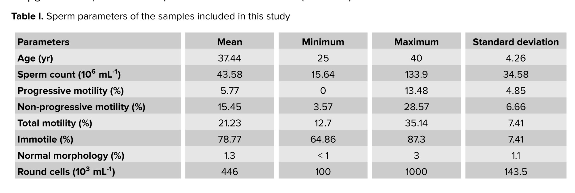
In this experimental study, 40 AT semen samples from men aged 22-40 yr were obtained from the Royan Institute for infertility workup. This study was performed from April and December 2023 at Royan Institute, Tehran, Iran. Recruitment was based on progressive motility < 30%, total motility < 42%, and normal morphology < 4% (Table I). Individuals with a history of antioxidant usage, drug consumption, systemic diseases, infections, or addiction were excluded. Samples were collected in sterile containers after 3-5 days of sexual abstinence. To be included in the study, semen samples had to contain fewer than 1 million leukocytes per ml. Semen parameters (count, motility, morphology) were analyzed according to World Health Organization (WHO) criteria (1).
2.2. Sample size
The sample size was determined by the following formula:
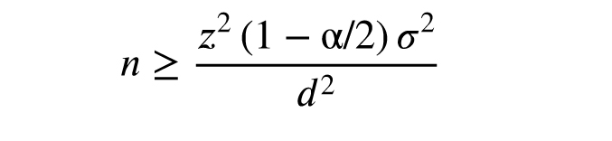
Where Z21-α: 1.96 (confidence interval 95%), σ (standard deviation): 2.6, and d (acceptable margin of error): 1.15 (19). According to this formula, at least 20 participants were needed.
2.3. Sample and antioxidant preparation
A simple washing method was used to prepare the samples. The semen samples were diluted with human tubal fluid culture medium containing 10% human serum albumin (Sigma, USA) and centrifuged at 300 g for 5 min. The supernatant was discarded, and the cell pellet was resuspended in human tubal fluid culture medium with albumin serum (Sigma, USA) (1). For antioxidant preparation, 2000 μL DMSO was added to 5 mg HT powder (Sigma, Germany) under sterile and dark conditions. Then, 25 μL of the antioxidant solution was aliquoted into 0.5 mL vials and stored at -20°C until use. In the first phase, samples were divided into groups with 0, 25, 50, 75, and 100 μg/mL HT concentrations and incubated at 37°C for 30, 45, and 60 min. Sperm count was maintained at 10 million/mL. Sperm motility and viability were assessed post-incubation to determine the optimal HT concentration and incubation time. In the second phase, using 25 and 50 μg/mL HT and 30 min incubation, MMP, ROS level, DNA fragmentation, and LPO were measured.
2.4. Sperm motility, viability, and morphology
Sperm motility was evaluated by CASA (Micropic Co., Spain). Curvilinear velocity cut-off values were as follows: rapid > 35 < moderate > 15 < slow > 10 > immotile; progression: Straightness > 80% determined according to WHO criteria (1). 10 μL of the semen sample was placed on the slide under the light microscope (Labomed, USA). For each sample, 2 slides and 6 fields were examined. In this system, in addition to total motility, progressive, non-progressive motility and immotile kinematic parameters were also analyzed, and the data were calculated as an average of all the observed spermatozoa. Sperm viability evaluation was performed using eosin-nigrosin (Sigma, Germany) staining. In this test, spermatozoa in red or pink color were considered dead, and white or light pink color were considered alive. To do this, 10 μL of eosin-nigrosin solution was mixed with 10 μL of the sample inside a microtube and given 30 sec. Afterward, 10 μL of the sample was placed onto a glass slide, and a smear was prepared and allowed to air-dry. The slides were then examined under a light microscope (Labomed, USA) at 1000× magnification, and at least 200 spermatozoa were evaluated (1). For sperm morphology assessment, Papanicolaou staining was performed. Smears were prepared from each sample and fixed. The staining solutions were arranged in 19 jars, and the fixed slides were placed in a staining rack and sequentially immersed in the appropriate solutions according to WHO guidelines. After staining, the slides were examined under a light microscope (Labomed, USA) at 1000× magnification, and a minimum of 200 spermatozoa were assessed (1).
2.5. Sperm DNA fragmentation
The sperm DNA integrity was evaluated by the sperm DNA fragmentation assay kit (IVF, Iran). To evaluate this, after preparing a suspension of 10-15 million spermatozoa, 50 μL of this suspension was added to a microtube containing 1% agarose (Sigma, Germany), which was melted at 95-100°C and was gently mixed. 25 μL of the above mixture was placed on a pre-covered slide cavity with a special gel, and the coverslip was put on it, they were then stored at a temperature of 4°C for 5 min. After gently removing the coverslips, the denaturation solution for 7 min and the lubricating solution for 15 min, respectively, were added to the slide at room temperature. After washing with deionized water for 5 min, they were dehydrated with ethanol (Sigma, Germany) 70%, 90%, and 100% for 2 min, and finally, stained with Wright (Sigma, Germany) color and washed. After drying at room temperature, they were observed with a light microscope (Labomed, USA, 1000x), and 200 spermatozoa were counted (20). In this test, sperm with intact or moderate damage to DNA have a large or medium halo around the head, while in the sperm with DNA fragmentation, a small halo is formed around the head or does not form at all or is degraded.
2.6. Intracellular ROS level
DCFH-DA (Sigma, Germany) test was used to evaluate the amount of hydrogen peroxide and DHE (Sigma, Germany) for superoxide by FACScalibur flow cytometry system (BD Biosciences, USA). To achieve this, 25 μM DCFH-DA and 1.25 μM DHE were separately added to 2, 1 mL samples, each containing 1-3 million spermatozoa. Then, samples were incubated at room temperature for 40 min for DCFH-DA and 20 min for DHE in a dark place. They were then examined using a flow cytometer system (BD Biosciences, USA) equipped with a 488-nm argon laser as a light source. Green fluorescence with a wavelength of 530-500 nm is related to DCFH, while red fluorescence with a wavelength of 590-700 nm is related to DHE. Data were analyzed using the software FlowJo (version 10, FlowJo, USA) and expressed as the percentage of fluorescent spermatozoa (21).
2.7. MMP
MMP was examined by the JC-1 staining and using the FACScalibur flow cytometry system (BD Biosciences, USA). For this purpose, the JC-1 color (Sigma, Germany) was melted at 37°C, and 1 μL was added to the 1 mL sample with a concentration of 3-5 million spermatozoa. After that, the samples were incubated at 37°C for 15 min in a dark place and centrifuged at 3000 rpm for 5 min. The supernatant was discarded, and the residual cells were washed with 1 mL of phosphate buffer saline. Finally, the samples were examined by the flow cytometer system, and the data were analyzed by software FlowJo (version 10, FlowJo, USA) (6).
2.8. LPO
Malondialdehyde (MDA) is one of the most important products of unsaturated fatty acid peroxidation that is used to evaluate oxidative stress conditions. Therefore, the peroxidation of lipid was measured by evaluating the amount of MDA through the thiobarbituric acid reaction and was read using an enzyme-linked immunosorbent assay plate reader (ELX800 ELISA reader; Bio-Tek Instruments, USA). Initially, 1 mL thiobarbituric acid (Sigma, Germany) was added to 500 μL of the sample. Then, it was placed in the boiling water bath for 1 hr and cooled by the water flow for 5 min. Finally, after the centrifugation, the supernatant was separated, and 250 μL of each sample was put in the microplate. The absorption was read at 532 nm wavelength (22).
2.9. Ethical Considerations
Informed written consent was obtained from all participants. This study was approved by the ethics committee of Tarbiat Modares University, Tehran, Iran (Code: IR.MODAREs.REC.1401.227).
2.10. Statistical Analysis
In this study, the distribution of numerical data was evaluated by the Shapiro-Wilk test. The data were expressed as the average standard deviation (Mean ± SD) and in some cases as the median, minimum, and maximum. Normal distribution data were evaluated using the one-way ANOVA test with Tukey’s post hoc test. Non-normal data distribution was compared using the Kruskal-Wallis test with Dunn’s post hoc test. The Repeated Measurement ANOVA test with Tukey’s post hoc test was used to determine the best time between different groups. The statistical analyses were performed by GraphPad Prism 5 (GraphPad Software Inc., USA). The significance level was considered as p < 0.05.
3. Results
3.1. Sperm motility and viability in experimental groups treated with HT
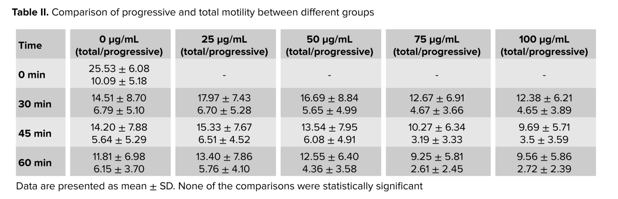
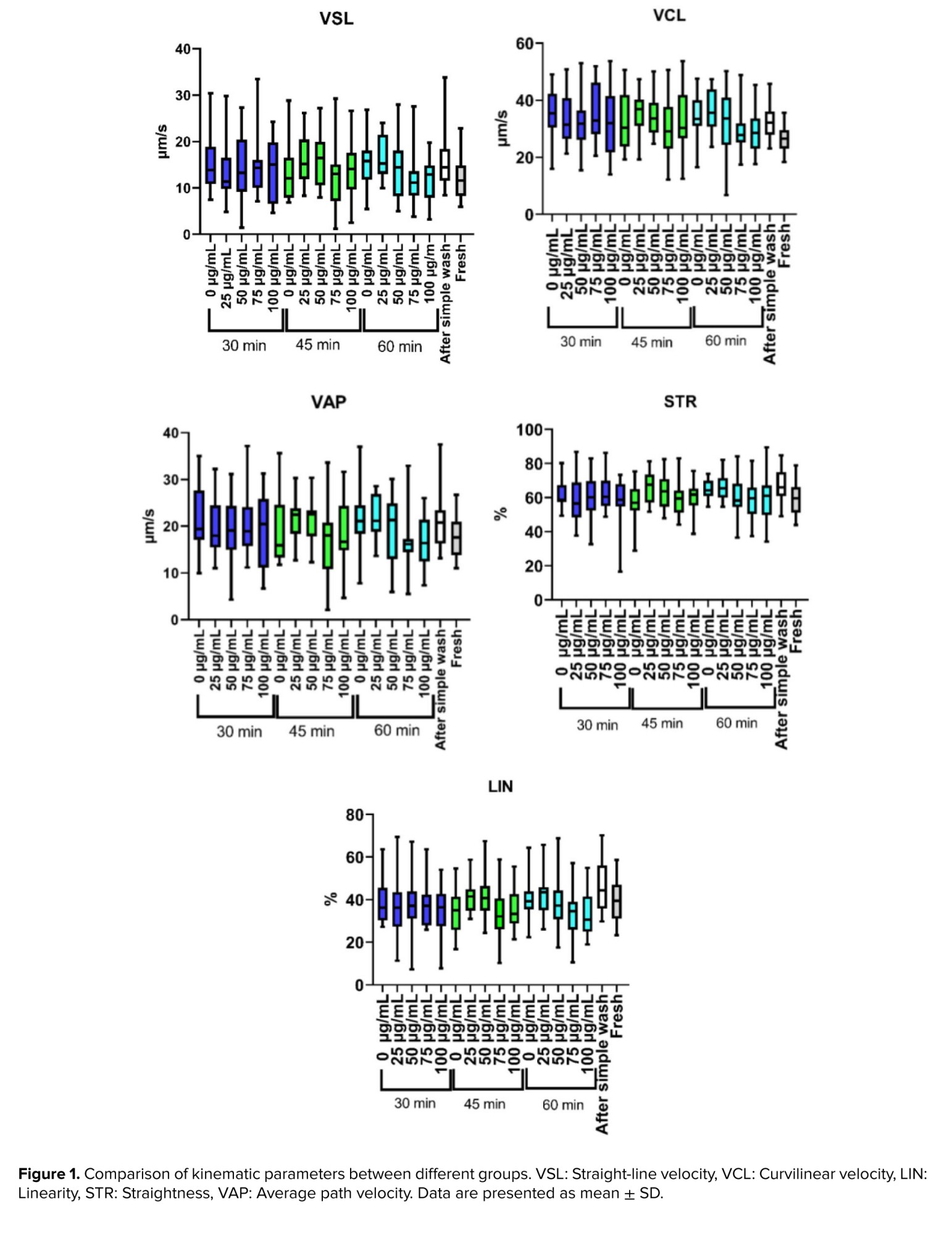
Sperm parameters included in this study are shown in table I. After incubation, sperm progressive and total motility were significantly decreased in different groups. Although there was an increasing trend in motility at concentrations of 25 µg/mL and 50 µg/mL compared to other groups, no significant differences were observed between the groups at different incubation times (Table II). In kinematic parameters, no significant improvement was observed in all experimental groups (Figure 1).
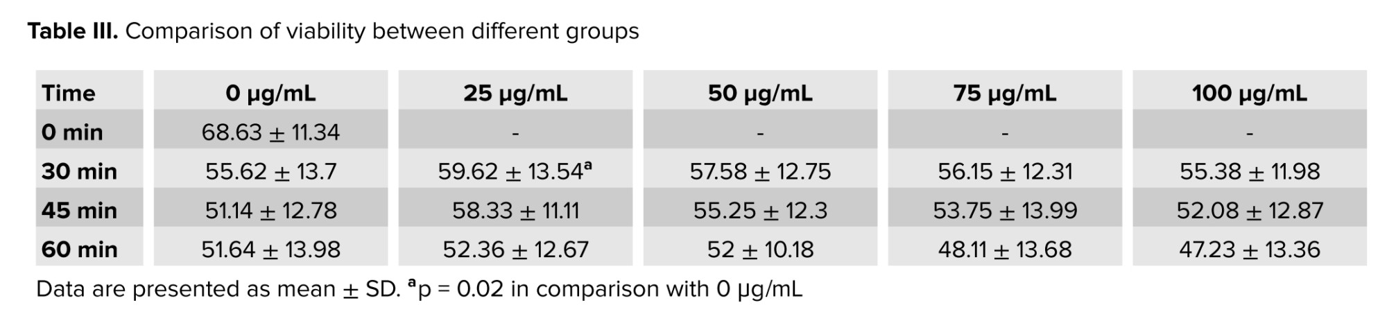
Similarly, sperm viability declined following incubation compared to the baseline. However, when comparing between groups, the viability at the concentration of 25 μg/mL after incubation for 30 min was significantly increased compared to the control group. 50 μg/mL HT had results close to the 25 μg/mL group (Table III). Based on the results of the first phase, the 2 concentrations of 25 and 50 μg/mL and 30 min of incubation were selected to assess the other parameters in the second phase.
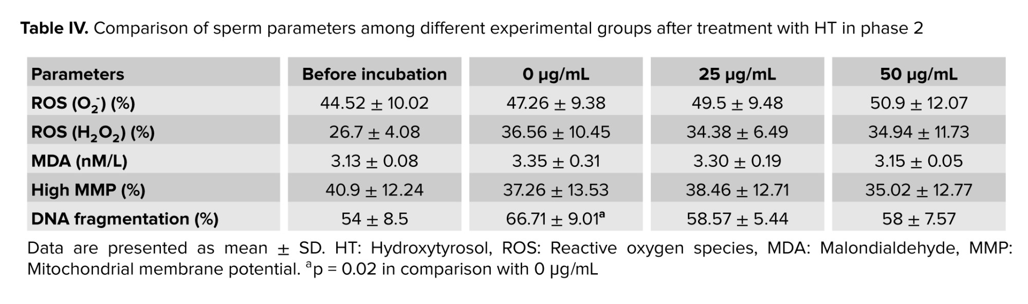
3.2. ROS level, LPO, MMP, and DNA fragmentation
No significant difference was observed in ROS levels, LPO, and MMP after incubation between different groups (Table IV). However, DNA fragmentation increased significantly at 0 μg/mL compared with pre-incubation levels (p = 0.02), whereas at both 25 and 50 μg/mL, it remained comparable to baseline values (Table IV).
4. Discussion
In the present study, no improvement was observed in the total and progressive motility between experimental groups compared to the control group after incubation. This aligns with previous findings of both incubation and cryopreservation on human and animal samples (9, 15, 23, 24). Conversely, it was shown that using HT during freezing preserves sperm motility in animal studies (14, 25, 26). Oxidative stress reduces sperm motility (27), and HT can potentially improve motility by reducing ROS through hydrogen donation and increasing intracellular antioxidant enzymes (8, 12). This contradiction in results with our findings could be due to the species of sperm because studies on human spermatozoa (9, 15) did not observe a significant impact on sperm motility in HT groups. Sperm kinetic and velocity parameters were also not increased after treatment with HT in line with our previous study (9). Our data showed variations in these parameters that may be attributed to differences in individual sample quality among AT patients, the sensitivity of sperm motion patterns to oxidative stress, and incubation time-dependent changes in sperm physiology. It should also be noted that prolonged in vitro incubation can itself promote oxidative stress due to continuous sperm metabolism and environmental exposure. In addition, higher antioxidant concentrations may slightly acidify the medium and potentially disrupt the physiological redox balance between ROS and antioxidants (28).
Along with our results about the effects on sperm viability, in the study of sperm incubation with 200 μg/mL (15), in the human frozen sperm at 100 μg/mL (9), at 10 μg/mL on the buck sperm (25) and in stallion sperm at 1.25, 2.5, 5, and 10 μg/mL (26), a significant improvement was observed in viability. HT may improve the viability of AT samples after incubation by reducing the level of ROS, which does this by counteracting and eliminating free radicals (12).
Our results showed that ROS levels after incubation were not decreased in any of the experimental groups compared to pre-incubation. In our previous study, ROS did not significantly reduce (9), which was consistent with the results of present study. It was also reported that HT with doses of 25 and 50 μM in low cryotolerance significantly reduced the H2O2 level compared to the control group. Still, they did not observe a decrease in the rate of O2- in low and high cryotolerance groups (14); however, the results related to the non-positive impact of HT in reducing O2- were in line with the results of our study.
Also, HT reduced the ROS level by neutralizing active oxygen in sperm or directly participating in the production of antioxidant enzymes, and thus improved the quality of pig sperm at 17°C (29). However, this contradiction in the results of various studies with our study can be due to the type of free radicals, the cell type, and the dose of antioxidants used. Also, the type of semen sample, that is normal vs. abnormal, is another source of contradictory findings. We used AT samples that needed more antioxidants compared to a normal sample. According to studies, high levels of polyphenols may abundantly remove oxygen species or act as pro-oxidants by reducing metal ions.
In assessing the level of MDA, consistent with our results, a recent study examining HT at doses of 50 and 100 μg/mL showed no significant reduction in LPO levels (9), while HT reduced LPO level in buck sperm (25). Also, evidence of decreased LPO in frozen-thawed ram sperm using HT suggests this reduction may be due to the antioxidants' capability to eliminate and prevent the formation of ROS (24). In animal studies, the sperm plasma membrane damage decreased after incubation in groups treated with HT (29, 30). It seems that the type of cell studied and the duration of cell contact with antioxidants may influence the results obtained in various studies.
In our study, MMP after incubation in experimental groups treated with HT did not show an improvement trend, which is in line with the results of the previous studies (9, 24). However, it was shown that MMP improved in low cryotolerance bull sperm (14), suggesting HT may not enhance mitochondrial function in human sperm.
Our data showed that DNA fragmentation increased after incubation. AT samples, due to the presence of morphologically abnormal spermatozoa, may produce ROS during incubation at 37°C, leading to increased DNA fragmentation. In contrast, the groups treated with HT showed reduced fragmentation rates, possibly due to the ROS-scavenging properties of HT. DNA fragmentation decreased after incubation with HT at both 25 and 50 μg/mL, aligning with studies on normal human sperm (15) and cryopreservation process (9), boar sperm at 17°C (29), and stallion sperm during freezing (26). However, Sharafi et al. did not observe a significant decrease in DNA damage in bull sperm (14), which could be due to differences in the study method and the species of sperm under investigation. In this study, DNA damage significantly increased in the control group after incubation compared to before incubation, while no damage to the plasma membrane or LPO was observed in the control group. These results could be due to the type of ROS that attacks the membrane and DNA. In the LPO phenomenon, peroxyl radicals remove electrons from lipids, leading to the formation of lipid peroxyl and lipid hydroperoxide, which are responsible for damaging DNA and sperm proteins. However, hydroxyl radical is the most reactive free radical with the potential for rapid and nonspecific reactions (31). As a phenolic antioxidant, HT neutralizes ROS, lowering oxidative stress that leads to DNA fragmentation in sperm. Sperm DNA is attacked and damaged by hydroxyl radicals, superoxide (32), products resulting from the LPO and oxidation of amino acids (33). Therefore, in DNA damage, free radicals are more abundant and stronger, whereas this is not the case in membranes. Another mechanism of HT may be related to metal chelating properties of HT to prevent hydroxyl radical and DNA damage (34).
Based on studies on the HT, the conflicting results obtained may have various causes, including the study design and examining sperm parameters, species differences, the use of different concentrations of antioxidants, normal or abnormal sperm cells, type of manipulation (cryopreservation or incubation), and even the duration of exposure of HT to the cell for evaluating sperm parameters have an impact on the obtained results.
In the present study, there were several limitations. The expression of genes related to the mechanism of HT effect, as well as the impact of HT on intracellular antioxidant enzymes, were not examined. AT samples were not investigated for their etiology, and evaluating sperm treated with this antioxidant on fertility and fertilization processes was not done in this study.
5. Conclusion
In conclusion, while HT treatment did not improve sperm motility or kinematic parameters, it notably enhanced sperm viability at concentrations of 25 μg/mL and 50 μg/mL after 30 min of incubation. Additionally, HT significantly reduced DNA fragmentation, indicating a protective effect on sperm DNA integrity without impacting oxidative stress markers such as ROS levels, LPO, or MMP. These findings suggest that HT may offer specific benefits in preserving sperm viability and DNA integrity under certain conditions.
Data Availability
Data supporting the findings of this study are available upon reasonable request from the corresponding author.
Author Contributions
I. Halvaei and A. Shahverdi designed the study. R. Shafieizade conducted the research. I. Halvaei, A. Shahverdi, and M. Hezavehei monitored, evaluated, and analyzed the results of the study. I. Halvaei, A. Shahverdi, and M. Hezavehei reviewed the article. All authors approved the final manuscript and take responsibility for the integrity of the data.
The study was conducted as a collaborative project between multiple institutions. Sampling was performed in Royan Institute, Tehran, Iran under Professor Shahverdi supervision and advanced tests were performed in Tarbiat Modares University, Tehran, Iran under Dr. Halvaei supervision.
Acknowledgments
The authors would like to thank Tarbiat Modares University, Tehran, Iran for financial support (grant no: MED-98697061). No artificial intelligence was used in the preparation of this paper. The authors express their gratitude to Dr. Vahid Esmaeli and Dr. Mina Sharbatoghli for their help during this study.
Conflict of Interest
The authors declare that there is no conflict of interest.
Asthenoteratozoospermia (AT) is a disorder that refers to men who produce sperm with abnormal morphology and low motility (1). Semen analysis of men with AT shows decreased sperm motility (progressive motility < 30% and total motility < 42%) and a high percentage of sperm with abnormal morphology (normal morphology < 4%) (1). Terato sperm and leukocytes in semen samples are major sources of reactive oxygen species (ROS) (2, 3). Also, it has been reported that AT semen samples have a high level of ROS compared to the normal ones (4). Elevated ROS levels can cause DNA damage, lipid peroxidation (LPO) of the sperm plasma membrane, and impair sperm function (5).
Antioxidants are substances that deal with free radicals’ damage by eliminating or preventing the formation of ROS, especially in abnormal samples (6).
The literature indicates that many antioxidants have been proposed to counteract the destructive effects of oxidative stress, showing positive effects on sperm parameters (7). Olive oil contains a wide range of important antioxidant compounds, such as polyphenols, which can be referred to as hydroxytyrosol (HT), that have significant antioxidant properties (8). HT is a compound of polyphenolic or phenolic plants with high antioxidant properties, found in the fruit and leaf of olive and olive oil (which is the main source), that is soluble in lipids. HT has important properties such as a cytoprotective agent (9, 10). This antioxidant remains relatively stable in cell culture medium, with about 75% remaining after 12 hr of incubation at 37°C. HT can pass through the plasma membrane and get into the cell due to its lipophilic properties and small size (11). This compound acts as an antioxidant in various ways, including donation of the hydrogen atom in the hydroxyl group in the ortho position, the creation of sustainable hydrogen bonds with free radicals, and the prevention of peroxidation of lipids (12). It also stimulates the activity of antioxidant enzymes such as catalase and superoxide dismutase, and counteracts ROS by activating cellular signaling pathways like nuclear factor estradiol, which codes for antioxidant response elements, including Phase 2 detoxifying enzymes (13). The cryoprotective roles of HT on animal and human spermatozoa have been reported earlier (9, 14). Kedechi et al. found that short incubation of HT has antioxidant effects in human normozoospermia (15). Animal studies have shown the beneficial effect of olive oil in ameliorating oxidative stress (16). It was found that supplementing the freezing extender with olive fruit extract significantly reduced oxidative stress induced by cryopreservation in buffalo semen (17). A recent study showed that using HT could improve sperm functionality under both cooled and cryopreserved storage conditions (18). There is a lack of studies assessing HT in human samples with defined pathological phenotypes. Existing human data focuses on short in vitro incubation of normal ejaculates, not clinically abnormal samples. To the best of our knowledge, the impact of HT on the incubation of AT samples has not been previously evaluated.
This study investigates the effect of HT on sperm incubation in 2 phases to ensure both optimization of treatment conditions and focused mechanistic evaluation. In the first phase, optimal antioxidant doses and appropriate incubation times for sperm motility and viability were determined. In the second phase, mitochondrial membrane potential (MMP), ROS levels, DNA fragmentation, and LPO of the plasma membrane were evaluated during incubation of AT samples.
2. Materials and Methods
2.1. Sample collection and analysis
In this experimental study, 40 AT semen samples from men aged 22-40 yr were obtained from the Royan Institute for infertility workup. This study was performed from April and December 2023 at Royan Institute, Tehran, Iran. Recruitment was based on progressive motility < 30%, total motility < 42%, and normal morphology < 4% (Table I). Individuals with a history of antioxidant usage, drug consumption, systemic diseases, infections, or addiction were excluded. Samples were collected in sterile containers after 3-5 days of sexual abstinence. To be included in the study, semen samples had to contain fewer than 1 million leukocytes per ml. Semen parameters (count, motility, morphology) were analyzed according to World Health Organization (WHO) criteria (1).
2.2. Sample size
The sample size was determined by the following formula:
Where Z21-α: 1.96 (confidence interval 95%), σ (standard deviation): 2.6, and d (acceptable margin of error): 1.15 (19). According to this formula, at least 20 participants were needed.
2.3. Sample and antioxidant preparation
A simple washing method was used to prepare the samples. The semen samples were diluted with human tubal fluid culture medium containing 10% human serum albumin (Sigma, USA) and centrifuged at 300 g for 5 min. The supernatant was discarded, and the cell pellet was resuspended in human tubal fluid culture medium with albumin serum (Sigma, USA) (1). For antioxidant preparation, 2000 μL DMSO was added to 5 mg HT powder (Sigma, Germany) under sterile and dark conditions. Then, 25 μL of the antioxidant solution was aliquoted into 0.5 mL vials and stored at -20°C until use. In the first phase, samples were divided into groups with 0, 25, 50, 75, and 100 μg/mL HT concentrations and incubated at 37°C for 30, 45, and 60 min. Sperm count was maintained at 10 million/mL. Sperm motility and viability were assessed post-incubation to determine the optimal HT concentration and incubation time. In the second phase, using 25 and 50 μg/mL HT and 30 min incubation, MMP, ROS level, DNA fragmentation, and LPO were measured.
2.4. Sperm motility, viability, and morphology
Sperm motility was evaluated by CASA (Micropic Co., Spain). Curvilinear velocity cut-off values were as follows: rapid > 35 < moderate > 15 < slow > 10 > immotile; progression: Straightness > 80% determined according to WHO criteria (1). 10 μL of the semen sample was placed on the slide under the light microscope (Labomed, USA). For each sample, 2 slides and 6 fields were examined. In this system, in addition to total motility, progressive, non-progressive motility and immotile kinematic parameters were also analyzed, and the data were calculated as an average of all the observed spermatozoa. Sperm viability evaluation was performed using eosin-nigrosin (Sigma, Germany) staining. In this test, spermatozoa in red or pink color were considered dead, and white or light pink color were considered alive. To do this, 10 μL of eosin-nigrosin solution was mixed with 10 μL of the sample inside a microtube and given 30 sec. Afterward, 10 μL of the sample was placed onto a glass slide, and a smear was prepared and allowed to air-dry. The slides were then examined under a light microscope (Labomed, USA) at 1000× magnification, and at least 200 spermatozoa were evaluated (1). For sperm morphology assessment, Papanicolaou staining was performed. Smears were prepared from each sample and fixed. The staining solutions were arranged in 19 jars, and the fixed slides were placed in a staining rack and sequentially immersed in the appropriate solutions according to WHO guidelines. After staining, the slides were examined under a light microscope (Labomed, USA) at 1000× magnification, and a minimum of 200 spermatozoa were assessed (1).
2.5. Sperm DNA fragmentation
The sperm DNA integrity was evaluated by the sperm DNA fragmentation assay kit (IVF, Iran). To evaluate this, after preparing a suspension of 10-15 million spermatozoa, 50 μL of this suspension was added to a microtube containing 1% agarose (Sigma, Germany), which was melted at 95-100°C and was gently mixed. 25 μL of the above mixture was placed on a pre-covered slide cavity with a special gel, and the coverslip was put on it, they were then stored at a temperature of 4°C for 5 min. After gently removing the coverslips, the denaturation solution for 7 min and the lubricating solution for 15 min, respectively, were added to the slide at room temperature. After washing with deionized water for 5 min, they were dehydrated with ethanol (Sigma, Germany) 70%, 90%, and 100% for 2 min, and finally, stained with Wright (Sigma, Germany) color and washed. After drying at room temperature, they were observed with a light microscope (Labomed, USA, 1000x), and 200 spermatozoa were counted (20). In this test, sperm with intact or moderate damage to DNA have a large or medium halo around the head, while in the sperm with DNA fragmentation, a small halo is formed around the head or does not form at all or is degraded.
2.6. Intracellular ROS level
DCFH-DA (Sigma, Germany) test was used to evaluate the amount of hydrogen peroxide and DHE (Sigma, Germany) for superoxide by FACScalibur flow cytometry system (BD Biosciences, USA). To achieve this, 25 μM DCFH-DA and 1.25 μM DHE were separately added to 2, 1 mL samples, each containing 1-3 million spermatozoa. Then, samples were incubated at room temperature for 40 min for DCFH-DA and 20 min for DHE in a dark place. They were then examined using a flow cytometer system (BD Biosciences, USA) equipped with a 488-nm argon laser as a light source. Green fluorescence with a wavelength of 530-500 nm is related to DCFH, while red fluorescence with a wavelength of 590-700 nm is related to DHE. Data were analyzed using the software FlowJo (version 10, FlowJo, USA) and expressed as the percentage of fluorescent spermatozoa (21).
2.7. MMP
MMP was examined by the JC-1 staining and using the FACScalibur flow cytometry system (BD Biosciences, USA). For this purpose, the JC-1 color (Sigma, Germany) was melted at 37°C, and 1 μL was added to the 1 mL sample with a concentration of 3-5 million spermatozoa. After that, the samples were incubated at 37°C for 15 min in a dark place and centrifuged at 3000 rpm for 5 min. The supernatant was discarded, and the residual cells were washed with 1 mL of phosphate buffer saline. Finally, the samples were examined by the flow cytometer system, and the data were analyzed by software FlowJo (version 10, FlowJo, USA) (6).
2.8. LPO
Malondialdehyde (MDA) is one of the most important products of unsaturated fatty acid peroxidation that is used to evaluate oxidative stress conditions. Therefore, the peroxidation of lipid was measured by evaluating the amount of MDA through the thiobarbituric acid reaction and was read using an enzyme-linked immunosorbent assay plate reader (ELX800 ELISA reader; Bio-Tek Instruments, USA). Initially, 1 mL thiobarbituric acid (Sigma, Germany) was added to 500 μL of the sample. Then, it was placed in the boiling water bath for 1 hr and cooled by the water flow for 5 min. Finally, after the centrifugation, the supernatant was separated, and 250 μL of each sample was put in the microplate. The absorption was read at 532 nm wavelength (22).
2.9. Ethical Considerations
Informed written consent was obtained from all participants. This study was approved by the ethics committee of Tarbiat Modares University, Tehran, Iran (Code: IR.MODAREs.REC.1401.227).
2.10. Statistical Analysis
In this study, the distribution of numerical data was evaluated by the Shapiro-Wilk test. The data were expressed as the average standard deviation (Mean ± SD) and in some cases as the median, minimum, and maximum. Normal distribution data were evaluated using the one-way ANOVA test with Tukey’s post hoc test. Non-normal data distribution was compared using the Kruskal-Wallis test with Dunn’s post hoc test. The Repeated Measurement ANOVA test with Tukey’s post hoc test was used to determine the best time between different groups. The statistical analyses were performed by GraphPad Prism 5 (GraphPad Software Inc., USA). The significance level was considered as p < 0.05.
3. Results
3.1. Sperm motility and viability in experimental groups treated with HT
Sperm parameters included in this study are shown in table I. After incubation, sperm progressive and total motility were significantly decreased in different groups. Although there was an increasing trend in motility at concentrations of 25 µg/mL and 50 µg/mL compared to other groups, no significant differences were observed between the groups at different incubation times (Table II). In kinematic parameters, no significant improvement was observed in all experimental groups (Figure 1).
Similarly, sperm viability declined following incubation compared to the baseline. However, when comparing between groups, the viability at the concentration of 25 μg/mL after incubation for 30 min was significantly increased compared to the control group. 50 μg/mL HT had results close to the 25 μg/mL group (Table III). Based on the results of the first phase, the 2 concentrations of 25 and 50 μg/mL and 30 min of incubation were selected to assess the other parameters in the second phase.
3.2. ROS level, LPO, MMP, and DNA fragmentation
No significant difference was observed in ROS levels, LPO, and MMP after incubation between different groups (Table IV). However, DNA fragmentation increased significantly at 0 μg/mL compared with pre-incubation levels (p = 0.02), whereas at both 25 and 50 μg/mL, it remained comparable to baseline values (Table IV).
4. Discussion
In the present study, no improvement was observed in the total and progressive motility between experimental groups compared to the control group after incubation. This aligns with previous findings of both incubation and cryopreservation on human and animal samples (9, 15, 23, 24). Conversely, it was shown that using HT during freezing preserves sperm motility in animal studies (14, 25, 26). Oxidative stress reduces sperm motility (27), and HT can potentially improve motility by reducing ROS through hydrogen donation and increasing intracellular antioxidant enzymes (8, 12). This contradiction in results with our findings could be due to the species of sperm because studies on human spermatozoa (9, 15) did not observe a significant impact on sperm motility in HT groups. Sperm kinetic and velocity parameters were also not increased after treatment with HT in line with our previous study (9). Our data showed variations in these parameters that may be attributed to differences in individual sample quality among AT patients, the sensitivity of sperm motion patterns to oxidative stress, and incubation time-dependent changes in sperm physiology. It should also be noted that prolonged in vitro incubation can itself promote oxidative stress due to continuous sperm metabolism and environmental exposure. In addition, higher antioxidant concentrations may slightly acidify the medium and potentially disrupt the physiological redox balance between ROS and antioxidants (28).
Along with our results about the effects on sperm viability, in the study of sperm incubation with 200 μg/mL (15), in the human frozen sperm at 100 μg/mL (9), at 10 μg/mL on the buck sperm (25) and in stallion sperm at 1.25, 2.5, 5, and 10 μg/mL (26), a significant improvement was observed in viability. HT may improve the viability of AT samples after incubation by reducing the level of ROS, which does this by counteracting and eliminating free radicals (12).
Our results showed that ROS levels after incubation were not decreased in any of the experimental groups compared to pre-incubation. In our previous study, ROS did not significantly reduce (9), which was consistent with the results of present study. It was also reported that HT with doses of 25 and 50 μM in low cryotolerance significantly reduced the H2O2 level compared to the control group. Still, they did not observe a decrease in the rate of O2- in low and high cryotolerance groups (14); however, the results related to the non-positive impact of HT in reducing O2- were in line with the results of our study.
Also, HT reduced the ROS level by neutralizing active oxygen in sperm or directly participating in the production of antioxidant enzymes, and thus improved the quality of pig sperm at 17°C (29). However, this contradiction in the results of various studies with our study can be due to the type of free radicals, the cell type, and the dose of antioxidants used. Also, the type of semen sample, that is normal vs. abnormal, is another source of contradictory findings. We used AT samples that needed more antioxidants compared to a normal sample. According to studies, high levels of polyphenols may abundantly remove oxygen species or act as pro-oxidants by reducing metal ions.
In assessing the level of MDA, consistent with our results, a recent study examining HT at doses of 50 and 100 μg/mL showed no significant reduction in LPO levels (9), while HT reduced LPO level in buck sperm (25). Also, evidence of decreased LPO in frozen-thawed ram sperm using HT suggests this reduction may be due to the antioxidants' capability to eliminate and prevent the formation of ROS (24). In animal studies, the sperm plasma membrane damage decreased after incubation in groups treated with HT (29, 30). It seems that the type of cell studied and the duration of cell contact with antioxidants may influence the results obtained in various studies.
In our study, MMP after incubation in experimental groups treated with HT did not show an improvement trend, which is in line with the results of the previous studies (9, 24). However, it was shown that MMP improved in low cryotolerance bull sperm (14), suggesting HT may not enhance mitochondrial function in human sperm.
Our data showed that DNA fragmentation increased after incubation. AT samples, due to the presence of morphologically abnormal spermatozoa, may produce ROS during incubation at 37°C, leading to increased DNA fragmentation. In contrast, the groups treated with HT showed reduced fragmentation rates, possibly due to the ROS-scavenging properties of HT. DNA fragmentation decreased after incubation with HT at both 25 and 50 μg/mL, aligning with studies on normal human sperm (15) and cryopreservation process (9), boar sperm at 17°C (29), and stallion sperm during freezing (26). However, Sharafi et al. did not observe a significant decrease in DNA damage in bull sperm (14), which could be due to differences in the study method and the species of sperm under investigation. In this study, DNA damage significantly increased in the control group after incubation compared to before incubation, while no damage to the plasma membrane or LPO was observed in the control group. These results could be due to the type of ROS that attacks the membrane and DNA. In the LPO phenomenon, peroxyl radicals remove electrons from lipids, leading to the formation of lipid peroxyl and lipid hydroperoxide, which are responsible for damaging DNA and sperm proteins. However, hydroxyl radical is the most reactive free radical with the potential for rapid and nonspecific reactions (31). As a phenolic antioxidant, HT neutralizes ROS, lowering oxidative stress that leads to DNA fragmentation in sperm. Sperm DNA is attacked and damaged by hydroxyl radicals, superoxide (32), products resulting from the LPO and oxidation of amino acids (33). Therefore, in DNA damage, free radicals are more abundant and stronger, whereas this is not the case in membranes. Another mechanism of HT may be related to metal chelating properties of HT to prevent hydroxyl radical and DNA damage (34).
Based on studies on the HT, the conflicting results obtained may have various causes, including the study design and examining sperm parameters, species differences, the use of different concentrations of antioxidants, normal or abnormal sperm cells, type of manipulation (cryopreservation or incubation), and even the duration of exposure of HT to the cell for evaluating sperm parameters have an impact on the obtained results.
In the present study, there were several limitations. The expression of genes related to the mechanism of HT effect, as well as the impact of HT on intracellular antioxidant enzymes, were not examined. AT samples were not investigated for their etiology, and evaluating sperm treated with this antioxidant on fertility and fertilization processes was not done in this study.
5. Conclusion
In conclusion, while HT treatment did not improve sperm motility or kinematic parameters, it notably enhanced sperm viability at concentrations of 25 μg/mL and 50 μg/mL after 30 min of incubation. Additionally, HT significantly reduced DNA fragmentation, indicating a protective effect on sperm DNA integrity without impacting oxidative stress markers such as ROS levels, LPO, or MMP. These findings suggest that HT may offer specific benefits in preserving sperm viability and DNA integrity under certain conditions.
Data Availability
Data supporting the findings of this study are available upon reasonable request from the corresponding author.
Author Contributions
I. Halvaei and A. Shahverdi designed the study. R. Shafieizade conducted the research. I. Halvaei, A. Shahverdi, and M. Hezavehei monitored, evaluated, and analyzed the results of the study. I. Halvaei, A. Shahverdi, and M. Hezavehei reviewed the article. All authors approved the final manuscript and take responsibility for the integrity of the data.
The study was conducted as a collaborative project between multiple institutions. Sampling was performed in Royan Institute, Tehran, Iran under Professor Shahverdi supervision and advanced tests were performed in Tarbiat Modares University, Tehran, Iran under Dr. Halvaei supervision.
Acknowledgments
The authors would like to thank Tarbiat Modares University, Tehran, Iran for financial support (grant no: MED-98697061). No artificial intelligence was used in the preparation of this paper. The authors express their gratitude to Dr. Vahid Esmaeli and Dr. Mina Sharbatoghli for their help during this study.
Conflict of Interest
The authors declare that there is no conflict of interest.
Type of Study: Original Article |
Subject:
Assisted Reproductive Technologies
Send email to the article author
| Rights and permissions | |
 |
This work is licensed under a Creative Commons Attribution-NonCommercial 4.0 International License. |
